UP Board Class 9 Science Notes For Chapter 1 Matter In Our Surroundings

Matter In Our Surroundings

In our surroundings, we see a large variety of objects with different shapes, sizes and textures. All objects everything in this universe is made up of material which scientists have named ‘matter’.

  • The air we breathe, the food we eat, stones, clouds, stars, plants and animals, even a small drop of water or a particle of sand— everything is matter. Matter can be seen, tasted, smelled or felt.
  • Matter can neither be created nor destroyed, it can only be changed from one form to another. In the modern day, scientists have evolved two types of classification of matter based on their physical and chemical properties. In this chapter, we shall learn about matter based on its physical properties.

Matter

Matter is anything that has mass and volume or we can say that anything that has mass, occupies space and can be felt by one or more sense organs is called matter.

Note: The SI unit of mass is kilogram (kg), volume is cubic metre (m3). The common unit of measuring volume is the litre (L) and 1L = 1 dm3, 1L = 1000 mL, 1 mL =1 cm3

Read and Learn  More Class 9 Science Notes

Classification Of Matter

  1. Early Indian philosophers classified matter into five basic elements, called the Panch-Tatva. These are air, water, earth, sky and fire. According to them everything living or non-living, was made up of these five basic elements.
  2. Nowadays, matter is classified according to its physical properties and chemical nature.

For example., solid, liquid and gas (based on particle arrangement or physical properties) or elements, compounds and mixtures (based on chemical nature).

Physical Nature Of Matter

If we study the physical composition of matter, we find that:

  1. Every matter is made up of certain particles which differ in shape, size and nature from other types of matter.
  2. The particles of matter are tiny (beyond our imagination).

Characteristics Of Particles Of Matter

Some important characteristics of particles of matter are as follows:

  1. Particles of matter have space between them.
  2. Particles of matter are in a state of continuous movement. This suggests that they possess some energy, called kinetic energy. As the temperature rises, the kinetic energy of the particles increases and hence, particles move faster.
  3. The particles of matter tend to diffuse, i.e. to intermix on their own with each other. They do so by getting into the spaces between the particles. The intermixing of particles of two different types of matter on their own is called diffusion.
  4. Particles of matter attract each other. A force of attraction exists between the particles, which is known as the intermolecular force of attraction. This force keeps the particles together. The strength of this force of attraction varies from one kind of matter to another.

Diffusion And Osmosis

Diffusion is the process in which molecules of a substance move from higher concentration to lower concentration and goes on until a uniform mixture is formed. In osmosis, the solvent molecules move from their lower concentration to higher concentration through a semipermeable membrane.

States Of Matter

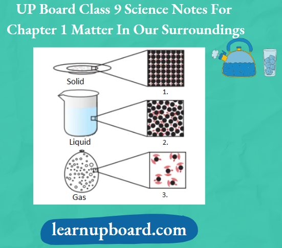
Matter around us exists in three different states which are solid, liquid and gas. These states of matter arise due to the variation in the characteristics of the particles of matter.

The Solid State: A Solid is defined as that form of matter which possesses rigidity, incompressible and hence, has a definite shape and a definite volume.

Some Important Properties Of Solid State Are As Follows:

  1. Solids have definite shapes, distinct boundaries and fixed volumes, i.e. have negligible compressibility.
  2. Solids tend to maintain their shape when subjected to outside force.
    1. Exception A rubber band changes shape under force and regains the same shape when the force is removed. If excessive force is applied, it breaks.
    2. Sugar and salt also take the shape of the container in which they are placed but are considered solids.
    3. This is because the shape of each sugar or salt crystal remains fixed.
  3. Solids either do not diffuse or diffuse at a very slow rate.
    1. Exception Sponge is compressible but considered as a solid. This is because a sponge has minute holes, in which air is trapped.
    2. When it is pressed, the air is expelled and we can compress it.
  4. Solids may break under force, but it is difficult to change their shape, so they are rigid.
  5. Generally, solids have higher densities as compared to their liquid or gaseous forms.
    1. Sugar, sand, rocks, stones, and metals like iron, copper, aluminium, gold, silver, etc., are examples of substances which exist in the solid state.

The mass per unit volume of a substance is called its density,

∴ Density =\(\frac{\text { Mass }}{\text { Volume }}=\frac{m}{V}\)

The Liquid State: Liquid is defined as that form of matter, which possesses a fixed volume, but has no fixed shape.

Some Important Properties Of the Liquid State Are As Follows:

  1. Liquids do not have a definite shape, i.e. they take up the shape of the container in which they are kept.
  2. Liquids flow and change shape, so they are not rigid but can be called fluid.
    1. Note: Fluid In science, the common name of gases and liquids is fluid.
  3. Solids, liquids and gases can diffuse into liquids. The gases from the atmosphere diffuse and dissolve in water. These gases, especially oxygen and carbon dioxide, are essential for the survival of aquatic animals and plants. Aquatic animals can breathe underwater due to the presence of dissolved oxygen in water.
  4. Liquids are almost incompressible.
  5. The attraction force between the particles of liquid is greater than that of gases but less than that of solids.
  6. The rate of diffusion of liquids is higher than that of solids. This is because, in the liquid state, particles move freely and have greater space between each other as compared to particles in the solid state.
  7. The density of a liquid is generally less than that of its solid form. Some exceptions are also there, for example., solid ice is lighter than water as it floats on water, i.e. the density of the solid form of water (ice) is less as compared to that of the liquid form of water. Water, milk, juice, oil, kerosene, petrol, alcohol, benzene etc., are examples of the substances which exist in the liquid state.

The Gaseous State: Gases can be defined as that form of matter which possesses high compressibility and hence, has neither definite shape nor definite volume.

Some Important Properties Of Gaseous State Are As Follows:

  1. Gases tend to flow as liquids do. Therefore, they are also considered as fluids.
  2. Gases show the property of diffusing very fast into other gases due to the high speed of particles and the large spaces between them.
    1. Due to the high diffusion tendency of gases, the smell of hot cooked food reaches us in seconds. The particles of the aroma of food mix with the particles of air spread, reaching us and even farther away.
  3. Gases are highly compressible. The Liquefied Petroleum Gas (LPG) cylinder used in our homes for cooking or the oxygen supplied to hospitals in cylinders is compressed gas.
    1. Compressed Natural Gas (CNG) is used as a fuel these days in vehicles. Due to its higher compressibility, large volumes of gas can be compressed into a small cylinder and transported easily.
  4. In a gaseous state, the particles move about randomly at high speed. Due to this random movement, gases exert pressure on the walls of the container, in which they are kept. Air is an example of a gaseous state. It is a mixture of gases like oxygen, nitrogen, carbon dioxide, inert gases, etc. Other examples of gases are hydrogen, ammonia, nitrogen dioxide, sulphur dioxide, etc.
  5. All living creatures need to breathe for survival. So, solids, liquids and gases can diffuse into liquids.
  6. The density of gases is minimal. A gas is much lighter than the same volume as a solid or a liquid.

Up Board Class 9 Science Notes For Chapter 1 Matter In Our Surroundings The Magnified Schematic Pictures Of The Three States Of Matter

Rigidity And Fluidity

Rigid means inflexible. A solid is a rigid form of matter, hence it does not require a container to keep it. Fluid is a material which can flow easily and requires a vessel to keep it. A liquid is a fluid form of matter which takes the shape of a container, while a gas is a fluid form of matter which fills the container.

Change Of States Of Matter

In your daily life, you come across various substances which exist in three states, i.e. solid, liquid and gas, for example. water, wax, ghee, etc. Water is the most commonly observed example that exists as ice (solid), water (liquid) as well as water vapour (gas).

Interconversion Of States Of Matter: The states of matter are interconvertible. The phenomenon of change of matter from one state to another and back to the original state by altering the conditions of temperature and pressure is called the interconversion of states of matter.

The following two factors (or any one of these) make it possible to convert one state of matter into another:

  1. Change in temperature
  2. Change in pressure

Terms Involved In Change Of State

The following terms are involved in a change of state:

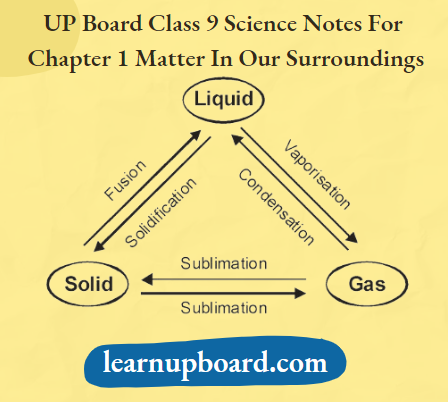
  1. Fusion or Melting and Melting Point
    1. The process of conversion of a matter from its solid state to its liquid state at specific conditions of temperature and pressure is called fusion/melting.
    2. The definite temperature at which a solid starts melting is called the melting point of that solid, for example., the melting point of ice is 0°C or 273.16 K. The Higher the melting point of a substance, the greater the force of attraction between its particles.
  2. Boiling and Boiling Point
    1. The process of conversion of a matter from its liquid state to vapours (gaseous state) at specific conditions of temperature and pressure is called boiling.
    2. It is a bulk phenomenon. The temperature at which a liquid starts boiling at the atmospheric pressure is known as its boiling point.
  3. Sublimation
    1. The process of changing of solid state directly into a gaseous state without passing through the liquid state upon heating and vice-versa on cooling is known as sublimation. for example., naphthalene, camphor, iodine, ammonium chloride, etc., are the solids that undergo sublimation.
  4. Vapourisation
    1. The process of conversion of a matter from its liquid state to a gaseous state at specific conditions of temperature and pressure is called vapourisation.
  5. Freezing and Freezing Point
    1. The process of conversion of matter from its liquid state to solid state at specific conditions of temperature and pressure is called freezing.
    2. It is a reverse process of fusion/melting. The definite temperature at which a liquid changes into a solid state by giving out heat energy at 1 atm is called the freezing point.
  6. Condensation
    1. The process of conversion of matter from its gaseous state to liquid state at specific conditions of temperature and pressure is called condensation. It is a reverse process of vapourisation.

Effect Of Change Of Temperature

When a solid is heated, the kinetic energy of its particles increases. Due to an increase in kinetic energy, the particles start vibrating with greater speed.

  • The energy supplied by the heat overcomes the forces of attraction between the particles.
  • The particles leave their positions and start moving more freely. At a certain stage (i.e. at a melting point), a solid melts and is converted into a liquid state.
  • At a certain temperature, a point is reached when the particles have enough energy to break free from the forces of attraction of each other. At this temperature (i.e. boiling point), the liquid starts changing into gas.
  • In contrast, by decreasing the temperature (by cooling), a gas can be converted into a liquid state and a liquid can be converted into a solid state.

The effect of change in temperature on the physical state may be summarised as:

Up Board Class 9 Science Notes For Chapter 1 Matter In Our Surroundings Effect Of Change Of Temperature On The Physical State

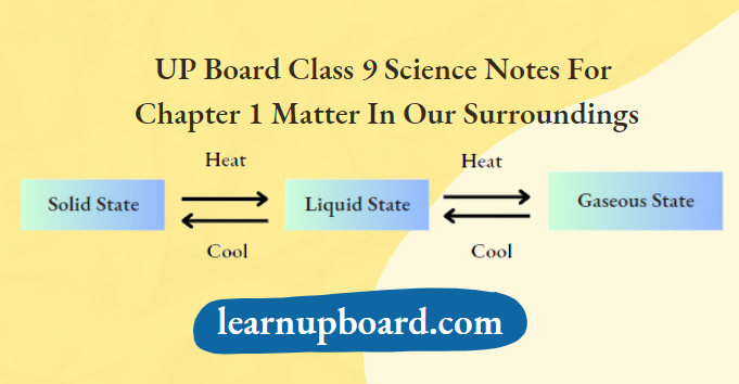
So, it can be concluded that the state of matter can be changed into another by changing the temperature.

Up Board Class 9 Science Notes For Chapter 1 Matter In Our Surroundings Interconversion Of The Three States Of Matter

Difference Between Gas And Vapour

A substance is said to be a gas if its boiling point is below room temperature, for example., O2, N2, CO2, etc. If the normal physical state of a substance is either a solid or a liquid, but gets converted into the gaseous state either on its own or by absorbing energy, the gaseous state is called the vapour state, for example., vapours of water in air.

Scales of Measuring Temperature

Three scales of measuring temperature are as follows:

1. Temperature on Kelvin scale

= Temperature on Celsius scale +273.16;

T(K)=t(°Q +273.16

Kelvin is the SI unit of temperature, 0 °C =273.16 K

For convenience, we take 0°C =273 K

2. Temperature on Celsius scale

= Temperature on Kelvin scale -273.16;

t(°C=T(K)-273.16

3. Temperature on Fahrenheit scale: Celsius and Fahrenheit temperatures are related to each other by the relation,

∴ \({ }^{\circ} \mathrm{F}=\frac{9}{5}\left({ }^{\circ} \mathrm{C}\right)+32\)

Question 1. Convert the temperature of 200°C to the Kelvin scale.
Answer:

We know that, the temperature on the Kelvin scale

Temperature on Celsius scale +273.16 = 200 + 273.16 = 473.16 K

Thus, a temperature of 200°C on the Celsius scale is equal to 473.16 K on the Kelvin scale.

Question 2. Convert the temperature of 450 K to the Celsius scale.
Answer:

We know that, the temperature on the Celsius scale

= Temperature on Kelvin scale – 273.16

= 450-273.16 =176.84°C

Thus, a temperature of 450 K on the Kelvin scale is equal to 176.84°C on the Celsius scale.

Latent Heat

When heat is given to a substance, its temperature increases. However, when heat is supplied to change the physical state of a substance, there is no increase in the temperature of a substance.

  • Thus, the heat energy which has to be supplied to change the state of a substance is called its latent heat. In actuality, the word latent’ means ‘hidden’.
  • Latent heat does not raise (or increase) the temperature. However latent heat is always supplied to change the state of a substance.

Latent Heat Is Of The Following Two Types:

Latent Heat Of Fusion (Solid To Liquid Change)

The amount of heat energy that is required to change 1 kg of a solid into liquid at atmospheric pressure and at its melting point is known as the latent heat of fusion. Particles in water at 0°C (273.16 K) have more energy as compared to particles in ice at the same temperature.

Latent Heat Of Vapourisation (Liquid To Gas Change)

The amount of heat energy that is required to convert 1 kg of a liquid into gas (at its boiling point) without any temperature rise is known as the latent heat of vapourisation. Particles in steam, i.e. water vapour at 373 K (100°C) have more energy than water at the same temperature.

Note: It has been found that burns caused by steam are much more severe than those caused by boiling water though both of them are at the same temperature of 100°C.

As particles in steam have absorbed extra energy in the form of latent heat of vapourisation. Thus, when steam falls on our skin and condenses to produce water, it gives more heat than boiling water.

Effect Of Change Of Pressure

The physical state of a substance can also be changed by changing the pressure. An increase in pressure brings the particles closer and increases the force of attraction between them, which brings about the change, for example., when high pressure is applied to a gas and its temperature is reduced, the gas is converted to a liquid, i.e. the gas is liquefied.

Hence, we can say that pressure and temperature determine the state of a substance, whether it will be solid, liquid or gas.

The pressure exerted by a gas is measured in the atmosphere (atm) unit. The pressure of air in the atmosphere is called atmospheric pressure.

Atmospheric pressure at sea level is taken as 1 atm which is also normal atmospheric pressure. As we go higher, atmospheric pressure decreases.

1 atm = 1.01 x 10⁵ Pa (Pa = Pascal, SI unit of pressure)

Up Board Class 9 Science Notes For Chapter 1 Matter In Our Surroundings By Applying Pressure Particles Of Matter Can Be Brought Close Together

Evaporation

The process of conversion of a liquid into its vapour state at any temperature below its boiling point is called evaporation. The particles of a liquid have different amounts of kinetic energy.

  • The particles present at the surface possess comparatively higher kinetic energy as compared to those present in the bulk.
  • Therefore, particles at the surface with higher kinetic energy can break away from the forces of attraction of other particles and get converted into vapour.
  • Water, when left uncovered, slowly changes into vapour. Wet clothes dry up, etc., are happen due to evaporation.

Factors Affecting Evaporation

The rate of evaporation of a liquid depends upon the following factors:

  1. Surface area Evaporation is a surface phenomenon, if the surface area is increased, the rate of evaporation increases, for example., while putting clothes for drying up, we spread them out.
  2. Temperature The rate of evaporation of a liquid increases with a temperature rise. With the increase of temperature, more particles get enough kinetic energy to go into a vapour state. That is why, evaporation is faster in a hot summer day than in winter or on a cloudy day.
  3. Humidity It is the amount of water vapour present in air. The air around us cannot hold more than a definite amount of water vapour at a given temperature. If the amount of water in air is already high, the rate of evaporation decreases. That is why, clothes dry up faster on a dry day than on a wet (rainy) day.
  4. Wind speed It is known that clothes dry faster on a windy day. This is because, with an increase in wind speed, the particles of water vapour move away with the wind, decreasing the amount of water vapour in the surroundings. That is why, the rate of evaporation of a liquid increases with increasing wind speed.

Note: The liquids which evaporate fast are called volatile liquids.

Evaporation Causes Cooling Effect

In an open vessel, the liquid keeps on evaporating. The particles of liquid absorb energy from the surroundings to regain the energy lost during evaporation. This absorption of energy from the surroundings makes the surroundings cold.

Some daily life examples of the cooling effect of evaporation are given below:

  1. When ice-cold water is kept in a glass tumbler for some time, water droplets are observed on its outer surface.
    1. Explanation This occurs as the water vapours present in the air come in contact with the glass tumbler, get cooled and condense to form these small water droplets.
    2. The formation of drops of water on the outside surface of a tumbler containing crushed ice shows the presence of water vapour in the air.
  2. Cotton clothes are used to wear during the summer season.
    1. Explanation Cotton is a good absorber of water, so it helps to absorb sweat from our bodies.
    2. As it is obvious, the person perspires more during summer due to the auto temperature control mechanism.
    3. Hence, wearing cotton clothes helps in the easy evaporation of sweat.
    4. When this sweat evaporates, it takes the latent heat of vapourisation from our body, which in turn, cools the body. Thus, a person feels comfortable.
  3. People sprinkle water on the roof or open ground on a hot sunny day.
    1. Explanation When water is sprinkled on a hot surface, it gets evaporates very quickly.
    2. As evaporated water leaves the surface cool due to the large latent heat of vapourisation of water, this technique is quite effective in summers for cooling the surface.
  4. Liquids like acetone (nail polish remover) or alcohol placed on your palm give you a feeling of cooling.
    1. Explanation Acetone and alcohol are volatile liquids. When kept on the palm, their particles gain energy from the palm or surroundings and evaporate causing the palm to feel cool.

Activity 1

Objective

To prove that matter is made up of tiny particles (and have intermodular space).

Materials Required

Beaker, water, salt or sugar, glass rod and marker.

Procedure

  1. Take a 100 mL beaker half filled with water and mark the initial water level with the help of a marker.
  2. Then, add a teaspoonful of sugar (or salt) into it and stir with the help of the glass rod.
  3. Mark the water level after the disappearance of the solute.

Up Board Class 9 Science Notes For Chapter 1 Matter In Our Surroundings When We Dissolve Salt In Water

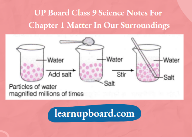
Question 1. What happens to the sugar when it is dissolved in water?
Answer: When sugar is dissolved in water, its crystals separate into very fine particles.

Question 2. Where does the sugar go?
Answer:

The sugar particles go into the spaces present between the particles of water and mix with them to form a sugar solution.

Activity 2

Objective

To prove that particles of matter:

  1. Are very small in size (particulate in nature).
  2. Move (diffuse) faster in a gaseous state as compared to solid or liquid states.
  3. Diffuse at a slower rate, if the density is higher.
  4. Diffuse faster at a higher temperature

Materials Required

Potassium permanganate, water, beakers, perfume, blue ink, honey and copper sulphate.

Observation

The water level does not change.

Explanation

  • Matter is not continuous and is particulate, i.e. it is made up of particles. When salt is dissolved in water the water level does not change.
  • It indicates that there are some vacant spaces among the particles of water. These are known as interparticle spaces. The particles of salt have occupied some of them.

Conclusion

Matter is made up of tiny particles and intermolecular spaces are present in between them. Check Yourself

Observation Table

Up Board Class 9 Science Notes For Chapter 1 Matter In Our Surroundings Difference Between Experiment Observation And Inference

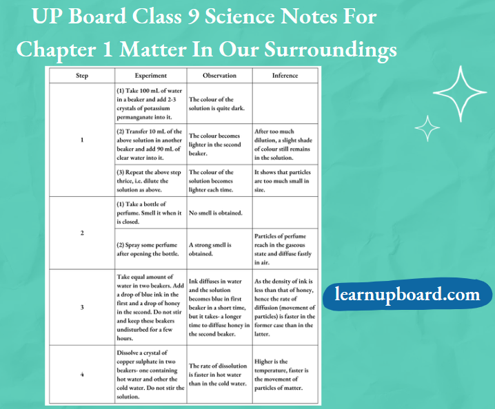
Question 1. What conclusion can you draw after adding 2-3 crystals of KMnO4 in water?
Answer:

After the addition of 2-3 crystals of KMn04 in water, it is concluded that a crystal of KMnO4 is made up of millions of tiny particles. They keep dividing themselves into smaller particles.

Question 2. When someone opens a bottle of perfume in one corner of a room, its smell spreads in the whole room quickly. Why?
Answer:

This happens because the particles of perfume (gas) move rapidly in all directions and mix with the moving particles of air in the room. They do so by getting into the spaces between the air particles.

Question 3. Why honey in step 3 dissolves at a slower rate?
Answer:

In step 3, honey dissolves at a slower rate because it is more viscous i.e. has more density and has strong intermolecular forces of attraction.

Question 4. From step 4, write the effect of temperature on diffusion.
Answer: Diffusion becomes faster at a higher temperature.

Question 5. The rate of diffusion is faster in gases, why?
Answer:

The molecules of gases have large intermolecular space between them and have higher speeds. So they diffuse faster.

Activity 3

Objective

To study the properties of solids and liquids.

Materials Required

Pencils, books, needles, thread, paper, hammers, some liquids (for example., water, oil, milk etc), containers of different shapes and the same volume.

Procedure

  1. Take a book, a needle and a piece of thread. Draw their sketch on paper with the help of a pencil. Observe their shapes and judge their volume. Now pull, drop and hammer these things one by one and record your observations.
  2. Take some liquids (50 mL each) and pour them into containers of different shapes. Put a 50 mL mark on these containers using a measuring cylinder from the laboratory.

Up Board Class 9 Science Notes For Chapter 1 Matter In Our Surroundings Water In Different Shapes Of The Container

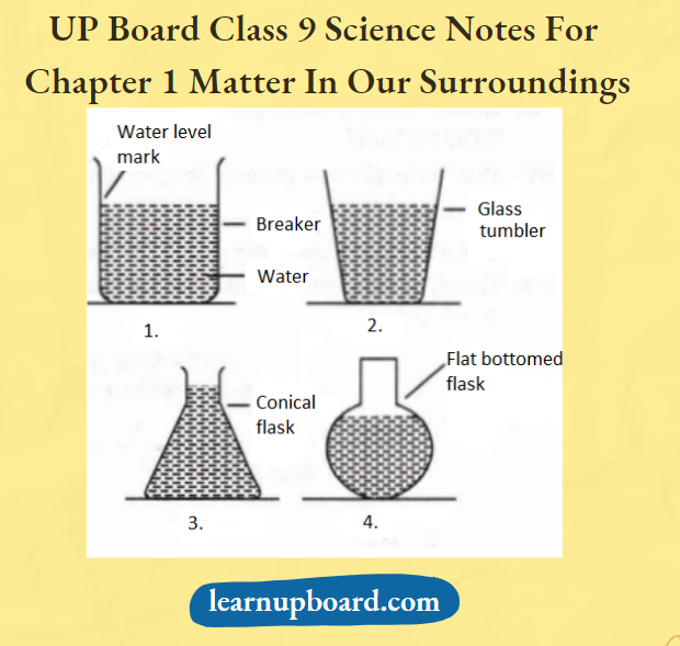
Observe the shapes of each liquid in each container. When 50 mL of liquid is poured into another container, does its volume change?

Observation

  1. All these things (books, needles and thread) have sharp boundaries, i.e. definite shapes and definite volumes. When pulled, dropped or hammered, these things remain unaffected or do not break.
  2. Volume of the liquids remains the same but its shape depends upon the shape of the container.

Conclusion

  • Solids have a definite shape and definite volume. Solids are hard and rigid and held together with greater force.
  • Liquids have fixed volumes but different shapes, as they acquire the shape of the container in which they are kept Liquids tend to flow, i.e. these are fluids.

Question 1. In which states of matter, the arrangement of particles is in the most ordered form?
Answer: In a solid state, the particles are arranged in the most ordered form.

Question 2. State one similarity between solids and liquids.
Answer: Both solids and liquids have a definite volume.

Question 3. What is the nature of shape and volume of liquids?
Answer: Liquids have indefinite shape and definite volume.

Activity 4

Objective

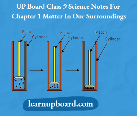
To show that gases can be compressed more easily than liquids and solids.

Materials Required

Three syringes, rubber corks, chalk and Vaseline.

Procedure

1. Take three 100 mL syringes and close their nozzles by rubber corks, as shown in the figure below.

Up Board Class 9 Science Notes For Chapter 1 Matter In Our Surroundings Compressing Gas Liquid And Solid On Applying Pressure

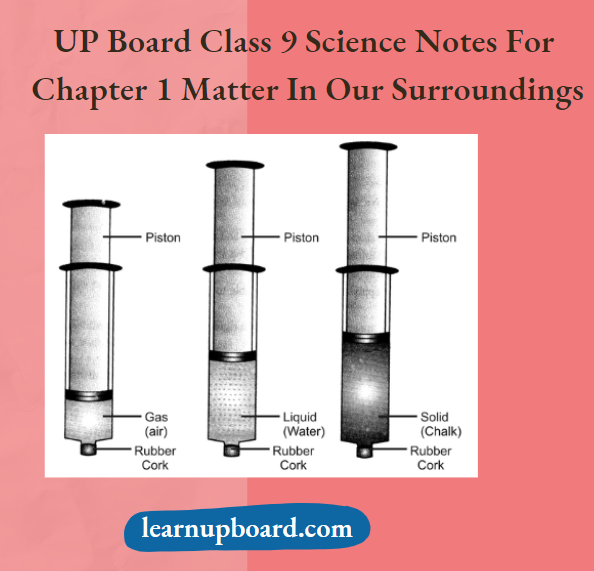
Compressing gas, liquid and solid on applying pressure

2. Remove the pistons from all the syringes.

3. Leaving the first syringe untouched, fill water in the second and pieces of chalk in the third.

4. Insert the pistons back into the syringes. You may apply some Vaseline on the pistons before inserting them into the syringes for their smooth movement.

5. Now, try to compress the content by pushing the piston in each syringe.

Observation

The piston of the first syringe can be moved very easily but less easily in the second and with the most difficulty in the third.

Conclusion

Gases are highly compressible as compared to solids and liquids.

Question 1. Why gases can be compressed more easily than liquids?
Answer:

Cases can be compressed more easily than liquids because of weak intermolecular forces of attraction between particles.

∴ \(\left(\text { Because, Compressibility } \propto \frac{1}{\text { Intermolecular forces of attraction }}\right)\)

Question 2. In this activity, what do you infer from your observations?
Answer: Gases are highly compressible as compared to solids and liquids.

Question 3. Why solids cannot be compressed?
Answer:

Solids cannot be compressed because constituent particles are very closely packed and the movement of constituent particles is restricted.

Question 4. Which form of water is highly compressible?
Answer: The gaseous form of water is highly compressible.

Activity 5

Objective

To show the effect of temperature on the physical state of matter.

Materials Required

Ice, beaker, laboratory thermometer, glass stirrer, burner and iron stand.

Procedure

  1. Take about 150 g of ice in a beaker and suspend a laboratory thermometer in it such that the bulb of the thermometer is in contact with the ice.
  2. Start heating the beaker on a low flame.
  3. Note the temperature when the ice starts melting.
  4. Note the temperature when all the ice has converted into water.
  5. The observations are recorded for the conversion of ice into water.
  6. Place a glass stirrer in the beaker and heat while stirring till the water starts boiling.
  7. Note the temperature when most of the water vaporised.
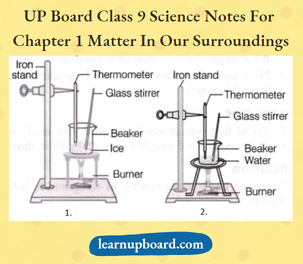
Up Board Class 9 Science Notes For Chapter 1 Matter In Our Surroundings When Most Of The Water Vaporised

Observation

  1. There is no change in temperature till all the ice melts though heating continues.
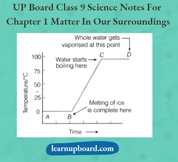
  2. Temperature remains constant at 0°C. Once the ice is converted to water, the temperature starts rising till the water begins to boil. Once the water starts boiling, the temperature remains constant at 100°C till all the water has changed to vapours.

Conclusion

  • During the change of state from solid to liquid or from liquid to gas, the temperature remains constant till all the solid has melted or all the liquid has vaporised.
  • The heat energy supplied is used up in overcoming the forces of attraction and hence, the thermometer does not show any temperature rise.

Question 1. What is the change in temperature during this activity?
Answer: The temperature remains constant during the complete melting and boiling process.

Question 2. Is melting an exothermic or endothermic process?
Answer: Melting is an endothermic process because heat is absorbed during this process.

Question 3. Under which conditions, we can boil water at room temperature?
Answer: We can boil water at room temperature under low pressure.

Question 4. Draw a temperature-time graph for the heating of ice.
Answer: The graph of temperature-time for the heating of ice is given below:

Up Board Class 9 Science Notes For Chapter 1 Matter In Our Surroundings Time Graph For The Heating Of Ice

Question 5. In an experiment for studying the effect of heating on ice, why do we use crushed ice?
Answer: Crushed ice will cover the thermometer bulb intimately and thus, would give the correct temperature.

Question 6. What do we call the temperature at which liquid starts boiling?
Answer: The temperature at which liquid starts boiling is called its boiling point.

Question 7. What would be the temperature when water starts boiling?
Answer: As the water starts boiling, the temperature will be 100°C or 373K.

Question 8. What is the temperature when all the water has boiled?
Answer: The temperature remains constant, i.e. 100°C during the complete boiling process.

Activity 6

Objective

To study the process of sublimation.

Materials Required

Camphor or ammonium chloride, China dish, funnel, cotton plug and burner.

Procedure

  1. Take some camphor or ammonium chloride. Crush it and put it in a China dish.
  2. Put an inverted funnel over the China dish.
  3. Put a cotton plug on the stem of the funnel and set the apparatus
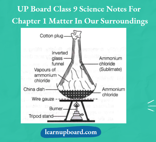
Up Board Class 9 Science Notes For Chapter 1 Matter In Our Surroundings Sublimation Of Ammonium Chloride.

Observation

Solid ammonium chloride changes into vapours without changing into a liquid state and gets condensed on the walls of the funnel.

Conclusion

A change of state directly from solid to gas without changing into liquid state or vice-versa is called sublimation.

Question 1. In an experimental set-up for sublimation, a perforated asbestos sheet is placed between the China dish and funnel. What is its purpose?
Answer: The asbestos sheet prevents direct heating of the funnel.

Question 2. What happens when solid ammonium chloride is heated?
Answer:

On heating, solid ammonium chloride changes into vapours without changing into a liquid state and gets condensed on the walls of the funnel,

Question 3. Name the solid substance which is obtained by cooling the vapour.
Answer: The solid substance obtained by cooling the vapour is known as sublimate.

Question 4. Which substance is sublimated in this activity?
Answer: Sublimate is pure ammonium chloride.

Activity 7

Objective

To study the factors which affect evaporation.

Materials Required

Test tubes, water, jar, China dish, thermometer and cupboard.

Procedure

  1. Take 5 mL of water in a test tube and keep it near a window or under a fan.
  2. Take 5 mL of water in an open China dish and keep it near a window or under a fan.
  3. Take 5 mL, of water in an open China dish and keep it inside a cupboard or on a shelf in your class.
  4. Record the room temperature.
  5. Record the time or days taken for the evaporation process in the above cases.
  6. Repeat the above three steps of activity on a rainy day and record your observations.

Observation

  1. In an open China dish kept near a window or on a shelf, water evaporates very fast as compared to a test tube.
  2. On sunny days, water evaporates very fast as compared to rainy days.

Conclusion

  • Surface area is increased the rate of evaporation increases.
  • If the temperature is increased, the rate of evaporation increases because with the increase of temperature, more particles get enough kinetic energy to go into the vapour state.
  • If wind speed is increased, the rate of evaporation increases because with the increase in wind speed, the particles of water vapour move away with the wind, decreasing the amount of water vapour in the surroundings.
  • If humidity is decreased, the rate of evaporation increases and vice-versa

Question 1. Why does water evaporate faster in a Chinese dish as compared to a test tube?
Answer:

China dish has more surface area as compared to a test tube. So, evaporation is faster in the case of China dishes.

Question 2. Why evaporation is fast when water is kept under a fan?
Answer:

When water is kept under a fan, the rate of evaporation increases because with the increase in wind speed, the particles of water vapour move away with the wind.

Question 3. What is the effect of humidity on evaporation?
Answer: If humidity is decreased, the rate of evaporation increases and vice-versa.

Question 4. What do you infer about the effect of temperature on evaporation?
Answer: If the temperature is increased, the rate of evaporation increases.

Question 5. Why evaporation is slow on a rainy day?
Answer: On a rainy day, humidity is increased, so the rate of evaporation decreases.

Matter In Our Surroundings Summary

Matter is anything that has mass and occupies volume. The SI unit of mass and volume is kilogram (kg) and cubic metre (m3), respectively.

  • Matter is classified based on their physical and chemical properties, i.e. physical properties (solid, liquid and gas) and chemical properties (elements, compounds and mixtures).
  • Every matter is made up of certain particles which differ in shape, size and nature.
  • The particles of matter tend to diffuse.
  • Solids have definite shapes, distinct boundaries and fixed volumes.
  • Liquids do not have a definite shape.
  • Gases have neither definite shape nor volume.
  • The state of matter can be interchanged by changing temperature or pressure.
  • At specific conditions of temperature and pressure; the conversion of a matter from its solid to its liquid state is called fusion.
  • The conversion of a matter from its liquid state to vapour (gaseous state) is called boiling.
  • It is a bulk phenomenon.
  • The conversion of a matter from its liquid to solid state is called freezing.
  • The conversion of a matter from its liquid to gaseous state is called vapourisation.
  • The conversion of matter from its gaseous to liquid state is called condensation.
  • The process of changing of solid state directly into a gaseous state without passing through the liquid state upon heating and vice-versa on cooling is called sublimation. The heat energy which has to be supplied to change the state of a substance is called latent heat.
  • The latent heat of vapourisation is the heat energy required to change 1kg of a liquid to gas at atmospheric pressure at its boiling point.
  • Latent heat of fusion is the amount of heat energy required to change 1 kg of solid into liquid at its melting point.
  • The process of conversion of a liquid into its vapour state at any temperature below its boiling point is called evaporation.
  • Apart from the solid, liquid and gaseous states, scientists have discovered two more states, i.e. plasma and Bose-Einstein condensate.

UP Board Class 9 Science Notes For Chapter 2 Is Matter Around Us Pure

Is Matter Around Us Pure

In chemistry, when we say a substance is pure, it means that the substance is made up of only one type of constituent particle. In other words, a substance is a pure single form of matter.

Depending upon the chemical composition, matter is classified into elements, compounds, (i.e. pure substances that are non-separable by physical methods) and mixtures (separable by physical methods like sublimation, etc).

Up Board Class 9 Science Notes For Chapter 2 Is Matter Around Us Pure Depending Upon The Chemical Composition Matter Is Classified Into Elements

Pure Substance

A substance that consists of only a single type of constituent particles is called pure substance, for example., gold, water, etc. Based on the nature of the constituent particles, a pure substance is of two types, i.e. elements and compounds.

1. Elements

The term element was first used by Robert Boyle in 1661. According to Antoine Laurent Lavoisier, a French chemist, (1743-94), ‘an element is a basic form of matter that cannot be broken down into simpler substances by chemical reactions’.

Read and Learn  More Class 9 Science Notes

An element is a pure substance. Till now 118 elements have been discovered, out of these 92 are natural elements and others are man-made. Based on variation in properties, elements can be broadly classified as metals, non-metals and metalloids.

  1. Metals
    1. A metal is an element that is malleable (i.e. can be hammered into thin sheets), ductile (i.e. can be drawn into wires), sonorous (i.e. make a ringing sound when hit), and conduct heat and electricity.
    2. They are lustrous (shine) and have silvery-grey or golden- yellow colour, for example., gold, silver, copper, iron, sodium, potassium, etc.
    3. Mercury is the only metal that is liquid at room temperature. Gallium and caesium because of their very low melting points remain in liquid state at a temperature slighdy above room temperature (303K).
  2. Non-metals
    1. A non-metal is an element that is neither malleable nor ductile and does not conduct heat and electricity.
    2. They display a variety of colours, for example., hydrogen, oxygen, iodine, carbon (coal, coke), bromine, chlorine, etc.
    3. Bromine is the only non-metallic element that exists in a liquid state at normal conditions of temperature and pressure.
  3. Metalloids
    1. Elements having intermediate properties between those of metals and non-metals are called metalloids, for example., boron, silicon, germanium, etc.

2. Compounds

A compound is a substance composed of two or more elements, chemically combined in a fixed proportion, for example., water (H2O), methane (CH4), carbon dioxide (CO2), ammonia (NH3), sodium chloride (NaCl), etc.

Mixtures

Mixtures are constituted by more than one kind of pure form, known as a substance. Most of the matter around us exist as mixtures of two or more pure components, for example., sea water, minerals, soil, etc, are all mixtures.

Types of Mixtures: Depending upon the nature of the components that form a mixture, we have two types of mixtures:

  1. Homogeneous Mixture
    1. A mixture in which the constituents are uniformly distributed throughout i.e. without any clear boundary of separation is called homogeneous mixture.
    2. Here, the constituents cannot be seen with naked eyes or under a microscope.
    3. Some of the examples of homogeneous mixtures are salt solution, sugar solution, air, soft drinks, petroleum, biogas, alloys, etc.
    4. Note Air is a homogeneous mixture of gas. Its two major constituents are oxygen (21 %) and nitrogen (78%) and other gases in small quantities.
  2. Heterogeneous Mixture
    1. A mixture that does not have uniform composition, i.e. has visible boundaries of separation between its constituents is called heterogeneous mixture.
    2. Here, the constituents of a heterogeneous mixture can be seen by naked eyes or under a microscope.
    3. Examples of heterogeneous mixtures are sugar and sand mixture, salt and sand mixture, polluted air, muddy water, etc.

Differences Between Compounds And Mixtures:

Up Board Class 9 Science Notes For Chapter 2 Is Matter Around Us Pure Differences Between Compounds And Mixtures

Solution

A homogeneous mixture of two or more substances is called a solution. A solution is sometimes also called a true solution.

  • Lemonade, soda water, salt solution, sugar solution, etc., all are examples of solutions.
  • In a solution, there is homogeneity at the particle level, i.e. the particles of dissolved substances are evenly distributed in the solution and are indistinguishable from one another.

There are two main components of a solution:

  1. Solvent (Dissolving Phase) The component (usually present in larger amounts) of the solution that dissolves the other component in it, is called the solvent.
  2. Solute (Dissolved Phase) The component (usually present in lesser quantity) of the solution that is dissolved in the solvent is called the solute.

Some Common Examples Of Solution:

  1. In sugar solution, sugar is the solute and water is the solvent.
  2. A solution of iodine in alcohol known as tincture of iodine, has iodine (solid) as the solute and alcohol (liquid) as the solvent.
  3. Aerated drinks like soda water, etc., are gas in liquid solutions. CO2 (gas) as solute and water (liquid) as solvent. Solid solutions (alloys) and gaseous solutions (air)

Alloys

  • Alloys are mixtures of two or more metals or a metal and a non-metal and cannot be separated into their components by physical methods.
  • But still, an alloy is considered as a mixture because it shows the properties of its constituents and can have variable composition, for example., brass is a mixture of approximately 30% zinc and 70% copper.

Properties Of A Solution

Some important properties of a solution are as follows:

  1. A solution is a homogeneous mixture.
  2. The particles of a solution are smaller than 1 nm (10-9 m) in diameter. Therefore, they cannot be seen by the naked eye.
  3. Due to very small particles, they do not scatter a beam of light passing through the solution. So, the path of light is not visible in a solution.
  4. A solution is stable, i.e. the solute particles do not settle down when left undisturbed. The solute particles cannot be separated from the mixture by the process of filtration.

Concentration Of A Solution

  • The concentration of a solution is the amount of solute present in a given amount (mass or volume) of solution, or the amount of solute dissolved in a given mass or volume of the solvent. In a solution, the relative proportion of the solute and solvent can be varied.
  • Depending upon the amount of solute present in a given amount of solvent, it can be classified as under:

Saturated solution A solution in which no more amount of solvent can be dissolved at a given temperature, is called a saturated solution. The amount of the solute present in the saturated solution at this temperature is called solubility.

Solubility =\(\frac{\text { Mass of solute }}{\text { Mass of solvent }} \times 100\)

Unsaturated solution If the amount of solute contained in a solution is less than the saturation level, it is called an unsaturated solution.

Concentration of solution

⇒ \(=\frac{\text { Amount of solute }}{\text { Amount of solution }}=\frac{\text { Amount of solute }}{\text { Amount of solvent }}\)

Expressing The Concentration Of A Solution: The methods by which the concentration of a solution can be expressed are:

Mass by mass percentage of a solution

⇒ \(=\frac{\text { Mass of solute }}{\text { Mass of solution }} \times 100\)

Mass by volume percentage of a solution

∴ \(=\frac{\text { Mass of solute }}{\text { Volume of solution }} \times 100\)

Question1. A solution contains 50 g of common salt in 450 g of water. Calculate the concentration of the solution.
Answer:

Concentration of solution = \(\frac{\text { Mass of solute }}{\text { Mass of solution }} \times 100\)

Mass of common salt (solute) = 50 g

Mass of water = 450 g

Mass of solution = 50 + 450 = 500 g

Concentration of solution = \(\frac{50}{500} \times 100=10 \%\)

Example 2. 4 g of a solute is dissolved in 40 g of water to form a saturated solution at 25°C. Calculate the solubility of the solute at 25°C.
Answer:

Solubility = \(\frac{\text { Mass of solute }}{\text { Mass of solvent }} \times 100\)

Mass of solute = 4g,

Mass of solvent = 40 g,

Solubility = \(\frac{4(\mathrm{~g})}{40(\mathrm{~g})} \times 100\)

= 10g

Suspension

A suspension is a heterogeneous mixture in which the solute particles do not dissolve, but remain suspended throughout the bulk of the medium, for example., a mixture of chalk powder in water, a mixture of sand in water, smoke coming out of a chimney of a factory.

Properties of Suspension

Some important properties of suspension are as follows:

  1. Suspension is a heterogeneous mixture.
  2. Its particles can be seen by the naked eye.
  3. Its particles scatter a beam of light passing through it and make its path visible (Tyndall effect).
  4. It is unstable, i.e. the solute particles settle down when suspension is left undisturbed. They can be separated by the process of filtration. When the particles settle down, the suspension breaks and it does not scatter light any more.

Note: The insoluble particles in a suspension are called ‘suspended particles’, whereas the solvent is referred to as ‘medium’.

Colloidal Solution

A colloid (or colloidal solution) is a mixture that is heterogeneous but appears to be homogeneous as the particles are uniformly spread throughout the solution, for example., milk, shaving cream, cheese, etc. Colloidal solutions are also called colloidal sols.

Properties Of A Colloid: Some important properties of a colloid are as follows :

  1. A colloid is a heterogeneous mixture.
  2. The size of particles of a colloid is too small to be individually seen by the naked eye.
  3. Colloids are big enough to scatter a beam of light passing through it and make its path visible.
  4. The colloids are quite stable. Particles do not settle down when a colloid is left undisturbed.
  5. Particles of colloid can pass through filter paper, therefore a colloid cannot be separated by filtration. However, they get separated by a special technique called centrifugation.

Common Examples Of Colloids

Colloids are classified according to the state (solid, liquid or gas) of the dispersion medium and the dispersed phase.

Types Of Colloids

Up Board Class 9 Science Notes For Chapter 2 Is Matter Around Us Pure Types Of Colloids

Tyndall Effect

The scattering of light by colloidal particles is known as the Tyndall effect. In a true solution, the solute particles are so small that they cannot scatter light falling on them.

  • In a colloidal solution, the particles are big enough to scatter light. Tyndall effect can also be observed in the following situations:
  • When a fine beam of light enters a room through a small hole (due to scattering of the beam of light by the particles of dust and smoke in the air).
  • When sunlight passes through the canopy of a dense forest (as the mist containing tiny droplets of water scatters it). The components of a colloidal solution are the dispersed phase and the dispersion medium.
  • The solute-like component or the dispersed particles in a colloid form is the dispersed phase and the component in which the dispersed phase is suspended, is known as the dispersion medium.

Up Board Class 9 Science Notes For Chapter 2 Is Matter Around Us Pure Demonstration Of Tyndall Effect

Physical And Chemical Changes

Physical Changes

The properties that can be observed and specified like colour, hardness, rigidity, fluidity, density, melting point, boiling point, etc., are physical properties.

  • The changes which occur without a change in composition and the chemical nature of the substance are called physical changes.
  • The interconversion of states is a physical change, for example., change of water in ice is a physical change because chemically, ice and liquid water both are the same.
  • Although ice, water and water vapour all look different and display different physical properties chemically they are the same.

Chemical Changes

In chemical changes, one substance reacts with another substance to change chemical composition. Chemical changes bring a change in the chemical properties of matter and a new substance is obtained.

  • A chemical change is also called a chemical reaction, for example., both water and cooking oil are liquid, but their chemical characteristics are different. They differ in odour and inflammability.
  • Oil burns in air whereas water extinguishes fire, i.e. it is the chemical property of oil that makes it different from water.

Note:

  • Burning is a chemical change.
  • During the burning of a candle, both physical and chemical changes take place.

Activity 1

Objective

To prepare homogeneous and heterogeneous mixtures.

Materials Required

Four beakers, copper sulphate powder, common salt, sulphur powder, iron filings, water, spatula, etc.

Procedure

  1. Label the beakers as A, B, C and D respectively.
  2. In beakers A and B, take 50 mL of water.
  3. Now, add one spatula of copper sulphate powder in beaker A and two spatula of copper sulphate powder in beaker B.
  4. Stir the solutions and observe the changes.
  5. Now, in beakers C and D, take equal quantity of common salt (sodium chloride).
  6. In beaker C, add iron filings and in beaker D, add sulphur powder.
  7. Mix them with the help of a glass rod and observe the changes.

Observation

In beakers A and B, the composition of the mixture is the same throughout but the intensity of the colour of the solutions is different. In beakers C and Z), the obtained mixture has physically distinct parts and a non-uniform composition.

Conclusion

Beakers A and B have homogeneous mixtures of variable composition. Beakers C and D have heterogeneous mixtures of variable composition.

Question 1. What are heterogeneous mixtures?
Answer: The mixtures in which composition are not uniform throughout and which have physically distinct parts are called heterogeneous mixtures.

Question 2. What name is given to a mixture having a uniform composition and no distinct parts?
Answer: Such mixtures are called homogeneous mixtures.

Question 3. On mixing iron filings with sulphur in a beaker, what type of mixture is obtained?
Answer: Heterogeneous mixture is obtained when iron filings are mixed with sulphur.

Question 4. If the substances present in the beaker as given in Q-3 are heated, what do you observe?
Answer: On heating iron with sulphur, we get black coloured iron
sulphide which is a compound, not a mixture as its properties are quite different from the properties of iron as well as sulphur.

Question 5. Give an example of homogeneous mixture.
Answer: Solution of sugar or salt in water.

Activity 2

Objective

To study the properties of solution, suspension and colloidal solution.

Materials Required

Three beakers, glass rod, copper sulphate crystals, chalk powder (or wheat flour) and few drops of milk or ink.

Procedure

  1. Label the beakers as A, B and C.
  2. In beaker A, take 20 mL of water and add copper sulphate crystals.
  3. In beaker B, take 20 mL of water and add chalk powder (or wheat flour).
  4. In beaker Q take 20 mL of water and add few drops of milk or ink.
  5. Stir the solutions with glass rod and observe the changes.
  6. Now, direct a beam of light from a torch through all the beakers containing the mixtures and observe from the front.
  7. Note your observations.
  8. Leave all the solutions undisturbed for a few minutes and observe the changes.
  9. Filter the solutions and observe whether there is a residue on the filter paper or not.

Observations

  1. In beaker A, solution of uniform composition is obtained. It does not affect the beam of light and no change is observed on keeping it undisturbed for a few minutes. Further, there is no residue on the filter paper in this case.
  2. In beaker B, solution of non-uniform composition is obtained. It scatters the beam of light, so its path become visible. On keeping undisturbed, the solute, i.e. chalk powder settle down and if we filter it, the chalk powder comes on filter paper as residue.
  3. In beaker C, solution of uniform composition is obtained, but it scatters the beam of light and thus, makes the path visible. If left undisturbed, these remain unaffected. Moreover, no residue left over the filter paper.

Conclusion

  1. Beaker A contains a true solution. True solutions are homogeneous and stable. They do not show the Tyndall effect and are not separated by filtration.
  2. Beaker B contains suspension. Suspensions are heterogeneous, opaque, unstable and exhibit the Tyndall effect. They are separated by filtration.
  3. Beaker C contains the colloidal solution. Colloids are also heterogeneous, translucent, stable and exhibit Tyndall effect. They are not separated by filtration.

Question 1. Why is the Tyndall effect not shown by true solutions?
Answer: This is because the particles of true solutions are very small in size (<1 nm) and hence, these are not able to scatter a beam of light.

Question 2. Give a technique through which colloids can be separated.
Answer: Colloids can be separated by centrifugation.

Question 3. Why do colloidal solutions exhibit the Tyndall effect?
Answer: Because the size of their particles is large enough to scatter light.

Question 4. Why filtration technique does not apply to the separation of a true solution?
Answer: Because the pore size of filter paper is much larger than the size of particles of true solutions.

Question 5. Which of the two will scatter light-soap solution or salt solution?
Answer: Soap solution will scatter light because soap solution is a colloid. A salt solution is a true solution so, it will not scatter light.

Activity 3

Objective

To demonstrate that the different substances in a given solvent have different solubilities.

Materials Required

Beakers, salt, sugar/barium chloride, water, burner, glass rod, etc.

Procedure

Up Board Class 9 Science Notes For Chapter 2 Is Matter Around Us Pure Preparing Saturated Solution Of Salt

  1. Take approximately 50 mL of water each in two separate beakers.
  2. Add common salt in the first beaker and sugar or barium chloride in the second beaker with continuous stirring.
  3. Continue the addition till it stops dissolving.
  4. When no more solute (salt/barium chloride) can be dissolved, heat the contents of the beakers.
  5. Start adding the solute again.
  6. Take a saturated solution at a certain temperature and cool it slowly.

Observations

  1. When no more salt or barium chloride can be dissolved in a solution or they stop dissolving, a saturated solution of salt or barium chloride is obtained.
  2. On heating, when more solute (i.e. salt or barium chloride) is added to the saturated solution, then this solution becomes unsaturated.
  3. When a saturated solution at a certain temperature is cooled, the solubility of a solute decreases and the amount of the solute which exceeds the solubility at the lower temperature crystallises out of the solution.

Conclusion

Different substances in a given solvent have different solubilities at the same temperature. In general, the solubility decreases as the solution is cooled and the extra amount crystallises out.

Question 1. What is a saturated solution?
Answer: A solution in which no more solute can be dissolved at any fixed temperature is called a saturated solution.

Question 2. What is an unsaturated solution?
Answer: A solution in which some more solute could be
dissolved at any fixed temperature is called an unsaturated solution.

Question 3. Write the effect of heating on a saturated solution.
Answer: If a saturated solution at a particular temperature is heated
to a higher temperature, then it becomes unsaturated.

Question 4. What happens when a saturated solution is allowed to cool?
Answer: If a saturated solution available at a particular temperature is cooled to a lower temperature, then some of its dissolved solutes will separate in the form of solid crystals.

Question 5. What is a supersaturated solution?
Answer: Any solution containing more solute than the required amount to prepare a saturated solution at any fixed temperature is called a supersaturated solution.

Question 6. How can you convert a saturated solution into an unsaturated solution?
Answer: By adding more solvent or by applying heat.

Summary

A substance that consists of only a single type of constituent particles is called a pure substance.

  • An element is a basic form of matter that cannot be broken down into simpler substances by chemical reactions.
  • A metal is an element that is malleable, ductile, and sonorous and conducts heat and electricity.
  • A non-metal is an element that is neither malleable nor ductile and does not conduct heat and electricity.
  • Metalloids are intermediate properties between those of metals and non-metals.
  • A compound can be defined as a substance composed of two or more elements, chemically combined in a fixed proportion.
  • Mixtures are constituted by more than one substance mixed in any proportion.

Depending upon the nature of the components that form a mixture, we have two types of mixtures:

  1. Homogeneous Mixture A mixture in which the constituents are uniformly distributed throughout i.e. without any clear boundary of separation is called a homogeneous mixture.
  2. Heterogeneous Mixture A mixture that does not have uniform composition, i.e. has visible boundaries of separation between its constituents is called a heterogeneous mixture.
  • A solution is a homogeneous mixture of two or more substances. There are two main components of a solution; solvent and solute.
  • The concentration of a solution is the amount of solute present in a given amount (mass or volume) of a solution, or the amount of the solute dissolved in a given mass or volume of the solvent.
  • Depending upon the amount of solute present in a given amount of solvent, it can be classified as under:
  1. Saturated Solution A solution in which no more amount of solvent can be dissolved at a given temperature, is called saturated solution.
  2. Unsaturated Solution If the amount of solute contained in a solution is less than the saturation level, it is called an unsaturated solution.

⇒ \(\text { Concentration of solution }=\frac{\text { Amount of solute }}{\text { Amount of solution }}\)

⇒ \(=\frac{\text { Amount of solute }}{\text { Amount of solvent }}\)

  • A suspension is a heterogeneous mixture in which the solute particles do not dissolve, but remain suspended throughout the bulk of the medium.
  • A colloid is a mixture that is actually heterogeneous, but appears to be homogeneous as the particles are uniformly spread throughout the solution.
  • The scattering of light by colloidal particles is known as Tyndall effect.
  • In physical changes, only physical properties of the substance changes.
  • In chemical changes, one substance reacts with another substance to undergo a change in chemical composition.

UP Board Class 9 Science Notes For Chapter 3 Atoms And Molecules

Atoms And Molecules

In the present chapter, we shall discuss about the various laws (which explains how atoms combine to form molecule), symbols and formulae of atoms and molecules and various ways of expressing their masses.

Laws Of Chemical Combination

Whenever reactants react together to form the products or the elements combine together to form a compound, they do this according to certain laws. These laws are called laws of chemical combination.

Antoine L. Lavoisier laid the foundation of chemical sciences by establishing two important laws of chemical combination which are as follows:

1. Law Of Conservation Of Mass

It states that ‘mass can neither be created nor be destroyed during a chemical reaction.5 This means that in any chemical reaction, the total mass of the reactants is equal to the total mass of the products and there is no change in mass during the chemical reaction.

Question 1. If 4.0 g of sodium carbonate reacts with 10 g of hydrochloric acid, it results in the formation of 2.5 g of carbon dioxide and 11.5 g of sodium chloride solution. Show that these results are by the law of conservation of mass.
Answer:

⇒ \(\underset{(4.0 \mathrm{~g})}{\text { Sodium carbonate }}+\underset{(10.0 \mathrm{~g})}{\text { Hydrochloric acid }} \longrightarrow \underset{(2.5 \mathrm{~g})}{\text { Carbon dioxide }}+\underset{(11.5 \mathrm{~g})}{\text { Sodium chloride }}\)

Here, total mass of reactants = 4.0 + 10 = 14 g

Total mass of products = 2.5 + 11.5 = 14 g

Since the reactants and products have the same mass, this means that there was no loss or gain of mass after the reaction. Hence, the data is in agreement with the law of conservation of mass.

Read and Learn  More Class 9 Science Notes

2. Law Of Constant Proportions/ Law Of Definite Proportions

According to this law, in a chemical substance (or compound), the elements are always present in definite proportions (or ratios) by mass.

For example., In a compound such as water, the ratio of the mass of hydrogen to the mass of oxygen is always 1:8, whatever the source of water. Thus, if 9 g of water is decomposed, 1 g of hydrogen and 8 g of oxygen are always obtained.

Similarly, carbon dioxide (CO2) always contains carbon and oxygen in the ratio of 3: 8. If a sample of CO2 contains 36 g of carbon then it is compulsory that the sample has 96 g of oxygen.

This is calculated as \(\frac{3}{8}=\frac{36}{x} ;\)

∴ \(x=\frac{36 \times 8}{3}=96 \mathrm{~g}\)

Question 2. Copper oxide was prepared by two different methods. In one case, 1.75 g of the metal gave 2.19 g of oxide. In the second case, 1.14 g of the metal gave 1.43 g of the oxide. Show that the given data illustrate the law of constant proportions.
Answer:

Case 1 Mass of copper = 1.75 g

And a mass of copper oxide = 2.19 g

So, mass of oxygen = Mass of copper oxide – Mass of copper

= 2.19-1.75 =0.44g

Now, in the first sample of the copper oxide compound.

Mass of copper: Mass of oxygen = 1.75: 0.44

⇒ \(\frac{1.75}{0.44}: 1\)

⇒ 3.93:1

Case 2 Mass of copper = 1.14 g

And, a mass of copper oxide = 1.43 g

So, mass of oxygen = Mass of copper oxide – Mass of copper

= 1.43 -1.14 =0.29 g

Now, in the second sample of the copper oxide compound.

Mass of copper: Mass of oxygen = 1.14: 0.29

⇒ \(\frac{1.14}{0.29}: 1\)

393:1 ≈ 4:1

From the above calculations, we can see that the ratio (or proportion) of copper and oxygen elements in the two samples of copper oxide compound is the same, i.e. 4:1. So, the given data verify the law of constant proportions.

Explanation Of Laws Of Chemical Combination: Dalton’s Atomic Theory

Dalton’s atomic theory explained the law of chemical combination. According to Dalton’s atomic theory, all matter (whether an element, a compound or a mixture), is composed of small particles, called atoms.

The Main Postulates Of Dalton’s Atomic Theory

  • Every matter is made up of very small particles, called atoms.
  • Atoms are indivisible particles which can neither be created nor be destroyed in a chemical reaction.
  • Atoms of a given element are identical in mass as well as in chemical properties.
  • Atoms of different elements have different masses and chemical properties.
  • Atoms combine in the ratio of small whole numbers to form compounds.
  • The relative numbers and kinds of atoms are constant in a given compound.

Atoms

Atoms are the smallest particles of an element which may or may not have independent existence but take part in a chemical reaction. These are the building blocks of all matter.

For example., atoms of hydrogen, oxygen, nitrogen etc., are not capable of independent existence whereas atoms of helium, neon etc., are capable of existing independently.

Size Of Atoms

Atoms are very small and their radius is measured in nanometres.

1/109 m = 1 nm or

lm = 109 nm

A hydrogen atom is the smallest atom and its radius is 0.1 nm.

Modern-Day Symbols Of Atoms Of Different Elements

In chemistry, symbols are the representation of an element. It is simple to use the symbol of an element rather than writing a whole word of an element. Dalton was the scientist who introduced symbols for representing elements for the first time.

UP Board Class 9 Science Notes For Chapter 3 Atoms And Molecules Some Elements Aas Proposed By Dalton

Symbols for some elements as proposed by Dalton

As Dalton’s symbols for elements were difficult to draw and inconvenient to use, modern symbols for the elements were introduced by J J Berzelius. These are defined as “a shorthand representation of the name of an element”.

  • In the beginning, the names of elements were derived from the name of the place where they were found for the first time.
  • Nowadays, it is the IUPAC (International Union of Pure and Applied Chemistry) that approves the names and symbols of the elements. Many of the symbols are the first one or two letters of the element’s name in English.
  • The first letter of a symbol is always written in capital letters and the second letter is a small letter.

For example., chlorine (Cl), zinc (Zn) and aluminium (Al). Symbols of some elements have been taken from their names in different languages such as Latin, German, Greek etc.

For example.,

  • Iron – Fe from Ferrum (Latin name)
  • Gold – Au from Aurum (Latin name)
  • Potassium – K from Kalium (Latin name)
  • Chlorine – Cl from Chloros (Greek name)
  • Cobalt – Co from Kobold (German name)
  • Sodium – Na from Natrium (Latin name)

Symbols For Some Elements

UP Board Class 9 Science Notes For Chapter 3 Atoms And Molecules Some Elements

Atomic Mass

According to Dalton, each element has a characteristic atomic mass. However, determining the mass of an individual atom was a relatively difficult task due to its very small size. Hence, their relative atomic masses were determined using the laws of chemical combinations and the compounds formed.

For this purpose, initially, 1/16 of the mass of an atom of naturally occurring oxygen was taken as a standard unit because of the following two reasons:

  1. Oxygen reacted with a large number of elements and formed compounds.
  2. This unit gave masses of most of the elements as whole numbers.

However, in 1961, carbon (C-12 isotope) was chosen as a standard reference for measuring atomic masses universally.

Relative Atomic Mass

It is defined as the number of times a given atom is heavier than 1/12th of the mass of 1 atom of carbon-12 (C-12) or it is the average mass of the atom as compared to l/12th the mass of one carbon-12 atom.

Atomic Mass Unit

It is defined as the mass unit equal to exactly 1/12th of the mass of one atom of the C-12 isotope. Earlier, it was abbreviated as amu but according to the latest recommendations of IUPAC, it is now written as ‘u’- unified mass.

Atomic Masses Of Few Elements

UP Board Class 9 Science Notes For Chapter 3 Atoms And Molecules Masses Of Few Elements

Note: Atoms of most of the elements are not able to exist independently. Atoms form molecules and ions. These molecules or ions aggregate in large numbers to form the matter that we can see, feel or touch.

Molecules

The smallest particle of an element or compound which is capable of independent existence and shows all the properties of that substance is called a molecule. In general, a molecule is a group of two or more atoms that are chemically bonded together. Atoms of the same element or different elements can join together to form molecules.

Molecules can be divided into two categories:

1. Molecules Of Elements

The molecules of an element contain the same type of atoms. Molecules of many elements are made up of only one atom of that element, for example., noble gases like argon (Ar), helium (He) etc.

The molecules of most of the non-metals are made up of more than one atom. for example., a molecule of oxygen (O2) consists of two atoms of oxygen and is known as a diatomic molecule, and ozone (O3) consists of three atoms of oxygen and is known as a triatomic molecule.

Atomicity

It is defined as the number of atoms present in a molecule. Based on atomicity, molecules can be classified as:

  1. Monoatomic molecules consist of only one atom.
    1. For example., He, Ne, Ar, Xe, Fe, Al etc.
  2. Diatomic molecules consist of two atoms.
    1. For example., H2, O2, N2, I2, Br2, Cl2, HCl, NaCl etc.
  3. Triatomic molecules consist of three atoms,
    1. For example., O3, CO2, NO2 etc.
  4. Tetra-atomic molecules consist of four atoms,
    1. For example., P4, H2O2 etc.
  5. Polyatomic molecules consist of more than four atoms,
    1. For example., CH4 (penta-atomic), S8 (octa-atomic) etc.

Atomicity of Some Elements (Non-metals):

UP Board Class 9 Science Notes For Chapter 3 Atoms And Molecules Chemical Atomicity Of Some Elements

2. Molecules Of Compounds

Atoms of different elements join together in definite proportions to form molecules of compounds.

UP Board Class 9 Science Notes For Chapter 3 Atoms And Molecules Molecules Of Some Compounds.

Prediction Of Number Of Atoms From Mass Ratio

To predict the number of atoms from the mass ratio, divide the given mass of each element by the atomic mass of the element and calculate the simplest ratio between the obtained moles, for example., we know that the mass ratio of nitrogen and hydrogen in an ammonia molecule is 14 : 3. The number of atoms of nitrogen and hydrogen present in the molecule of ammonia can be calculated as,

Edit Post “UP Board Class 9 Science Notes For Chapter 3 Atoms And Molecules” ‹ Learn UP Board — WordPress

Thus, in an ammonia molecule, one N and three H-atoms are present hence, the formula of ammonia is NH3.

Ions

When atoms, groups of atoms or molecules lose or gain electron(s) they become charged. These charged species are known as ions. Atoms in solution generally exist in the form of ions. These can be negatively or positively charged and thus can be categorised into two groups.

Cations

The positively charged ions are known as cations, for example., Na+, K+, Ca2+, Al3+ etc. These are formed when elements lose electrons. Usually, metals form cations.

Anions

The negatively charged ions are known as anions, for example., Cl, Br, O2-, N3- etc. These are formed when elements gain electrons. Usually, non-metals form anions.

Poly-atomic Ion

A group of atoms carrying charge and acting as a single entity is known as a polyatomic ion. It carries a fixed charge, for example., NO3 (nitrate ion), CO2-3 (carbonate ion) and SO2-4 (sulphate ion) etc.

Some Ionic Compounds

UP Board Class 9 Science Notes For Chapter 3 Atoms And Molecules Some Ionic Compounds

Note: Ionic compounds are formed by cations and anions, for example., sodium chloride or common salt (NaCl) consisting of a positively charged sodium ion (Na+ cation) and negatively charged chloride ion (Cl anion).

Valency

The combining power (or capacity) of an element is called its valency. Valency can be used to find out how the atoms of an element will combine with the atom(s) of another element to form a chemical compound. The valency of an ion is equal to the charge of the ion.

Names, Symbols And Valency Of Some Ions

UP Board Class 9 Science Notes For Chapter 3 Atoms And Molecules Names Symbols And Valency Of Some Ions

Note These elements show more than one valency. Here, the Roman numeral written in brackets shows their valency.

Writing Chemical Formulae

The shortest way to represent a compound with the help of symbols and the valency of elements is known as a chemical formula. The chemical formula of a compound shows its constituent elements and the number of atoms of each combining element.

In ionic compounds, the charge on each ion is used to determine the chemical formula of a compound.

There are some rules for writing the chemical formula:

  1. The valencies or charges on the ion must be balanced.
  2. When a compound consists of a metal and a non-metal, the symbol of the metal is written first and on the left whereas of non-metal on its right, for example., calcium oxide (CaO), sodium chloride (NaCl), iron sulphide (FeS), copper oxide (CuO) etc., where oxygen, chlorine, sulphur are non-metals and are written on the right, whereas calcium, sodium, iron and copper are metals and are written on left.
  3. When a compound is formed with polyatomic ions, the ion is enclosed in a bracket before writing the number to indicate the ratio, for example., Ca(OH)2. In case the number of polyatomic ions is one, the bracket is not required, for example., NaOH.

Formulae Of Simple Compounds

To write the chemical formula for simple compounds :

  1. Write the symbols of constituent elements and their valencies as shown below.
  2. Write the symbol of the cation first followed by the symbol of the anion.
  3. Then criss-cross their charges or valencies to get the formula.
  4. The positive and negative charges must balance each other and the overall structure must be neutral.

Note The simplest compounds made up of two different elements are also called binary compounds.

Hydrogen sulphide

Atoms And Molecules Hydrogen Sulphide

Note: When the subscript is number 1, subscript is not written.

Carbon tetrachloride

Atoms And Molecules Carbon Tetrachloride

Magnesium chloride

Atoms And Molecules Magnesium Chloride

Calcium oxide

Atoms And Molecules Calcium Oxide

Note: When the valency of both elements is numerically equal, the subscripts are not written.

Aluminium oxide

Atoms And Molecules Aluminium Oxide

Sodium nitrate

Atoms And Molecules Sodium Nitrate

Potassium carbonate

Atoms And Molecules Potassium Carbonate

Sodium carbonate

Atoms And Molecules Sodium Carbonate

We use brackets when we have two or more of the same polyatomic ions in the formulae, for example.,

Aluminium Hydroxide

Atoms And Molecules Aluminium Hydroxide

Ammonium sulphate

Atoms And Molecules Ammonium Sulphate

All subscripts must be reduced to the lowest term (except for molecule or covalent compound) for example., Tin oxide

Atoms And Molecules Tin 4 Oxide

Molecular Mass

The molecular mass of a substance is the sum of the atomic masses of all the atoms in a molecule of the substance. It is, therefore, the relative mass of a molecule expressed in atomic mass units (u).

For example., the relative molecular mass of water (H2O) is 18 u, which can be calculated as

the atomic mass of hydrogen = 1u

the atomic mass of oxygen = 16u

H2O contains two hydrogen atoms and one oxygen atom.

Therefore, the molecular mass of water is=2 × 1 + 1 × 6 = 18u

Question 3. Calculate the molecular mass of the following substances.

  1. Ammonia
  2. Hydrochloric acid
  3. Phosphorus molecule
  4. Hydrogen molecule
  5. Oxygen molecule
  6. Sulphur dioxide

Answer:

The molecular mass of ammonia (NH3)

= 1 × 14 + 3 × 1 =17 u

The molecular mass of hydrochloric acid (HCl)

= 1 × 1 + 1 × 35.3

=36.5 u

The molecular mass of phosphorus molecule (P4)

= 4 × 31

= 124 u

The molecular mass of hydrogen molecule (H2)

= 2 × 1

= 2u

The molecular mass of oxygen molecule (O2)

= 2 × 16

= 32 u

The molecular mass of sulphur dioxide (SO2)

= 32 + 2 × 16

= 64 u

Formula Unit Mass

It is the sum of the atomic masses of all atoms present in a formula unit of a compound. Formula unit mass is calculated in the same manner as we calculate the molecular mass, for example., formula unit mass for sodium chloride (NaCl)

= 1 × 23 +1 × 35.5

= 58.5 u

Activity 1

Objective

To understand that there is no change in mass when a chemical reaction takes place. (To understand the law of conservation of mass experimentally).

Procedure

1. Take any one of the following sets, X and Y of chemicals

  1. Copper sulphate, 1.25 g Sodium carbonate, 1.43 g
  2. Barium chloride, 1.22 g Sodium sulphate, 1.53 g
  3. Lead nitrate, 2.07 g Sodium chloride, 1.17 g

2. Prepare separate solutions of any one pair of substances listed under Xand Yeach in 10 mL water.

3. Take the solution of Y in a conical flask and the solution of X in a small test tube.

4. Hang the test tube in the flask carefully. Put a cork on the flask and weigh it.

UP Board Class 9 Science Notes For Chapter 3 Atoms And Molecules Small Test Tube Containing Solution Of X

In (1), on weighing, its weight is (1.25 + 1.43 + x) g = (2.68 + x)g

In (2), on weighing, its weight is (1.22 + 1.53 + x)g= (2.75 + x)g

In (3), on weighing, its weight is (2.07+ 1.17+ x)g= (3.24 + x)g

x = combined weight of the set-up (conical flask, test tube, cork etc).

5. Now, tilt and swirl the flask, so that the solutions X and F get mixed. We should put a cork on the mouth of the flask so that no gas can pass out if formed.

6. Weigh again.

Observation

The sum of the weights of the products formed is the same as before the mixing of reactants.

In the reaction flask, the following chemical reactions take place (in respective cases):

  1. CuSO4 + Na2CO3 → Na2SO4 + CuCO3
  2. BaCl2 + Na2SO4 → BaSO4 + 2NaCl
  3. Pb(NO3)2 + 2NaCl → 2NaNO3 + PbCl2

Conclusion

The combined mass of the flask and its contents does not change which means mass remains conserved in the reaction. i.e. mass can neither be created nor be destroyed in chemical reactions.

Question 1. Which law is verified by this activity?
Answer: The law of conservation of mass is verified by this activity.

Question 2. When 20 g of BaCl2 is mixed with 10.6 g of Na2SO4, it produces 8.2 g of NaCI and some amount of BaSO4. What is the mass of BaSO4 produced?
Answer:

Total mass of reactants = 20+ 10.6

= 30.6 g

So, a mass of products should be 30.6 g

∴ Mass of BaSO4 = (30.6 – 8.2)

= 22.4 g

Question 3. What role does this law play in the balancing of chemical equations?
Answer: Chemical equations are balanced to satisfy this law.

Question 4. Who gave the law of conservation of mass in chemical reactions?
Answer: Antoine Lavoisier

Question 5. Matter can neither be created nor be destroyed is the law of…
Answer: Matter can neither be created nor be destroyed is the law of conservation of mass.

Activity 2

Objective

To understand how atoms of different elements join together in definite proportion to form molecules of compounds.

Procedure

1. The ratio by number of atoms for a water molecule can be found as follows:

UP Board Class 9 Science Notes For Chapter 3 Atoms And Molecules The Ratio By Number Of Atoms For A Water Molecule

Thus, the ratio by number of atoms for water is H: O = 2: 1

2. The ratio by number of atoms for ammonia molecule can be found as follows:

UP Board Class 9 Science Notes For Chapter 3 Atoms And Molecules Prediction Of Number Of Atoms From Mass Ratio

Thus, the ratio by number of atoms for ammonia is N: H = 1:3

3. The ratio by number of atoms for carbon dioxide molecule can be found as follows:

UP Board Class 9 Science Notes For Chapter 3 Atoms And Molecules The Ratio By Number Of Atoms For Carbon Dioxide Molecule.

Thus, the ratio by number of atoms for carbon dioxide molecule is C: O = 1: 2.

Question 1. What is the atomic mass of nitrogen?
Answer: The atomic mass of nitrogen is 14.

Question 2. What is the ratio of N and H by mass in ammonia molecules?
Answer: The ratio of N and H by mass in ammonia molecule is 14 : 3.

Question 3. Find the ratio of C and O by mass in CO2.
Answer: The ratio of C and O by mass in CO2 is 3: 8.

Question 4. What is the simplest ratio by several atoms of C and O for CO2?
Answer: The simplest ratio for CO2 is 1: 2.

Question 5. Write the molecular formula of ammonia.
Answer: Ammonia is NH3

Atoms And Molecules Summary

Antoine L Lavoisier laid the foundation of chemical sciences by establishing two important laws of chemical combination which are:

  1. Law of Conservation of Mass It states that “mass can neither be created nor destroyed in a chemical reaction”.
  2. Law of Constant Proportion It states that “in a pure chemical substance, the elements are always present in definite proportions by mass”. It was given by J Proust and is also known as the law of definite proportions.
  • Dalton’s Atomic Theory
    1. It states that matter is made up of very small indivisible particles called atoms.
    2. Atoms of a given element are identical but those of different elements have different masses and chemical properties.
    3. The major drawback of this theory is that atoms are no longer considered indivisible. Discoveries show that atoms are made up of electrons, protons and neutrons.
  • Atom It is the smallest particle of matter which takes part in a chemical reaction.
  • Symbols of elements are derived from one or two letters of the names of the elements in English, Greek, Latin, German etc. First letter is written in capital and the second one is small, for example., iron: Fe (from Ferrum).
  • Relative Atomic Mass It is defined as the number of times a given atom is heavier than 1/12th of the mass of 1 atom of carbon-12.
  • Atomic mass unit (amu) now called unified mass (u) is defined as the mass of l/12th of the mass of one atom C-12 isotope.
  • Molecule It is an atom or group of bonded atoms that exist independently.
  • Molecules of Elements These are made up of atoms of only one kind. for example., Ar, He, O2, O3, etc.
  • Molecules of Compounds These are made up of atoms of different elements joined together in fixed ratios, for example., H2O, CH4, etc.
  • Atomicity It is defined as the number of atoms present in a molecule of an element or a compound. Monoatomic (He, Ne etc.) diatomic (H2, HCl etc.) triatomic (O3, H2Oetc.) and polyatomic (P4, S8 etc.) molecules consist of one, two, three and more than three atoms respectively.
  • Ions are the charged species and can be positively or negatively charged. Positive charged ions are called cations (for example Na+, K+ etc) and negative charged ions are called anions (for example., Cl, O2- etc). Polyatomic ions consist of a group of atoms that carry a net charge on them. (for example., OH, SO2-4 etc).
  • Ionic Compounds These compounds are made up of cations and anions, for example., NaCl (Na+, Cl).
  • Valency It is the combining capacity of an element and it is equal to charge in the case of ions.
  • Molecular Mass It is the sum of atomic masses of all the atoms present in a molecule. It is expressed in atomic mass unit (u). For example., H2O = 2 × 1 + 16 = 18 u

UP Board Class 9 Science Notes For Chapter 4 Structure Of The Atom

Structure Of The Atom

We have learned that atoms and molecules are the fundamental building blocks of matter. The existence of different kinds of matter around us is due to different types of atoms and molecules present in them.

  • Dalton assumed that an atom is indivisible, i.e. it has no constituent particles. But, a series of experimental evidence revealed that an atom is not the smallest particle. Some other particles smaller than the atom are also present which are called sub-atomic particles, i.e. electrons, protons, and neutrons.
  • The atoms of different elements differ in the number of electrons, protons, and neutrons.
  • In this chapter, we will describe how electrons, protons, and neutrons were discovered and the various models that have been proposed to explain how these particles are arranged within the atom.

Charged Particles In Matter

The particles that carry an electric charge are called charged particles. Generally, on rubbing two objects together, they become electrically charged. It means that some charged particles are present within the atom or the atom is made up of some charged particles. Two such particles are electrons and protons.

Read and Learn  More Class 9 Science Notes

Discovery Of Electrons

  • It was known by 1900, that the atom was not a simple, indivisible particle but contained at least one sub-atomic particle—the electron, which was identified by J. J. Thomson when he performed a cathode ray experiment using a discharge tube.
  • In the experiment, a gas at low pressure was taken in a discharge tube made up of glass At the ends of the discharge tube two electrodes (metal plates) were placed, connected to a battery for high voltage supply.
  • The electrode connected to the negative end was known as the cathode and that to the positive is the anode. During this experiment, he found a beam of negatively charged particles, called cathode rays, as they originated from the cathode. These negatively charged particles were called electrons.

UP Board Class 9 Science Notes For Chapter 4 Structure Of The Atom Discovery Of Electrons

Electrons are negatively charged particles and are denoted by ‘e‘ The charge present on an electron is equal to -1.6 × 10⁻¹⁹ Coulomb.

Since this charge is considered to be the smallest, therefore, charge on e is taken as -1. The mass of an electron is equal to 9.1 × 10⁻³¹ kg.

Discovery Of Protons

An atom is electrically neutral but the formation of cathode rays has shown that all the atoms contain negatively charged electrons. So, atoms must also contain some positively charged particles to balance the negative charge of electrons. This was the basis of the discovery of protons.

  • Before the identification of electrons, E. Goldstein in 1886, discovered the presence of new radiations known as canal rays or anode rays. These rays were positively charged radiations which are seen moving from the anode towards.
  • Cathode in a specially designed discharge tube (with a porous cathode), when a high voltage is applied across the electrodes. A porous cathode is used to provide the path for passing anode rays. It led to the discovery of another sub-atomic particle, the proton.

UP Board Class 9 Science Notes For Chapter 4 Structure Of The Atom Hole In Cathode

Protons are positively charged particles and are denoted by ‘p+’. The charge present on a proton is equal to +1.6 × 10⁻¹⁹ Coulomb and it is considered as +1.

The mass of a proton is equal to 1.6 × 10⁻²⁷ kg. The mass of a proton is approximately 2000 times that of an electron.

Conclusion

The mass of a proton is taken as one unit and its charge is (+1), whereas the mass of an electron is considered to be negligible and its charge is (-1). It seems that an atom is composed of protons and electrons, mutually balancing their charges.

The Structure Of An Atom

According to Dalton’s atomic theory, the atom was indivisible and indestructible. Now, the discovery of two fundamental particles (electrons and protons) inside the atom, led to the failure of this aspect of Dalton’s theory. To know the arrangement of electrons and protons within an atom, many scientists proposed various atomic models.

Thomson’s Model Of An Atom

  • J.J. Thomson was the first scientist to propose a model for the structure of an atom. Thomson’s model of an atom was similar to Christmas pudding. The electrons in a sphere of positive charge were like currants (dry fruits) in a spherical Christmas pudding.
  • It can also be compared to a watermelon, in which, the positive charge in an atom is spread all over like the red edible part, while the electrons are studded in the positively charged sphere, like the seeds in the watermelon.

UP Board Class 9 Science Notes For Chapter 4 Structure Of The Atom Thomsons Model Of An Atom

The following are the postulates of this model:

  1. Electrons are embedded in the sphere of positive charge.
  2. The negative and positive charges are equal in magnitude. Therefore, the atom as a whole is electrically neutral.
  3. The mass of an atom is assumed to be uniformly distributed throughout the atom.

Limitations of Thomson’s Model of an Atom

Limitations of J.J. Thomson’s model of an atom are:

  1. J.J. Thomson’s model could not explain the experimental results of other scientists such as Rutherford, as there is no nucleus in the atomic model proposed by Thomson.
  2. It could not explain the stability of an atom, i.e. how positive and negative charges could remain, so close together.

Rutherford’s Model Of An Atom

Ernest Rutherford designed an experiment to know how the electrons are arranged within an atom. He bombarded fast-moving a-particles (these are doubly charged helium ions having a mass of 4 u) on a thin sheet of gold foil. He selected a gold foil because he wanted a layer as thin as possible. This gold foil was about 1000 atoms thick.

The following observations were made by Rutherford:

  1. Most of the fast-moving a-particles passed straight through the gold foil.
  2. Some of the a-particles were deflected by the foil by small angles.
  3. Very few a-particles (one out of 12000) appeared to rebound.

Based on his experiment, Rutherford concluded that:

  1. Most of the space inside the atom is empty because most of the a-particles pass through the gold foil without getting deflected.
  2. Very few particles were deflected from their path, indicating that the positive charge of the atom occupies very little space.
  3. A very small fraction of a-particles were deflected by 180° (i.e. they rebound), indicating that all the positive charge and mass of the atom were concentrated in a very small volume within the atom.

UP Board Class 9 Science Notes For Chapter 4 Structure Of The Atom Rutherfords Alpha Particles Scattering Experiment

Based on his experiment, Rutherford put forward the nuclear model of an atom, having the following features:

  1. There is a positively charged, highly dense center in an atom, called a nucleus. Nearly, the whole mass of the atom resides in the nucleus.
  2. The electrons revolve around the nucleus in a circular path.
  3. The size of the nucleus (10-15m) is very small as compared to the size of the atom (10-10m).

Note: Rutherford suggested that his model of the atom was similar to that of the solar system. In the solar system, the different planets revolve around the Sun. Similarly, in an atom, the electrons are revolving around the nucleus. So, these electrons are also called planetary electrons.

Limitations Of Rutherford’s Model Of An Atom

Limitations of Rutherford’s model of an atom are:

  1. Any charged particle when accelerated is expected to radiate energy. To remain in an ill circular orbit, the electron. Electrons would need to undergo acceleration. Therefore, it would radiate energy.
    1. Thus, the revolving electron would lose energy and finally fall into the nucleus. If this were so, the atom should be highly unstable. Therefore, matter would not exist, but we know matter exists. It means that atoms are quite stable.
    2. Thus, it could not explain the stability of an atom when charged electrons are moving under the attractive force of positively charged nucleus.
  2. Rutherford’s model could not explain the distribution of electrons in the extra nuclear portion of the atom.

UP Board Class 9 Science Notes For Chapter 4 Structure Of The Atom Limitations Of Rutherfords Model

Bohr’s Model Of An Atom

To overcome the objections raised against Rutherford’s model of the atom, Neils Bohr put forward the following postulates about the model of an atom:

  1. Atom consists of a positively charged nucleus around which electrons revolve in discrete orbits, i.e. electrons revolve in certain permissible orbits and not just in any orbit.
  2. Each of these orbits are associated with certain value of energy. Hence, these orbits are called energy shells or energy levels. As the energy of an orbit is fixed (stationary), orbit is also called stationary state.
  3. Starting from the nucleus, energy levels (orbits) are represented by numbers (1, 2, 3, 4, etc.) or by alphabets (K, Z, M, Netc.).
  4. The electrons present in the first energy level (Z^) have the lowest energy. Energies increases on moving towards outer energy levels.
  5. The energy of an electron remains the same as long as it remains in a discrete orbit and it does not radiate energy while revolving.
  6. When energy is supplied to an electron, it can go to higher energy levels. Wfiile an electron falls to lower energy level, when it radiate energy.

UP Board Class 9 Science Notes For Chapter 4 Structure Of The Atom A Few Energy Levels In An Atom

Neutrons (n)

  • Neutron is another sub-atomic particle, discovered by J. Chadwick in 1932. It is represented by n. Neutrons are electrically neutral particles and are as heavy as protons (i.e. their mass is 1.67493 × 10⁻²⁷kg) which is equal to that of protons.
  • Neutrons are present in the nucleus of all atoms except hydrogen. The mass of an atom is given by the sum of the masses of protons and neutrons present in the nucleus.

Distribution Of Electrons In Different Orbits (Shells)

The distribution of electrons into different orbits of an atom was suggested by Bohr and Bury. For writing the number of electrons in different energy levels or shells, some rules are followed. These are:

1. The maximum number of electrons present in a shell is given by the formula 2n2, where, n is the orbit number or energy level, 1, 2, 3,….

Therefore, the maximum number of electrons in different shells is as follows:

First orbit or K-shell = 2 × (1)2 =2

Second orbit or Z-shell = 2 × (2)2 =8

Third orbit or Mshell = 2 × (3)2 = 18

Fourth orbit or Af-shell = 2 × (4)2 =32 and so on.

2. The maximum number of electrons that can be accommodated in the outermost orbit is 8.

3. Electrons are not accommodated in a given shell unless the inner shells are filled (i.e. the shells are filled in a stepwise manner).

UP Board Class 9 Science Notes For Chapter 4 Structure Of The Atom Atomic Structure Of The First Twenty Elements

Valency

The electrons present in the outermost shell of an atom are known as the valence electrons. They govern the chemical properties of atoms. The atoms of elements having completely filled outermost shell means which has eight electrons show little chemical activity, i.e. they are highly stable. Such elements are called inert elements.

  • It means, their valency is zero. Of these inert elements, the helium atom has two electrons in its outermost shell and all other elements have atoms with eight electrons in the outermost shell.
  • The tendency to react with atoms of the same or different elements to form molecules is an attempt to attain fully-filled outermost shell. It means, atoms react with other atoms in order to attain fully-filled outermost shell.
  • An outermost-shell, which had eight electrons is called an octet. Atoms would thus react, so as to achieve an octet in the outermost shell. This was done by sharing, gaining or the loss of electrons. The number of electrons lost or gained or shared by an atom to become stable or to achieve an octet in the outermost shell is known as valency of that element.
  • In other words, it is the combining capacity of the atom of an element with the atom(s) of other element(s) in order to complete its octet.

The valencies of elements of some groups are described below:

  1. Hydrogen (H), lithium (Li), sodium (Na) and potassium (K) atoms contain one electron each in their outermost shell, therefore, each one of them can lose one electron to become stable. Hence, their valency is 1.
  2. The valency of each of Mg, Ca and Be is 2 because all of these have 2 valence electrons and they can lose these 2 electrons to make the octet of electrons in the outermost shell or to become stable.
  3. The valency of boron and aluminium is 3 because each has 3 valence electrons.
  4. The valency of carbon and silicon is 4 because each has 4 valence electrons. Nitrogen and phosphorus each has 5 valence electrons, so their valency is 3 because they can gain 3 electrons (instead of losing five electrons) to become stable. Hence, their valency is determined by subtracting five electrons from the octet, i.e. 8-5 = 3. However, P can also share 5 electrons, hence it shows a valency of 5 along with 3.
  5. Oxygen and sulfur each have 6 valence electrons, therefore, their valency is 2 because they can gain 2 electrons or share 2 electrons to complete their octet.
  6. Similarly, fluorine and chlorine each has 7 valence electrons, their valency is 1 because they can gain 1 electron or share 1 electron to complete their octet.
  7. All the inert elements, i.e. He, Ne, Ar, etc., have filled outermost shells. Therefore, their valency is zero.

Note: For metals, valency = Number of valence electrons and for non-metals, valency = 8 – number of valence electrons.

Composition of Atoms of the First Twenty Elements with Electron Distribution in Various Shells:

Struccture Of The Atom Composition of Atoms of the First Twenty Elements With Electron Distribution In Various Shells

Atomic Number And Mass Number

Atomic Number

It is defined as the number of protons present in the nucleus of an atom. All the atoms of the same element have the same number of protons in their nuclei and hence, they have the same atomic number. It is denoted by Z and written as a subscript to the left of the symbol, for example., \({ }_2^4 \mathrm{He},{ }_3^7 \mathrm{Li} \text {, }\) Z =2 and 3 for He and Li respectively.

Note: In a neutral atom, atomic number = number of protons = number of electrons

Mass Number

It is defined as the sum of several protons and neutrons present in the nucleus of an atom. Protons and neutrons together are called nucleons. The mass number is denoted by A. Mass number = Number of protons + a number of neutrons, e.g. 2 He, 3 Li, A -4, and 7 for He and Li respectively.

Number of neutrons = Mass number – atomic number

(∵ Atomic number = Number of protons)

The mass number is written as a superscript to the left of the symbol.

In the notation for an atom, the atomic number, mass number, and symbol of the element are to be written as:

Struccture Of The Atom Mass Number And Ssymbol Of The Element

Question 1. An atom of an element A may be written as \({ }_{12}^{24} \mathrm{~A} \text {. }\)

  1. What does the superscript 24 indicate?
  2. What does the subscript 12 indicate?
  3. What are the number of protons, neutrons, and electrons in atom A?
  4. Write the symbol of an ion formed by an atom of element A.

Answer:

24 is the mass number of atom A.

12 is the atomic number of atom A.

Number of protons =12 Number of neutrons =12 Number of electrons = 12

Electronic configuration of given atom =\(\begin{aligned}
& K L M \\
= & 2, 8,2.
\end{aligned}\)

It can lose two electrons (and attain a stable configuration), therefore the symbol of its ion is A2+.

Different Atomic Species

Isotopes

These are defined as the atoms of the same element, having the same atomic number but different mass numbers, e.g. there are 3 isotopes of the hydrogen atoms, namely protium (1H1), deuterium (2H1), and tritium (3H1).

  • In other words, it can be said that isotopes have same number of protons but differ in the number of neutrons. Each isotope of an element is a pure substance.
  • Since, chemical properties of elements largely depend on their electronic configuration or outermost electrons and as the isotopes of an element have similar electronic configuration, therefore, isotopes of an element have same chemical properties.
  • We know that, masses of isotopes of elements are different. Since, physical properties such as density, light scattering etc., depend on mass therefore, these are different for isotopes of an element.

Average Atomic Mass

If an element has no isotopes, the mass of its atom would be the same as the sum of masses of protons and neutrons in it. But if an element occurs in isotopic forms, then from the percentage of each isotopic form, the average mass is calculated as:

The average atomic mass of an element [(Atomic mass of isotope 1 × percentage of isotope 1) + (Atomic mass of isotope 2 × percentage of isotope 2) +… ] e.g. the two isotopic forms of chlorine atom with masses 35u and 37u occur in the ratio of 3: 1.

Therefore, the average atomic mass of a chlorine atom can be calculated as:

The average atomic mass of a chlorine atom

⇒ \(\left(35 \times \frac{75}{100}+37 \times \frac{25}{100}\right)\)

⇒ \(\left(\frac{105}{4}+\frac{37}{4}\right)=\frac{142}{4}=35.5 \mathrm{u}\)

Here, 35.5 u is not the atomic mass of any one atom of chlorine but it shows that its given amount contains both the isotopes and their average atomic mass is 35.5 u.

Note: The fractional atomic masses of elements are due to the existence of their isotopes having different masses.

Applications Of Isotopes

  1. An isotope of uranium (U-235) is used as a fuel for the production of electricity in nuclear reactors.
  2. U-238 is used to determine the age of very old rocks and even the age of the earth.
  3. An isotope of cobalt (Co-60) is used in the treatment of cancer.
  4. An isotope of carbon (C-14) is used to determine the age of old specimens of wood or old bones of living organisms.
  5. An isotope of iodine (1-131) is used in the treatment of goiter.

Isobars

Atoms of different elements with different atomic numbers but the same mass number are known as isobars. In other words, isobars are the atoms of different elements that have the same number of nucleons (protons + neutrons) but differ in the number of protons, for example., \({ }_{18}^{40} \mathrm{Ar} \text { and }{ }_{20}^{40} \mathrm{Ca}\) are isobars.

Since isobars have different atomic numbers as well as different electronic configurations. Thus, they also have different chemical properties.

Question 2. Consider the following pairs,

⇒ \((1) ${ }_{26}^{58} A,{ }_{28}^{58} B$
(2) ${ }_{35}^{79} \mathrm{X},{ }_{35}^{80} \mathrm{Y}$\)

  1. Which of the above pairs are isotopes and isobars?
  2. What factors are responsible for the change in superscripts, 79, 80 (in case II), though the element is the same?
  3. Give the nuclear composition of H A.

Answer:

Isobars:

  1. \(\text { Isobars: }{ }_{26}^{58} A \text { and }{ }_{28}^{58} B \text { Isotopes: }{ }_{35}^{79} X \text { and }{ }_{35}^{80} Y\)
  2. X and Y are pairs of isotopes. Isotopes have the same number of protons but differ in the number of neutrons (hence, their mass number differs, from each other because mass number is the sum of several protons and neutrons).
  3. Number of protons = 26, Number of electrons = 26 Number of neutrons = 58 – 26 = 32

Activity 1

Objective

To show the presence of charged particles in matter.

Procedure

  1. Comb dry hair.
    1. Bring the comb near the small pieces of paper.
  2. Rub a glass rod with a silk cloth and bring the rod near an inflated balloon.

Observation

The comb will attract the pieces of paper. Rod will also attract the inflated balloon. In both cases, it is concluded that on rubbing two objects together, they become electrically charged.

Conclusion

This shows that an atom is divisible and consists of charged particles.

Question 1. What happens when we rub two objects?
Answer: On rubbing two objects together, they become electrically
charged.

Question 2. What conclusion can you draw from the above activity?
Answer: The activity shows that an atom is divisible and consists of
charged particles.

Question 3. Name the charged particles present in an atom.
Answer: Electrons and protons are the charged particles, present in an
atom.

Question 4. Name the neutral particle present in an element.
Answer: A neutron is a neutral particle present in an element.

Question 5. What happens if two oppositely charged substances are placed near each other?
Answer: They attract each other.

Activity 2

Objective

To understand the composition of atoms of the first twenty elements.

Procedure

Make a static atomic model displaying the electronic configuration of the first twenty elements.

Electronic Configuration Of Some Elements:

Struccture Of The Atom Electronic Configuration Of Some Elements

Question 1. What is the number of valence electrons in an atom of element having atomic number =16?
Answer: The electronic configuration of elements is 2, 8, 6. So, the number of
valence electrons is 6.

Question 2. Name the atom that shows two types of valencies.
Answer: P shows a valency of 3 and 5.

Question 3. The electronic configuration of an element Vis \(\begin{aligned}
& K L M N \\
& 2,8,8,1
\end{aligned}\). Name this element Y.

Answer: The element Y is potassium.

Question 4. There are four atoms A, B, C, and D with atomic numbers 9, 11,13, and 15 respectively. Which atom contains less than 8 electrons in an L-shell?
Answer: The electronic configuration of A (9) is 2, 7. So, atom A contains less than 8 electrons in the L-shell

Question 5. Calcium has twenty electrons which occupy K, L, M, and N-shells. Which of these is/are incomplete?
Answer: The electronic configuration of Ca is\(\begin{aligned}
& K L M N \\
& 2,8,8,2
\end{aligned}\). As N-shells can accommodate a maximum of 8 electrons thus, N-shells are incomplete.

Structure Of The Atom Summary

Discovery Of Electrons

  • J.J. Thomson in 1990 discovered cathode rays (or electrons) originating or emitting from the cathode in a gas discharge tube. Electrons are the fundamental particles of all atoms.
  • Cathode rays travel in a straight line. In the presence of an electric field, these get deflected towards the positive electrode. They produce fluorescence when strike on the walls of the discharge tube.
  • The charge and mass of electrons are 1.6 ×10⁻¹⁹ C and 9.11 × 10⁻³¹ kg respectively.

Discovery Of Protons

  • E. Goldstein in 1886, discovered the presence of new radiations known as canal rays or anode rays passing through holes or ‘canals’ of the cathode and moving towards the cathode in a discharge tube.
  • Anode rays consist of positively charged particles, known as protons.
  • Protons have a charge, equal in magnitude but opposite in sign to that of electrons. Its mass is about 1840 times that of the electron.

Thomson’s Model of an Atom: Postulates are:

  • The mass of an atom is assumed to be uniformly distributed throughout the atom.
  • An atom is considered to be a sphere of uniformly distributed positive charge in which electrons are embedded.
  • The negative and positive charges balance each other, therefore, the atom as a whole is neutral.

Rutherford’s Model of an Atom After performing a-particle experiment, he suggested that:

  • There is a positively charged, highly dense center in an atom, called the nucleus. Nearly the whole mass of the atom resides in it.
  • The electrons revolve around the nucleus in circular paths. The size of the nucleus is very small compared to the size of the atom.

Bohr’s Model of an Atom: Postulates are:

  • Only certain special orbits called discrete orbits or energy levels of electrons are allowed inside the atom.
  • While revolving in discrete orbits, the electrons do not radiate energy.
  • The orbits are represented by the letters K, L, M, and N or the numbers 1,2,3, and 4.

UP Board Class 9 Science Notes For Chapter 4 Structure Of The Atom Thomsons Bohrs Model

Neutrons

In 1932, J. Chadwick discovered another sub-atomic particle called neutrons. They are electrically neutral and are as heavy as protons. They are present in the nucleus of all atoms, except hydrogen.

Bohr and Bury Scheme for Distribution of Electrons in Different Energy Levels The maximum number of electrons in an energy level is equal to 2n2 where ‘n’ is the energy level of orbits or shells.

Valency It is the combining capacity of an element with the atom(s) of another element (s) to complete its octet.

Atomic Number It is defined as the number of protons present in the nucleus of an atom. It is also equal to the number of electrons in the case of a neutral atom. It is denoted by Z and written as a subscript, for example., 6C.

Mass Number It is defined as the sum of the numbers of protons and neutrons in the nucleus. It is denoted by A.

Isotopes They have the same atomic number but different mass numbers or the same number of protons but different numbers of neutrons, for example., 1H1,1H2, 1H3. Their chemical properties are the same due to the same atomic number.

Isobars They have different atomic numbers but the same mass number. Their physical and chemical properties are different, for example., \({ }_{18}^{40} \mathrm{Ar},{ }_{20}^{4 a} \mathrm{Ca} .\)

UP Board Class 9 Science Notes For Chapter 5 The Fundamental Unit Of Life

The Fundamental Unit Of Life

All organisms including plants and animals are composed of cells. Every cell arises from a pre-existing cell. These cells become specialised to perform different specialised functions after division.

Cell is the basic fundamental structural and functional unit of living organisms. In this chapter, we will study the complex structure of a cell, its various organelles and their functioning inside the cell.

Discovery Of Cell

Robert Hooke (1665), examined a thin slice of cork under a primitive microscope. He observed that cork consists of small box-like structures resembling honeycomb. He called these boxes cells. The substance called cork comes from the bark of a tree.

Cell is a Latin word for a little room. The basic characteristics of cells are as follows:

  1. They can replicate independently.
  2. They contain hereditary information.
  3. They can perform all the life-sustaining activities on their own.
  4. They show similar chemical composition and metabolic activities.

Read and Learn  More Class 9 Science Notes

Major Landmarks Related To Cell Discovery:

UP Board Solutions For Class 9 Science Chapter 5 The Fundamental Unit Of Life Major Landmarks Related To Cell Discovery

Cellular Composition In Different Organisms

Based on the number of cells present in different organisms, they are classified into two types:

  1. Unicellular organisms (having single cell)
  2. Multicellular organisms (having many cells).

Differences Between Unicellular And Multicellular Organisms

UP Board Solutions For Class 9 Science Chapter 5 The Fundamental Unit Of Life Differences Between Unicellular And Multicellular Organisms

Note:

  • Every multicellular organism starts its life as a single cell (i.e. zygote), which divides and forms many cells. All cells thus, come from pre-existing cells.
  • The invention of magnifying lenses made the discovery of single-celled microscopic organisms possible.

Microscopes

These are high-resolution instruments. They are used for observing the fine details of very minute objects, for example., cells. With the help of a microscope, the size of a small cell can be magnified up to 300-1500 times.

A simple microscope, which is often used in schools is a compound microscope. It uses sunlight for the illumination of objects to be seen, so it is called a light microscope. An electron microscope is used to observe complex structures of the cell.

Shape Of Cells

Some cells have fixed shapes (for example., most plant and animal cells), while some cells like WBCs and Amoeba keep changing their shapes.

Fixed-shaped cells may be of various types like elliptical (for example., fat cell), spherical (for example., ovum), spindle-shaped (for example., smooth muscle cell), knobbed thread (for example., sperm), discoidal (for example., RBC), elongated (for example., nerve cell), etc.

The following figures depict some cells from the human body:

UP Board Solutions For Class 9 Science Chapter 5 The Fundamental Unit Of Life Vanious Cells From The Human Body

Size Of Cells

The size of the cell varies significantly from the smallest cell of Mycoplasma (0.1-0.5 pm) to very large egg cells of the Ostrich (18 cm). The longest cells of the human body are the nerve cells, which may reach upto a length of 90 cm.

Functions Of Cells

Each living cell can perform some basic functions that characterise living organisms.

  1. The shape and size of cells are related to the specific function they perform.
  2. Multicellular organisms like human beings perform these functions by division of labour. Different parts of the human body are specialised to perform different functions such as the heart is made to pump blood, the stomach to digest food, the kidney to filter urine etc.
  3. Division of labour is also seen within a single cell. Every cell possesses certain specific components known as cell organelles. These enable it to survive and perform special functions.
  4. Cell organelles together with protoplasm constitute the basic unit of life called the cell. Each kind of cell organelle performs a specific function. For example, obtaining nutrition, respiration, clearing waste material or forming new progeny. Mitochondria is the organelle responsible for providing energy to the cell.

Note: The same cellular organelles are found in all the cells regardless of their function and the type of organism they are found in.

Structural Organisation Of A Cell

Microscopic studies revealed that every cell possesses three basic features in common, i.e. plasma membrane, nucleus and cytoplasm. Due to the presence of these features, all activities inside the cell and interaction of the cell with its environment are possible.

Plasma Membrane Or Cell Membrane

This is the outermost living, thin and delicate covering of cells. It separates the contents of the cell from its external environment.

  • The presence of lipids and proteins (as phospholipids) provides flexibility to the plasma membrane. It enables cells to engulf food and other materials from the external environment.
  • This process is called endocytosis, for example., Amoeba acquires food through this process, with the help of finger-like projections called pseudopodia.

Functions Of Plasma Membrane

  1. It allows the entry and exit of some selective materials in and out of the cell. The cell membrane, therefore, acts as a semipermeable, selectively permeable, partially permeable and differentially permeable membrane.
  2. It helps to maintain the shape of the cell.
  3. It acts as a mechanical barrier and protects the internal contents of the cell from leaking out.
  4. It protects against microbes and foreign substances.
  5. It gets modified to perform different functions, for example., microvilli in the intestine of human beings for absorption.
  6. Its semipermeability enables the cell to maintain cellular homeostasis. Amongst all the functions listed above, the transport of substances is the most important function.

It is, therefore, discussed in detail below:

Transport Across The Membrane

Transportation of substances across the membrane may take place with the expenditure of energy (active transport) and without the expenditure of energy (passive transport).

Transport Across The Membrane By Diffusion

The spontaneous movement of a substance (solid, liquid or gas) from a region of its higher concentration to a region of its lower concentration is called diffusion.

  • For example, CO2 (cellular waste, which needs to be excreted out) accumulates in higher concentrations inside the cell. In the cell’s external environment, the concentration of CO2 is lowered compared to the inside of the cell.
  • Due to this difference in the concentration, CO2 moves out of the cell by the process of diffusion. Similarly, O2 enters the cell by the process of diffusion, when the level or concentration of O2 inside the cell decreases.
  • Diffusion is faster in gases than in liquids and solids. It plays an important role in gaseous exchange between the cells and also between the cell and its -external environment.

In addition to gaseous exchange, diffusion also helps an organism in obtaining nutrition from the environment.

Transport Across The Membrane By Osmosis

The movement of water molecules through a selectively permeable membrane along the concentration gradient is called osmosis.

  • The movement of water across the plasma membrane is also affected by the amount of substance dissolved in water.
  • Osmosis is thus, also defined as the movement of water molecules from a region of its higher concentration to a region of lower concentration through a semipermeable membrane.
  • Unicellular freshwater organisms and most plant cells tend to gain water through osmosis.

Absorption of water by plant roots is also an example of osmosis. The process of osmosis can be seen in a cell placed in a solution of different concentrations (such as hypotonic, isotonic and hypertonic).

  1. Hypotonic Solution The medium or solution surrounding the cell has a high water concentration as compared to the inside of the cell (or the outside solution is very diluted).
    1. The cell gains water and swells up via endosmosis. This happens because the water molecules are free to pass through the cell membrane in both directions. More water however enters the cell than that leaving it.
  2. Isotonic Solution The medium surrounding a cell has the same concentration of water as that present inside the cell.
    1. Water crosses the cell membrane in both directions, but the amount moving in remains the same as the amount moving out. So there is no overall movement of water. As a result, no overall change is observed and the cell size remains the same.
  3. Hypertonic Solution The medium surrounding a cell has a lower concentration of water than the cell (i.e. outside solution is very concentrated).
    1. Water crosses the cell membrane in both directions, but this time more water leaves the cell than enters it. As a result, the cell protoplasm gets shrunk (exosmosis).

Cell Wall

It is a tough, non-living covering outside the plasma membrane. It is found in plant and fungal cells. It is freely permeable. It is mainly made up of cellulose, a complex substance that provides structural strength to plants.

Functions of Cell Wall

  1. The cell wall permits the cells of plants, fungi and bacteria to withstand hypotonic conditions without bursting.
    1. In hypotonic media, the cells tend to take up water by osmosis. The cell swells up, building up pressure against the cell wall. The wall exerts an equal pressure against the swollen cell.
    2. Cell walls help plant cells to tolerate greater changes in surrounding medium than animal cells.
  2. It has narrow pores, called pits. Through them, fine strands of cytoplasm (or cytoplasmic bridges) called plasmodesmata can cross the cell walls. Plant cells interact with each other through these cytoplasmic channels.

Plasmolysis

It is the phenomenon, in which a living plant cell loses water through osmosis when kept in a hypertonic solution. It results in either the shrinkage or contraction of protoplasm away from the cell wall.

Nucleus

It is popularly called the brain of a cell. It is composed of a double-layered covering called a nuclear membrane. It has numerous pores called nuclear pores. They transfer the materials from inside the nucleus to the cytoplasm.

The nucleus contains chromosomes. They are visible as rod-shaped structures only when the cell is about to divide. It encloses a liquid ground substance called nucleoplasm. It contains nucleolus and chromatin material.

The nucleolus is a more or less round structure found inside the nucleus. It does not have a covering of membranes. It is known as the factory of ribosomes.

Chromatin is an entangled network of long, thread-like structures. It condenses to form chromosomes during cell division.

UP Board Solutions For Class 9 Science Chapter 5 The Fundamental Unit Of Life Eukanyotic Nucleus

Chromosomes contain information for the inheritance of features from parents to the next generation in the form of DNA (Deoxyribonucleic Acid). Chromosomes are composed of two components, i.e. DNA and protein.

The DNA molecules contain information necessary for constructing and organising cells. The functional segments of DNA are called genes. The nucleus also contains RNA that directs protein synthesis.

Functions Of Nucleus

  1. The nucleus plays an important role in cellular reproduction. In this process, a cell divides to form two new cells.
  2. It determines the cell’s development and maturity by directing the chemical activities of the cell.
  3. It helps in the transmission of hereditary traits from parents to offspring.
  4. It controls all metabolic activities of cells. If it is removed, the protoplasm dries up.

In some organisms like bacteria, the nuclear region of the cell is poorly defined because of the absence of a nuclear membrane. The nuclear region in these organisms contains only nucleic acid. Such an undefined nuclear region is called a nucleoid.

Organisms whose cells lack a nuclear membrane are called prokaryotes (pro = primitive, eukaryote ≈ karyon = nucleus).

UP Board Solutions For Class 9 Science Chapter 5 The Fundamental Unit Of Life Pnokanyotic Cell

  • Prokaryotes also lack cytoplasmic organelles. Most functions are thus performed by poorly developed parts of the cytoplasm.
  • For example, the chlorophyll in photosynthetic prokaryotic bacteria is associated with membrane vesicles. Plastids are not observed in it as in photosynthetic eukaryotes.
  • The organisms with cells having a well-defined nucleus enclosed in a nuclear membrane are called eukaryotes. Eukaryotic cells are further categorised into plant and animal cells. These are also different from each other in many ways.

UP Board Solutions For Class 9 Science Chapter 5 The Fundamental Unit Of Life Difference Between Plant And Animal

Differences Between Prokaryotic And Eukaryotic:

UP Board Solutions For Class 9 Science Chapter 5 The Fundamental Unit Of Life Differences Between Proka Ryotic And Eukaryotic Cell

Cytoplasm

The large region of each cell enclosed by the cell membrane is called cytoplasm. It is the fluid content present inside the plasma membrane. It contains many specialised cell organelles, each of which performs a specific function for the cell.

Fundions Of Cytoplasm

  1. It helps in the exchange of material between cell organelles.
  2. It acts as a storehouse of vital molecules such as amino acids, glucose, vitamins, iron etc.
  3. It acts as the site for certain metabolic pathways such as glycolysis etc.

Cell Organelles

Large and complex cells need a lot of chemical activities to support their complicated structure and function.

  • To keep these activities separated from each other, these cells use membrane-bound structures.
  • These structures perform specialised functions within themselves and are called cell organelles. This is the main characteristic feature that differentiates eukaryotic cells from prokaryotic cells.

Endoplasmic Reticulum (ER)

It is a large network of membrane-bound tubes and sheets. It extends from the outer nuclear membrane into the cytoplasm. It looks like long tubules of round and oblong bags (vesicles). The ER membrane is similar in structure to the plasma membrane. lt occurs in three forms, i.e. cisternae, vesicles and tubules. Depending upon the nature of its membrane, ER is of two types:

  1. Rough Endoplasmic Reticulum (RER) It contains ribosomal particles, due to which its surface is rough. The ribosomes are the site of protein synthesis. RER is mainly formed of cisternae.
  2. Smooth Endoplasmic Reticulum (SER) It helps in the manufacture of fat molecules or lipids. It is formed of vesicles and tubules. Its surface is smooth due to the absence of ribosomes. ER appears in varying forms in different cells. It always forms a network system of vesicles and tubules.

Functions Of Endoplasmic Reticulum

  1. Ribosomes present in all active cells act as sites for protein synthesis. Proteins manufactured here are transported throughout the cell by the endoplasmic reticulum.
  2. Fat and lipid molecules manufactured by SER help in building cell membranes. This process is called membrane biogenesis.
  3. Some other proteins and lipids synthesised by ER function as enzymes and hormones.
  4. SER plays a crucial role in the detoxification of poisons and drugs in the liver cells of vertebrates (a group of animals).
  5. It forms a network system, providing channels for the transport of materials, especially proteins. It transports between various regions of the cytoplasm or between the cytoplasm and the nucleus.
  6. It functions as a cytoplasmic framework. It provides a surface for some of the biochemical activities of the cell.
  7. It gives mechanical support to the cells.

Golgi Apparatus

It consists of a system of membrane-bound, fluid-filled vesicles, large spherical vacuoles and smooth, flattened cisternae. These are stacked parallel to each other. Each of these stacks is called a cistern.

The Golgi apparatus (or dictyosomes) arises from the membrane of smooth ER. Therefore, it constitutes another portion of a complex cellular membrane system.

UP Board Solutions For Class 9 Science Chapter 5 The Fundamental Unit Of Life Ultnastnuctune Of Golgi Appanatus

The material that is synthesised near the Endoplasmic Reticulum (ER) is packaged and dispatched to various parts of the cell through the Golgi apparatus.

Functions Of Golgi Apparatus

  1. Golgi apparatus stores modifies and packs products in vesicles.
  2. It is involved in the formation of lysosomes.
  3. It forms complex sugars from simple sugars in some cases.
  4. It is involved in the synthesis of cell wall and plasma membrane.

Note: The scientist, who described the Golgi apparatus for the first time was Camillo Golgi. Most of his investigations were concerned with the nervous system. His greatest work was a revolutionary method of staining individual nerve and cell structures.

This method is called ‘black reaction’. It uses silver nitrate solution to trace the most delicate ramification of cells. He shared the Nobel Prize in 1906 with Santiago Ramon y Cajal for their work on the structure of the nervous system.

Lysosomes

  • These are a kind of waste disposal system of the cell. Lysosomes are membrane-bound sacs that are filled with digestive enzymes. These enzymes are made by the rough endoplasmic reticulum. Lysosomes are also called the suicidal bags of a cell.
  • During the disturbance in cellular metabolism or when the cell gets damaged, lysosomes may burst and the enzymes can digest their cell. They are absent in RBCs.

Functions Of Lysosomes

  1. They help to keep the cell clean by digesting any foreign material that enters the cell as well as worn-out cellular organelles. Hence, called scavengers and cellular housekeepers.
  2. They remove foreign material by breaking it into small pieces through their powerful digestive enzymes. These enzymes can break down all organic materials.
  3. During starvation, the lysosomes digest stored food contents by autophagy and supply energy to the cell.

Mitochondria

Mitochondria were first observed by Kolliker in 1880. It is a double membrane-bounded cell organelle. The outer membrane is very porous. The inner membrane is deeply folded into finger-like projections called cristae. It creates a large surface area for ATP-generating chemical reactions.

UP Board Solutions For Class 9 Science Chapter 5 The Fundamental Unit Of Life lnternal Structure Of Mitochondria

The space between the outer and inner membrane is called intermembranous space. The mitochondrion is a self-replicating (semiautonomous) organelle. It is the largest organelle in animal cells.

Functions Of Mitochondria

  1. It generates energy for various activities of the cell. It is known as the powerhouse of the cell. Mitochondria are sites of cellular respiration. They release energy required by the cell in the form of ATP (Adenosine Triphosphate). This ATP is known as the energy currency of the cell.
  2. Whenever the cell requires energy, the ATP molecule breaks down. It generates energy to be used for metabolic activities of the body.
  3. Mitochondria are strange organelles in the sense that they have their DNA and ribosomes. Hence, they can make some of their proteins.
  4. They provide intermediates for the synthesis of various chemicals like fatty acids, steroids, amino acids etc.

Plastids

  • These are found only in plant cells. The internal organisation of plastids contains numerous membrane layers embedded in a material called the stroma.
  • Plastids are similar to mitochondria in external structure. They are double-layered. They contain their DNA and ribosomes.

Types Of Plastids

Plastids are of three types:

1. Chloroplasts These are the plastids containing chlorophyll (a green pigment). They give a green colour to the plant. Chloroplasts also contain various yellow or orange pigments in addition to chlorophyll. It is a semiautonomous organelle. Chloroplasts are also known as the kitchen of cells.

Function These are important for photosynthesis in plants.

UP Board Solutions For Class 9 Science Chapter 5 The Fundamental Unit Of Life lnternal Structure Of Chloroplast

Note: Photosynthetic bacteria do not contain chloroplasts. They contain light-absorbing pigments and reaction centres, which make them capable of converting light energy into chemical energy.

2. Leucoplasts These are the white or colourless plastids. They can change into other types of plastids.

Function Leucoplasts store materials such as starch (amyloplasts), oils (elaioplasts) and protein granules (aleuroplasts).

3. Chromoplasts These are coloured plastids (except green).

Function Chromoplasts impart colour to flowers and fruits. They are rich in carotenoid pigments and lipids.

Vacuoles

These are the storage sacs for solid or liquid contents. In animal cells, vacuoles are small-sized, but in plants, the vacuoles are large-sized. Some may occupy 50-90% of the total cell volume. The vacuole is bounded by a membrane called the tonoplast.

Functions Of Vacuoles

  1. Vacuoles are full of cell sap and provide turgidity and rigidity to cells in plants.
  2. Many substances like amino acids, sugars, organic acids and proteins are stored in vacuoles.
  3. In Amoeba, consumed food items are stored in food vacuoles.

Activity 1

Objective

Observation of plant cells.

Materials Required

Onion, knife, forceps, watch glass, water, dilute glycerine, safranine (safranin) solution, thin camel hair paint brush, dropper, mounting needle, glass slide, coverslip, blotting paper and microscope.

Procedure

Cut a small piece from an onion bulb. Separate a fleshy scale with the help of forceps and remove a transparent peel (called epidermis) from the inner concave side of the scale.

  • Place it immediately in the watch glass containing a small quantity of water. Pour 2-3 drops of safranin solution into the watch glass. After 5-10 minutes, remove the stain, pour water into the watch glass and wash it again.
  • Place a drop of water or dilute glycerine in the middle of a glass slide. Transfer a small piece of stained onion and peel onto it using a fine camel hair paintbrush.
  • Place a cover slip over the peel with the help of a mounting needle, avoiding the appearance of air bubbles. Wipe out excess liquid by blotting paper. Now observe it under the compound microscope.

Observation

The peel is found to have several similar elongated rectangular cells joined with each other. Each cell consists of an outer cell wall, a filler material or cytoplasm with a covering of plasma membrane containing a small oval nucleus.

Question 1. Why glycerine is used in this experiment?
Answer:

Glycerine is used in this experiment because it acts as a preservative and has a high refractive index.

Question 2. Why safranin is used in this experiment?
Answer: Safranin is an azo dye commonly used for plant microscopy, especially to stain lignified tissues such as the xylem of plants, etc

Question 3. Why was the sample washed again with water after beeping in safranin solution for 5-10 minutes?
Answer: The sample was washed again with water to remove the extra stain of safranin.

Question 4. What will happen, if air bubbles appear while placing a cover slip?
Answer: If air bubbles appear, then the structure of the sample will not be observed properly.

Activity 2

Objective

Temporary mounts of cells from different sources. Prepare temporary mounts of onion peel, leaf peel and onion root tip. Study the shapes, sizes and internal structures of the cells.

Materials Required

Onion, knife, forceps, watch glass, water, dilute glycerine, safranine (safranin) solution, thin camel hair paint brush, dropper, mounting needle, glass slide, coverslip, blotting paper and microscope.

Procedure

Repeat the procedure described in Activity 1 for all three types of tissues onion peel, leaf peel and onion root tip.

Observation

The cells from different sources are of different shapes and sizes besides having certain specific structures.

Question 1. What conclusion can be drawn from this activity?
Answer: The cells from different sources are of different shapes and sizes, but have the same structure.

Question 2. Describe briefly the basic structure of cells as observed in this activity.
Answer: Cells consist of an outer cell wall having cytoplasm, plasma membrane and a nucleus.

Question 3. Why blotting paper is used in this activity?
Answer: Blotting paper wipes out excess liquid.

Question 4. What is the shape of cells in onion peel?
Answer: The cells of the onion peel are elongated and rectangular.

Activity 3

Objective

To observe the phenomenon of osmosis in a typical cell

Skill Developed

Observation skills, problem-solving and critical thinking.

Time Required

1 hour 30 minutes.

Materials Required

Containers dilute hydrochloric acid, eggs, concentrated sugar/salt solution and water.

Procedure

  1. Place an egg in dilute hydrochloric acid.
  2. The outer shell starts to dissolve in the acid and the egg membrane appears clearly. A thin outer skin now encloses the egg.
  3. Place the egg into a container containing distilled water.
  4. Record the observation.
  5. Place another similar egg into a container containing a concentrated salt/sugar solution.
  6. Record the observation.

Observation

  1. The egg in distilled water swells up because water moves into the egg by osmosis (in this case solution inside the egg is more concentrated).
  2. The egg in the concentrated solution shrinks because water moves out of the egg, into the concentrated sugar/salt solution (in this case solution outside the egg is more concentrated)

UP Board Solutions For Class 9 Science Chapter 5 The Fundamental Unit Of Life Endosmosis And Exosmosis

Question 1. Name the process that made the egg swell.
Answer:

The egg swelled up due to the process of endosmosis. The inward flow of a fluid through a semipermeable membrane towards a fluid of greater concentration is called endosmosis.

Question 2. Differentiate between exosmosis and endosmosis.
Answer:

Exosmosis The passage of water molecules out of the cell into a concentrated solution surrounding it. Endosmosis The passage of water molecules into the cytoplasm of a cell from a less concentrated solution surrounding it.

Question 3. What happens to an egg, when it is kept in hydrochloric acid?
Answer: Hydrochloric acid dissolves the outer shell of the egg and the thin semi-permeable membrane of an egg becomes visible.

Question 4. Name the process of diffusion through a semi-permeable membrane that occurs during certain conditions.
Answer: Osmosis is a special case in which diffusion takes place through a selectively permeable membrane.

Activity 4

Objective

To observe osmosis in raisins or apricots.

Time Required

1 hour 30 minutes.

Materials Required

Water, raisins or apricots, sugar or salt solution and beakers.

Procedure

  1. Take two beakers and label one as ‘water’ and the other as ‘concentrated’.
  2. Take some water in the beaker labelled ‘water’ (beaker A)
  3. Put some raisins or apricots in this beaker A containing water for some time.
  4. Add salt or sugar-concentrated solution in a beaker marked as ‘concentrated’ (beaker B).
  5. Now add raisins or apricots to a concentrated beaker.
  6. Observe the raisins or apricots in both beakers.

Observation

UP Board Solutions For Class 9 Science Chapter 5 The Fundamental Unit Of Life Water And Concentrated Beakers

  1. The raisins in beaker A swell up as water moves inside from outside due to the low concentration of water inside the cell. This process is called endosmosis.
  2. The raisins in beaker B shrink because water moves out of the cell. Since the solution outside the cell is concentrated and the water concentration is lower than that present inside the cell, the water moves outside the cell and this process is called exosmosis.

Question 1. What does a concentrated solution mean?
Answer:

A concentrated solution is a solution with a high amount of solutes. When a large amount of solute (like sugar) is mixed in a solvent (like pure water) the solution becomes a concentrated solution (for example., sugar solution).

Question 2. Why did the raisins in Beaker A swell?
Answer:

  • In beaker A, raisins were kept in water. The concentration of water outside the cell was more than the concentration of water inside the cell.
  • Therefore, water moved from the region of high concentration to the region of low concentration (inside raisin) and caused swelling of the raisins.

Question 3. Why did the raisins in Beaker B shrink?
Answer: In beaker B, raisins were kept in a concentrated solution, where water concentration was lower than that of the cell. Hence, water moved from the cell to outside leading to the shrinkage of the cell (exomosis).

Question 4. What is the primary requirement of osmosis?
Answer: For osmosis, the presence of a semi-permeable membrane is the primary requirement.

Activity 5

Objective

To study plasmolysis using the cells of Rheo leaf.

Time Required

1 hour 30 minutes (approximately).

Materials Required

Microscope, glass slide, cover-slip, Rheo leaf, salt/sugar solution, water and burner.

Procedure

  1. Mount an epidermal peel of a freshly plucked Rheo leaf in a drop of water on a glass slide.
  2. Cover it with a cover slip and observe it under a microscope.
  3. Remove the cover slip and add a drop of concentrated sugar/salt solution. Place the coverslip again onto the slide.
  4. Observe the slide again under a microscope after 1 or 2 minutes.
  5. Record your observation.
  6. Remove the cover-slip and wipe out the salt/sugar solution, add one or two drops of water and observe the slide under a microscope after 1-2 min.
  7. Record your observation.
  8. Repeat the same experiment with a leaf that was kept in boiling water for a few minutes.
  9. Record your observation.

Observation

UP Board Solutions For Class 9 Science Chapter 5 The Fundamental Unit Of Life Protoplasm

  1. When the peel is mounted first in water, pink colour is uniformly filling up the cells.
  2. When sugar/salt solution is added, the pink colour (protoplasm) of the cell appears in small areas of the cell, cell, i.e. it has undergone plasmolysis (shrinking of cell protoplasm due to outward movement of water).
  3. When water is added to replace the sugar/salt solution, the pink colour/protoplasm fills the cell again.
  4. In the second leaf, which was kept in boiling water for a few minutes, no effect of sugar/salt solution was seen.

Question 1. Name the phenomenon which occurs due to shrinkage or contraction of the content away from the cell wall.
Answer: During osmosis, when a living plant cell shrinks or contracts with its contents away from the cell wall, the phenomenon is called plasmolysis.

Question 2. The boiled leaf showed no change with the sugar/salt solution. Why?
Answer: The cells of the leaf died when the leaf was kept in boiling water for a few minutes.

Question 3. Can osmosis occur in dead cells?
Answer: No, osmosis can occur only in living cells.

Question 4. What happens to the cell when kept in an isotonic solution?
Answer: In an isotonic solution, the concentration of the medium was the same as that of the cell, there is no net movement of water across the cell membrane. Thus, the cell will stay the same size.

Activity 6

Objective

Study of human cheek cells.

Materials Required

Sterilised toothpick or ice-cream spoon, glass slide, needle, cover-slip, water or dilute glycerine, dropper, methylene blue, blotting paper and microscope.

Procedure

  1. Scrape a small piece of membrane from inside of your cheek lightly with the help of a clean sterilised toothpick or an ice-cream spoon.
  2. Mount the membranous scraping in a drop of water or dilute glycerine over a clean glass slide. Spread the scraping with the help of a needle.
  3. Put a drop of methylene blue over it. Wait for two minutes.
  4. Now put a cover-slip gently over the slide avoiding entry of air bubbles.
  5. Replace the stain by pouring water drop on one side and soaking the stain from the sides using blotting paper.
  6. Observe it under the microscope first with normal or low power (1 Ox) and then with high power (45x).

Observation

Several polygonal flat (squamous) cells are present in the scraped membrane. Each cell has a distinct boundary of plasma membrane, a central rounded or oval nucleus, many small dotted mitochondria, small vacuoles and cytoplasm.

UP Board Solutions For Class 9 Science Chapter 5 The Fundamental Unit Of Life Human Cheek Cells

Question 1. What is the purpose of putting methylene blue on the cell?
Answer: Methylene blue is poured on the slide to stain the cells.

Question 2. What is the shape of the observed cells?
Answer: The observed cells are polygonal, flat or squamous shaped.

Question 3. Why it is necessary to avoid air bubbles?
Answer: Air bubbles will not give the correct image to the observer.

Question 4. Why after staining, water is poured over the cell?
Answer: Water is poured after staining to remove the excess stain. Blotting paper is used for soaking this stain and water mixture.

The Fundamental Unit Of Life Summary

A cell is the basic structural and functional unit of all living organisms. It was discovered by Robert Hooke in the year 1665.

  • Unicellular organisms are those organisms which are made up of a single cell only, for example., Amoeba, Chlamydomonas, bacteria, etc.
  • Multicellular organisms are organisms made up of many cells. These cells group and assume different functions in the body to form various body parts, for example., plants and animals.
  • Prokaryotic cells are cells lacking a well-defined nucleus enclosed by a nuclear membrane, for example., bacteria and cyanobacteria.
  • Eukaryotic cells are those having a well-defined nucleus enclosed in a nuclear membrane, for example., plant cells and animal cells. Plant cells possess a cell wall and a vacuole that occupies most of the space. It lacks centrosome and centrioles.
  • Animal cells do not have cell walls, they possess highly complex Golgi bodies, centrioles, etc.
  • Structurally, a cell mainly consists of a plasma membrane, cytoplasm and nucleus. Cell organelles such as Golgi bodies, mitochondria, etc, are also present in cytoplasm.
  • The plasma membrane is the outermost covering of the cell that is composed of proteins and lipids.
  • It permits the entry and exit of some materials. It maintains the shape of the cell, acts as a mechanical barrier and protects the internal contents of a cell. Transport of substances across plasma membrane may take place by diffusion, i.e. process of movement of solutes or osmosis, i.e. process of movement of water.
  • The nucleus is properly called as brain of the cells. It controls all functions of a cell. It also determines the development of cells by directing the chemical activities of cells.
  • Cytoplasm is the fluid content present inside the plasma membrane that contains many specialised cell organelles and acts as a site for metabolic pathways such as glycolysis.
  • The endoplasmic reticulum is a large network of membrane-bound tubules and sheets. It plays an important role in protein and lipid synthesis.
  • Mitochondria is known as the powerhouse of cells that releases energy required by the cell in the form of ATP.
  • Golgi apparatus consists of a system of membrane-bound vesicles called cisternae. It helps in the formation of lysosomes and in storing and packaging various molecules in a cell.
  • Lysosomes are the waste disposal systems of a cell also called suicidal bags of cells.
  • Plastids are found in plant cells as chloroplasts, chromoplasts and leucoplasts.
  • Vacuoles are storage sacs of solids and liquids.

UP Board Class 9 Science Notes For Chapter 6 Tissues

Tissues

All living organisms are composed of cells. In unicellular organism, a single cell performs all the basic functions, but in multicellular organisms, different functions are performed by different cells. Cells specializing in one function group together and form tissues.

  • A group of cells similar in structure that work together to achieve a particular function forms a tissue. These cells are arranged and designed, so as to give the highest possible efficiency of the function they perform. All cells of a tissue have a common origin.
  • A tissue may be a simple or complex type. Blood, phloem, and muscles are all examples of tissues. The structural and functional organization of cells in plants and animals is different. Plants remain stationary while animals move as per their needs.
  • Each pursues different feeding methods and is differently adapted. In this chapter, we will study various types of tissues found in plants and animals along with their respective functions.

Plant Tissues

Based on dividing capacity, plant tissues can be classified into two fundamental types as follows:

UP Board Class 9 Science Chapter 6 Tissues Classified Into Two Fundamental Types

1. Meristematic Tissue

The tissues in which cells always keep dividing giving rise to new cells are called meristematic tissues. These tissues are responsible for the growth of plants. Plants grow only in those regions where meristematic tissues are present, for example., root and shoot tips.

Read and Learn  More Class 9 Science Notes

  • It is also called growth tissue. Cells forming this tissue are very active and have dense cytoplasm, thin cellulose walls, and prominent nuclei. They lack vacuoles. The new cells produced by meristem are initially like those of meristem.
  • Their characteristics change once they grow and become differentiated as components of other tissues. Meristematic tissue is classified based on the regions, where they are present.

UP Board Class 9 Science Chapter 6 Tissues Location Of Meristematic Tissues In Plant Body

  1. Apical Meristem
    1. These are present at the growing tips of stems and roots. This helps increase the length of the stems and the roots. It acts as a pro-meristem having actively dividing cells, giving rise to other meristems.
  2. Intercalary Meristem
    1. These are present at the base of the leaves or internodes (on either side of the node) of twigs. It helps in the longitudinal growth (elongation) of plants.
  3. Lateral Meristem (Cambium)
    1. These are present on the lateral sides of stems and roots. It helps in increasing the girth of the stem and root.

2. Permanent Tissue

  • This tissue is formed from the cells of meristematic tissue when they lose their ability to divide and have attained a permanent shape, size, and function by the process called differentiation.
  • As a result of differentiation, the meristematic tissues tend to form different types of permanent tissues as follows:

Simple Permanent Tissue

It is made up of only one type of cells, i.e. the cells forming these tissues are similar in structure and function.

Simple permanent tissue is further classified as:

Parenchyma: A few layers of cells form the basic packing tissue. They are present in the cortex and pith of stems and roots in the mesophyll of leaves.

Characteristics: These are simple living cells with little specialization and thin cell walls.

UP Board Class 9 Science Chapter 6 Tissues Parenchyma Tissue

Simple Permanent Tissue Functions

  • It serves as a food storage tissue.
  • This tissue provides support to plants.
  • When the parenchyma cell contains chlorophyll in some situations, it performs photosynthesis. Such type of parenchyma tissue is called chlorenchyma.
  • In aquatic plants, large air cavities are present in parenchyma cells to give buoyancy to plants, which helps them to float. Such type of parenchyma tissue is called aerenchyma.
  • Parenchyma of stems and roots also stores nutrients and water.

Collenchyma: These tissues are generally found in leaf stalks below the epidermis and leaf midribs.

Collenchyma Characteristics

  • Cells are living, elongated, and irregularly thickened at the corners due to the deposition of pectin.
  • They have very little intercellular spaces.

Collenchyma Functions

  • It provides mechanical support and elasticity (flexibility) to plants.
  • It also allows easy bending in various parts of a plant (leaf and stem) without breaking.

UP Board Class 9 Science Chapter 6 Tissues Collenchyma Tissue

Sclerenchyma

This type of tissue is present in stems, around vascular bundles, in the veins of leaves, and in the hard covering of seeds and nuts.

Sclerenchyma Characteristics

  • The cells of sclerenchymatous tissue are dead. The cells are long and narrow in appearance.
  • Cell walls are thickened due to lignin (a chemical substance) deposition, which acts as cement and hardens them.
  • Due to the presence of thick walls, there is no internal space between the cells.

UP Board Class 9 Science Chapter 6 Tissues SclerenchymaTissue

Sclerenchyma Functions

  • It is known to be the chief mechanical tissue, that makes plants hard and stiff, for example., the husk of the coconut is made up of sclerenchymatous tissue.
  • It forms a protective covering around seeds and nuts. It gives rigidity, flexibility, and elasticity to the plant body.

Protective Tissues

  • The protective tissue i; meant to protect the plants from undue loss of water.
  • Thus, they retain adequate water in them. The two types of protective tissues present in plants are:

UP Board Class 9 Science Chapter 6 Tissues Protective Tissues

Protective Tissues Epidermis

  • The outermost layer, i.e. epidermis in plants is made up of a single layer of cells. It protects all parts of the plant.
  • On the aerial parts of the plant, epidermal cells often secrete a waxy, water-resistant layer on their outer surface. It protects against loss of water, mechanical injury, and invasion by microbes.
  • Cells of epidermal tissue form a continuous layer. They have no intercellular spaces due to their protective role. Most epidermal cells are relatively flat. The outer wall and side walls are thicker than the inner wall.
  • Epidermal cells of the leaf bear small pores known as stomata. These are enclosed by two kidney-shaped cells called guard cells.

Stomata are responsible for the exchange of gases with the atmosphere and for the process of transpiration (loss of water in the form of water vapors).

UP Board Class 9 Science Chapter 6 Tissues Guard Cells And Epidermal Cells

  • Epidermal cells of the roots bear long hair-like outgrowths called root hairs. They greatly increase the total absorptive surface area and help the roots to absorb water and nutrients from the soil.
  • In the case of desert plants, the epidermis of the aerial parts has a thick waxy coating of cutin (a chemical substance with waterproof quality) on its outer surface. It prevents water loss.

Protective Tissues Cork

It is the strip of secondary meristem, which replaces the epidermis of older stems. Cells of cork are dead, compactly arranged, and have no intercellular spaces. It forms the bark of the tree (several layers thick). A chemical called suberin is present in their walls. It makes them impervious to gases and water.

Complex Permanent Tissue: It is made up of more than one type of cells having a common origin. Regardless of different appearances, all the cells coordinate to perform a common function.

Types of complex permanent tissue are:

  1. Xylem
  2. Phloem

Both of them are conducting tissues and constitute vascular bundles. This is a distinctive feature of complex plants. It provides them with the possibility of surviving in the terrestrial environment.

Complex Permanent Tissue Xylem

It is responsible for the transport of water and minerals from roots to other parts of the plant. The cells of the xylem have thick walls and many of them are dead. Xylem consists of various types of elements, which are as follows:

UP Board Class 9 Science Chapter 6 Tissues Xylem.

1. Tracheids

  • These are dead, long, tubular structures with tapering ends.
  • They transport water and minerals vertically.

UP Board Class 9 Science Chapter 6 Tissues Tracheids And Vessels

2. Vessels

  • Long, tube-like structures, formed by a row of cells, placed end to end.
  • These are also dead cells with lignified walls.
  • They also help in the conduction of water.

3. Xylem parenchyma

  • These are only living cells of the xylem with thin cell walls.
  • It stores food and helps in the sideways conduction of water.

UP Board Class 9 Science Chapter 6 Tissues Xylem Parenchyma

4. Xylem fibres

  • They are elongated dead cells with tapering ends and thick cell walls.
  • These are fibers associated with the xylem and supportive of the functioning of the xylem.

Phloem

It transports food from leaves to other parts of the plant. Materials can move in both directions in it. All phloem cells are living except phloem fibers.

UP Board Class 9 Science Chapter 6 Tissues Section Of Phloem.

Phloem is made up of the following four types of elements:

  1. Sieve tubes
    1. These are tubular cells with perforated walls.
    2. They have a thin layer of cytoplasm.
  2. Companion cells
    1. These are small elongated cells having thin walls that are not perforated and have active cytoplasm. They help sieve tubes in the translocation of food.
  3. Phloem fibers
    1. They are thick-walled sclerenchyma cells that provide mechanical strength to the tissue.
  4. Phloem parenchyma
    1. They are thin-walled cells that help in the storage and slow lateral conduction of food.

Differences Between Meristematic And Permanent Tissues:

UP Board Class 9 Science Chapter 6 Tissues Differences Between Meristematic And Permanent Tissues

Animal Tissues

Based on the functions they perform, animal tissues are classified into four basic types namely epithelial, connective, muscular, and nervous tissue.

UP Board Class 9 Science Chapter 6 Tissues Animal Tissues

1. Epithelial Tissue

The covering or protective tissues in the animal body are epithelial tissues. It is the simplest protective tissue of the animal body. It covers most organs and cavities of the body.

  • It forms a barrier to keep different body systems separated from each other. In these tissues cells are tightly packed and form a continuous sheet.
  • There is almost no intercellular space between them. They have a very small amount of cementing material between them.
  • The epithelium is separated from underlying tissue by an extracellular fibrous basement membrane containing collagen.
  • Based on the shape of the cells and their arrangement, epithelial tissues are further classified as follows:

Squamous Epithelium

Squamous epithelial tissue constitutes the skin that protects the body. It is further categorized as:

Simple Squamous Epithelium

  • It is single-layered and closely fitted. The cells are very thin and flat and appear as tiles over a floor.
  • It forms a delicate lining of blood vessels and lung alveoli, where substance transport occurs through a selectively permeable membrane.
  • It also covers the esophagus and the lining of the mouth.

Stratified Squamous Epithelium

  • It is found on the outer side of the skin as it is highly resistant to mechanical injury and is water-proof.
  • Cells are arranged in many layers to prevent their wear and tear

Cuboidal Epithelium

  • It is made up of cube-shaped cells, which have round nuclei.
  • It forms the lining of kidney tubules and ducts of salivary glands, where it provides mechanical support. It also forms the germinal epithelium of gonads.
  • It also helps in absorption, excretion, and secretion.

UP Board Class 9 Science Chapter 6 Tissues Cuboidal

Columnar Epithelium

  • It is made up of tall, pillar-like cells, with elongated nuclei.
  • It is usually found in the inner lining of the intestine, where absorption and secretion occur.
  • It facilitates movement across the epithelial barrier.

Ciliated Columnar Epithelium

  • When columnar epithelial cells possess cilia (hair-like projections), it is called ciliated columnar epithelium.
  • The cilia can move. Their movement pushes substances like mucus forward.
  • It is found in the respiratory tract and also lines oviducts, sperm ducts, kidney tubules, etc.

UP Board Class 9 Science Chapter 6 Tissues Columnar

Glandular Epithelium

  • Gland cells secrete substances at the epithelial surface.
  • Sometimes, a portion of epithelial tissue folds inward. This results in the formation of a multicellular gland. Its tissue is called glandular epithelium.

Functions of Epithelial Tissue

  1. It protects the underlying cells from drying, injury, infections, and also from harmful effects of chemicals.
  2. It plays a vital role in regulating the exchange of materials between the body and the external environment and between different body parts.
  3. It helps in the absorption of water and nutrients and in the diffusion of gases.
  4. It helps in the elimination of waste products from the body.

2. Connective Tissue

  • This tissue is specialized to connect various body organs. For example., it connects two or more bones muscles to bones, binds different tissues together, and also gives support to various parts of the body.
  • The cells of connective tissue are loosely packed, living, and embedded in an intercellular matrix that may either be jelly-like, fluid, dense, or rigid. The nature of the matrix differs in concordance with the function of the particular connective tissue.

Various types of connective tissues are:

Connective Tissue Blood

  • It is a fluid connective tissue that links different parts of the body. It helps to maintain the continuity of the body. It contains a fluid matrix called plasma and blood cells such as RBCs (Red Blood Corpuscles or Cells), WBCs (White Blood Corpuscles), and platelets suspended in it.
  • Plasma also contains proteins, salts, and hormones. Blood transports nutrients, gases, hormones, and vitamins to various tissues of the body. It carries excretory products from tissues to excretory organs. It also conducts heat and regulates body temperature.

Properties shown by different blood cells in the body are as follows:

  • RBCs Help in the transport of respiratory gases, oxygen, and carbon dioxide with the help of hemoglobin to and from the various parts of our body. The average lifespan of RBCs is 120 days.
  • WBCs Also called leucocytes, fight diseases by producing antibodies.
  • Blood platelets Also called thrombocytes, help in the clotting of blood.

UP Board Class 9 Science Chapter 6 Tissues Types Of Blood Cells

Connective Tissue Bone

  • It is a very strong and non-flexible tissue. It is porous, highly vascular, mineralized, hard, and rigid. Its matrix is made up of proteins and is rich in salts of calcium and phosphorus.
  • It forms the framework that supports the body. It also anchors the muscles and supports the main organs.

UP Board Class 9 Science Chapter 6 Tissues Compact Bone

Ligaments: They connect one bone to another bone. A ligament is very elastic and has considerable strength. It contains very little matrix. Ligaments strengthen joints and permit normal movement. Their overstretching leads to sprain.

Tendons: They are strong and inelastic structures, which join skeletal muscles to bones. These are composed of white fibrous tissues with limited flexibility, but great strength.

Cartilage: It is a specialized connective tissue having widely spaced cells. It has a solid matrix called chondrin which is composed of proteins and sugars.

Cartilage provides smoothness to the bone surfaces at the joints. It is present in the nose, ear, trachea, and larynx. We can fold the cartilage of the ears, but we cannot bend the bones in our arms.

UP Board Class 9 Science Chapter 6 Tissues Hyaline Cartilage.

Areolar Tissue: It is a supporting and packing tissue found between the organs lying in the body cavity. It is located between skin and muscles, around blood vessels and nerves, and in the bone marrow.

It is a loose and cellular tissue. It fills the space inside the organs and supports internal organs. It helps in the repair of tissues.

UP Board Class 9 Science Chapter 6 Tissues Areolar Tissue

Adipose Tissue: It serves as a fat reservoir, and keeps visceral organs in position. It acts as an insulator due to the storage of fats. It is located below the skin in between the internal organs.

UP Board Class 9 Science Chapter 6 Tissues Adipose Tissue

3. Muscular Tissue

It consists of elongated cells, called muscle fibers. This tissue is responsible for the movement in our body. It contains a special type of proteins called contractile proteins which causes the movement of muscles by contraction and relaxation. Different types of muscular tissues are given below:

Striated Muscles

  • The muscles present in our limbs which move or stop as per our will, are called striated muscles. These are also called as voluntary muscles as we can move them by conscious will. Mostly these are attached to bones and help in body movement, e.g, muscles of limbs.
  • Hence, they are also called as skeletal muscles. The cells constituting their muscles are long, cylindrical, unbranched, and multinucleate (having many nuclei). Under microscope, striated muscles show alternate light and dark bands or striations.
    Thus, they are also known as striated muscles.
    Nuclei

UP Board Class 9 Science Chapter 6 Tissues Striated Muscles.

Unstriated Muscles Or Smooth Muscles

  • The muscles which we cannot move as per our will are called unstriated or smooth muscles.
  • They are also called involuntary muscles. For example, movement of food in the alimentary canal, contraction, and relaxation of blood vessels, iris of the eye, and muscles present in ureters and in bronchi of the lungs.
  • The cells constituting these muscles are long, with pointed ends (spindle-shaped) and uninucleate (single nucleus).
  • These muscles do not show any dark or light bands. Hence, they are also called unstriated muscles.

UP Board Class 9 Science Chapter 6 Tissues Smooth Muscle

Cardiac Muscles: These are involuntary muscles present only in our heart. They perform rhythmic contraction and relaxation throughout life.

The cells constituting cardiac muscles are cylindrical, uninucleate, and branched. Cardiac muscles have stripes of light and dark bands.

UP Board Class 9 Science Chapter 6 Tissues Cardiac Muscle

4. Nervous Tissue

  • The tissue that receives a stimulus and transmits it from one part of the tissue to another, are nervous tissue.
  • The cells that constitute nervous tissue are called nerve cells or neurons. These are highly specialized for receiving a stimulus and then transmitting it very rapidly from one place to another within the body itself.
  • The brain, spinal cord, and nerves are composed of nervous tissues.

An individual nerve cell or a neuron may be upto a metre long and is composed of three major parts:

  1. Cell body It consists of cytoplasm, nucleus, and cell membrane.
  2. An axon is a single long conducting fiber extending from the neuron. It transmits impulses away from the cell body.
  3. Dendrites These are short-branched fibers of neurons, which receive nerve impulses. Nucleus

UP Board Class 9 Science Chapter 6 Tissues Unit Of Nervous Tissue

Note: Synapse is a region of the union of axons of one neuron with the dendrite of the next. This allows the transfer of nerve impulses generated to and fro in the body.

  • Many nerve fibers bound together by connective tissue make up a nerve. Nerve impulse allows us to move our muscles according to our will.
  • Combination of nerve and muscle tissue in animals is of fundamental importance as causes rapid movement in response to stimuli.

Differences Between Plant Tissues And Animal Tissues

UP Board Class 9 Science Chapter 6 Tissues Difference Between Plant Tissues And Animal Tissues

Activity 1

Objective

Apical meristem causes growth in the length of the plant.

Time Required

Five days or a week.

Materials Required

Two jars of the same size, two onion bulbs, water, scissors, and a measuring scale.

Procedure

  1. Take two glass jars and fill them with water.
  2. Now, take two onion bulbs and place one in each jar.
  3. Observe the growth of roots in both the bulbs for a few days.
  4. Measure the length of roots on days 1, 2, and 3.
  5. On day 4, cut the root tips of the onion bulb in jar 2 by about 1 cm. After this, observe the growth of roots in both the jars and measure their lengths each day for five more days, and record the observations in a table.

Observation

UP Board Class 9 Science Chapter 6 Tissues Observation Table Growth Of Roots In Onion Bulbs

Result/Conclusion

When root tips are removed from the onion of jar 2, it is observed that the growth of roots is more in the onion of jar 1, because apical meristem which is responsible for the increase in the length of roots is present only at the growing tips of roots. Thus, when we remove root tips, the apical meristem is also lost and the roots stop growing.

Question 1. What is meristematic tissue?
Answer: Meristematic tissues are those tissues that contain cells that are capable of dividing and forming new cells throughout their life.

Question 2. What will happen if the apical meristem is damaged or cut?
Answer: When apical meristem is damaged or cut, the growth or length of the growing tissue will be retarded.

Question 3. Why did the roots of the bulb of jar 2 stop growing after the fourth day?
Answer: Due to the removal of the apical portion of the roots in jar 2, the growth stops.

Question 4. What is the function of meristematic tissue?
Answer: It is responsible for increasing the length of the plant.

Question 5. Which of the two onions has longer roots? Why?
Answer: The onion in jar 1 has longer roots, as the growth of roots continues in it. This is because apical meristem which is responsible for the increase in the length of roots is present at the growing tips of roots.

Activity 2

Objective

To understand the structure, location, and arrangement of various types of cells of permanent tissue in plants.

Time Required

30-45 minutes.

Materials Required

A plant stem, blade, safranin, glycerine, coverslip, and a microscope.

Procedure

  1. Take a plant stem and with the help of your teacher, cut it into very thin slices or sections.
  2. Now, stain the slices with safranin. Place one needy cut section on a slide and put a drop of glycerine.
  3. Cover the cut section of the plant stem with a cover slip and observe the arrangement of cells under a microscope.
  4. Draw the figure you observed and label the parts.

Observation

The section of the stem appears as drawn below:

UP Board Class 9 Science Chapter 6 Tissues Sections Of A Stem

Result/Conclusion

The section of the stem depicts that various types of cells are arranged in specific ways. All cells are different in structure. The epidermis forms the outermost layer followed by the cortex. Vascular bundles are present encircled by the cortex.

Question 1. Which chemical is used to stain the plant sections in this activity?
Answer: Safranin is used to stain the plant sections in this activity.

Question 2. What is the role of glycerine in the above activity?
Answer:

Glycerine forms a protective covering on the plant section. This forms a barrier between the air and the plant section. Thus, the mount does not get dry and remains fresh for some time.

Question 3. Name the tissues that comprise vascular bundles.
Answer: The xylem and phloem comprise vascular bundles.

Question 4. What is the function of a vascular bundle?
Answer: The vascular bundle acts as a conducting tissue, i.e. they conduct water and food to all parts of the plant.

Activity 3

Objective

To understand the role of epidermis in plants. Or To understand the location and function of stomata.

Time Required

30-45 minutes.

Materials Required

Leaf of Rhoeo, petri dish, water, safranin, slides, coverslip, and microscope.

Procedure

  1. Take a freshly plucked leaf of the Rhoeo plant.
  2. Stretch and break it by applying pressure.
  3. While breaking it, keep it stretched gently so that some peel or skin projects out from the cut.
  4. Remove this peel and put it in a petri dish filled with water.
  5. Add a few drops of safranin to it.
  6. Wait for a couple of minutes and then transfer it onto a slide. Gently place a cover slip over it.
  7. Observe it under a microscope.

Observation

The cells appear as shown below:

UP Board Class 9 Science Chapter 6 Tissues Guard Cells And Epidermal Cells

Result/Conclusion

  1. The section shows the outermost layer called the epidermis. It is a single layer of cells with stomata embedded in it. It protects against water loss and mechanical injury.
  2. Stomata are small pores present here and there in the epidermis of the leaf. These are enclosed by two kidney-shaped cells called guard cells.

Question 1. Name the outermost layer of cells in old plants.
Answer: The outermost layer of cells in old plants is called epidermis.

Question 2. Name the cells that enclose the stomata.
Answer: Stomata are enclosed by two kidney-shaped cells called guard cells.

Question 3. State the location of stomata in plants.
Answer: Stomata are small pores present in the epidermis of the leaf.

Tissues Summary

Tissues are group of cells that are similar in structure and work together to achieve a particular function, for example., blood, phloem, and muscles.

  • Tissues are broadly classified into plant and animal tissues.
  • On the basis of dividing capacity, plant tissues can be classified into two fundamental types, i.e. meristematic tissue and permanent tissue.
  • Meristematic tissues divide actively throughout life. They are found in growing regions of plants like root and shoot tips. These tissues are mainly of three types, i.e. apical meristem, intercalary meristem, and lateral meristem.
  • Apical meristem are present at the growing tips of stems and roots. These are helpful in increasing the length of the stems and the roots. Intercalary meristems are present at the base of the leaves or internodes of the twigs.
  • Lateral meristems are present on the lateral sides of stems and roots. It helps in increasing the girth of the stem or root.
  • Permanent tissue is formed from the cells of meristematic tissue when they lose their ability to divide and have attained a permanent shape, size, and function by the process called differentiation. These are mainly of two types, i.e. simple and complex permanent tissue. “
  • Parenchyma tissue forms the basic packing tissue. These tissues are present in the cortex, the pith of stem, roots and also in the mesophyll of leaves.
  • Collenchyma cells are living, elongated, and irregularly thickened at the corners, generally found in leaf stocks below the epidermis. These provide mechanical support and elasticity to plant tissues.
  • Sclerenchyma tissue is present in stems around vascular bundles, in veins of leaves, and in hard covering of seeds and nuts. These provide strength and enable the plant to bear various stresses.
  • Complex permanent tissues are made up of more than one type of cell.
  • The xylem is a vascular and mechanical conducting tissue. It is responsible for transport of food from roots to other parts of a plant.
  • Phloem is a vascular tissue, responsible for transport of food from leaves to other parts of the plant.
  • Animal tissues are classified on the basis of the functions they perform, i.e. epithelial, connective, muscular, and nervous tissue. Epithelial tissue is a protective tissue. It is tightly packed. It is present in the skin and lining of the mouth.
  • Squamous epithelium cells are flat, it form the delicate lining of the esophagus and mouth. It may be several layers thick as in skin, known as stratified squamous epithelium.
  • Columnar epithelium cells are tall, pillar-like, and have elongated nuclei. It is usually found in the inner lining of the intestine, where absorption and secretion occur.
  • Ciliated columnar epithelium cells have cilia, hair-like projections found on the outer surface of columnar epithelial cells found in the trachea, bronchi, etc.
  • Glandular epithelium cells acquire additional specialization known as gland cells that can secrete substances at the epithelial surface.
  • Connective tissue connects various body organs, i.e. blood, bone, tendon, areolar, adipose, cartilage, etc.
  • Muscular tissue consists of elongated cells and is responsible for movement. Striated muscles are mostly attached to bones and help in body movement. Unstriated muscles cannot be moved according to will. Cardiac muscles present in the heart, show rhythmic contraction and relaxation throughout life.
  • Nervous tissue enables the body to respond to stimuli. They transmit stimuli from one place to another within the body, through neurons.
  • Neuron forms the functional unit of nervous tissue.

UP Board Class 10 Biology Notes For Control And Coordination

Control And Coordination

Control is the faculty to regulate an activity, start it, slow down it, expedite it, or stop it. Coordination is the orderly working of different parts to smoothly carry out an activity.

  • For example, during the eating of food, the nose smells it, saliva moistens the food, teeth masticate it and the tongue tastes it as well as moves it for mastication and swallowing.
  • However, if the nose is blocked due to a bad cold, the food will appear bland or tasteless showing the requirement of coordination between smell and taste.
  • Major controls and coordination are achieved through movements. Movements or changes in position are of two types-growth movements and nongrowth movements.
  • Growth movements mostly occur in plants, for example., sprouting of seed to form seedlings. Animals perform mostly nongrowth movements, for example., running, playing, buffalo chewing cud, and shouting.
  • Plants also perform many non-growth movements for example., folding and drooping of leaves in Sensitive Plant on being touched.
  • Movements usually occur in response to changes in the external or internal environment. They are carefully controlled and coordinated. For example, while talking to your classmates in the class you whisper. In the playground, you shout at your classmates.
  • In multicellular organisms, control and coordination are achieved using specialized tissues and organs. In animals, this is performed by nervous tissue and muscular tissue.

Additional control and coordination are achieved through the endocrine system. In plants, only the endocrine system operates in control and coordination.

Animals Nervous System

What is the system to detect information from the environment? What are receptors?

The nervous system is a system of nervous organs, nerves, and a network of electrically conducting neurons that runs throughout the body of the animal for controlling and coordinating body activities. Body activities are performed in response to internal and external stimuli.

  • Stimulus is a detectable change in environment, factor, agent, or chemical that brings about a response in the organism.
  • The response is the specific reaction of an organism to a specific stimulus.
  • Receptors are cellular structures that are capable of receiving specific stimuli and generating impulses to be picked up by sensory or afferent nerves.
  • An impulse is an electrical signal that travels along the length of nerve fiber for the passage of information.
  • Effectors are muscles, glands, cells, tissues, or organs that respond to a stimulus received through nerve impulses. The message to effectors is sent through motor or efferent nerves.
  • Sense and Sense Organ. The faculty by which an organism perceives the change in external or internal environment is called sense, for example., smell, hearing, touch.
    • The organ which contains sensory receptors for obtaining information about the environment is called a sense organ, for example., the eye, or ear.
    • In humans and higher animals, there is the brain that functions as a super center for the control and coordination of various activities. The basic unit of the nervous system is neurons or nerve cells.

Neuron or Nerve Cell

A neuron or nerve cell is the structural and functional unit of the nervous system. It can receive, conduct, and transmit impulses. Neurons appear like miniature branched trees.

It can reach a length of 90-100 cm. A neuron has three parts body, dendrites, and axon. Dendrites and axons are collectively called neurites.

  1. Cell Body (Cyton). It is the widest part of neuron which is polygonal with a stellate outline. There is a prominent centrally located nucleus, various cell organelles (except centrosome), and two special structures, Nissl granules and neurofibrils. Nissl granules are ribosomes containing particles. Neurofibrils are cytoskeletal fibrils (microtubules, microfilaments, and neurofilaments).
  2. Dendrites. They are short, fine, branched, protoplasmic outgrowths of the cell body. Nissl granules and neurofibrils are present. Dendrites pick up sensations and transmit the same to the cell body.
  3. Axon. It is a long fiber-like process of the cell body that is specialized to carry impulses away from the cell body. It contains neurofibrils but lacks Nissl granules. Axon is branched terminally to form knobbed ends called boutons. They are meant for transmission of impulses to another neuron, cell, gland, muscle, or organ.

Axon is covered by one or two sheaths. The ensheathed axon is known as a nerve fiber. The single or the outer sheath is called neurilemma (= neurolemma).

  • It is made up of Schwann cells. When two layers are present, the inner layer is made of lipid or myelin. Such nerve fibers are called myelinated or medullated.
  • At intervals, nonmyelinated areas occur. They are called nodes of Ranvier. Myelinated or medullated nerve fibers are more efficient in the transmission of impulses than nonmedullated ones.

Animals Nervous System Nerve Cell Or Neuron

Animals Nervous System Difference Between Axon And Dendrite

Types Of Neurons

  • Depending upon the Impulne enameled motor or Inlerneuron, Scnnory or account lemon picks up Nenmny Impulse iVom lorcplt^i or sense organ and immimilii llm same towards brain or spinal cord.
  • Motor or efferent neuron carries a message from the brain or spinal cord to muscle, gland, organ, or other effector.

Nerves

What is a nerve? Name different types of nerves.

  • They are elongated thread-like structures of the nervous system which are formed by grouping and regrouping of nerve fibers inside the sheath of connective tissue.
  • Connective tissue sheaths keep the nerve fibers insulated from one another. Like neurons, based on their functioning, nerves are of three types -sensory, motor, and mixed. Mixed nerves contain both sensory and motor fibers.

Differences between Sensory and Motor Nerve Fibres

Animals Nervous System Difference Between Sensory And Motor Nerve Fibres

Transmission of Nerve Impulse

Define impulse. Name the chemical that helps in the transmission of impulses.

An impulse is a self-propagated electrical signal that travels along the length of the neuron for the passage of a message. At rest, a neuron is polarized with a positively charged exterior and a negatively charged interior.

  • As soon as it receives a stimulus, the neuron region undergoes depolarisation with the outer surface becoming negatively charged and the inner surface becoming positively charged. This happens due to the opening of ion channels causing entry of Na+ ions inside the nerve.
  • The depolarized region functions as a local current or stimulus which depolarises the next part of the nerve while itself becoming polarised. The process continues till the impulse reaches the end of the neuron.
  • From here impulse jumps to the next neuron, muscle, or other ‘target organ with the help of chemicals (for example.,. acetylcholine) called neurotransmitters.

Animals Nervous System Transmission Of Nerve Impulse

Synapse

It is a narrow communicating junction between two neurons. The axon terminal of one neuron forms a presynaptic Knob. The dendrite tip of the next neuron is expanded but depressed to form postsynaptic depression.

  • The narrow space between the two is called the synaptic cleft. The presynaptic knob possessed a number of neurotransmitters containing synaptic vesicles.
  • The remembrance of the postsynaptic depression possesses several chemoreceptor sites.

Animals Nervous System Transmission Across Synapse

As the impulse reaches the presynaptic knob, it activates the synaptic vesicles. They reach the presynaptic membrane and burst open. Ncurotransmittcr (for example., acetylcholine) is released.

Neurotransmitter molecules come in contact with chemoreceptor sites of the postsynaptic membrane.

The latter becomes depolarized to generate an electric current or impulse. The impulse travels across the synapse only in one direction from the axon terminal to the dendrite tip.

Neuromuscular Junction

What is a neuromuscular junction? Give its function.

It occurs at the junction between the muscle (or some other effector organ) and the axon end of the motor neuron. The axon end bears a motor end plate of knobbed branches.

  • A knob or bouton of the motor end plate comes in near contact with depression called a sole plate on the surface of the muscle fiber. As the motor impulse reaches the synaptic knob, it activates the synaptic vesicles.
  • They come to the surface and burst releasing neurotransmitters (for example., acetylcholine). The neurotransmitter coming in contact with the sole plate receptors brings about excitation that causes the muscle to contract.

Animals Nervous System Neurotransmitter Junction

Activity 2.1

Taste Vs Smell

Put some sugar in your mouth. It tastes sweet. Rinse mouth with water. Block your nose with your thumb and index finger. Put some sugar again in your mouth. There is very little sweetness indicating that our taste is largely governed by smell.

Nerve Actions

There are three types of nerve actions, involuntary and reflex, Voluntary nerve action is under the control or command of the brain, for example., talking, and writing.

  • Involuntary nerve action operates without the command of the brain as per the requirement of sensation generated in internal organs, for example., peristalsis.
  • Reflex action is nerve-mediated automatic mechanical and immediate response to a stimulus for protection and functional efficiency, eg., closing of eyes when strong light is flashed over them, pulling of hand from a hot surface before the brain feels the pain.

Control And Coordination Long Answer Type Questions

Question 2. Mention the part of the brain involved in the following:

  1. Walking in a straight line.
  2. Picking up a pencil.
  3. Blood pressure.
  4. A question is asked by the teacher in the class.
  5. Change in size of the pupil in response to the intensity of light.

Answer:

  1. Walking in a straight line. Cerebellum.
  2. Picking up a pencil. Cerebrum and cerebellum.
  3. Blood Pressure. Medulla oblongata.
  4. The question being asked by the teacher. Temporal lobe and frontal lobe of the cerebrum.
  5. Changing pupil. Superior corpora quadrigemina of midbrain.

Question 5.

  1. Define nerve impulse. Name the structure that helps to conduct a nerve impulse
    1. Towards cell body
    2. Away from the cell body.
  2. Why have organisms adapted to use electrical impulses to transmit messages? State two limitations of the use of electrical impulses.

Answer:

  1. Nerve Impulse. It is a progressive electrochemical wave that develops in response to stimulus and travels along a nerve fiber to stimulate or inhibit the action of another nerve, muscle, or gland.
    1. Dendrite
    2. Axon.
  2. Electrochemical impulses are very fast. There is no chance of spillover or after-effects. This allows the animal body to use the same nerve time and again.

Impulse Limitations.

  1. Electrochemical impulse is unidirectional.
  2. It is short-acting, also in the presence of stimulus only,
  3. It does not reach every cell of the body,
  4. After the generation and transmission of an electrical impulse, the nerve cell will take some time to receive and transmit a fresh electrochemical impulse.

UP Board Class 10 Biology Notes For Coordination In Plants

Coordination In Plants

What are movements? How do they differ from locomotion? What are tropic movements?

Plants are devoid of nerves and muscles. They are fixed. Even then they show movements though locomotion is absent. These movements may be directional (tropic) or nondirectional (nastic), growth, or turgor-based.

  • The movements are quite slow in most of the cases. It may take a few days to notice the change in position or movement. However, some turgor-based movements are quite fast and easily discernible.

Turgor-Based Plant Movements

  • The plants have certain areas that have special cells that can shrink or swell with the loss or gain of water. They show reversible movements, for example., opening or closing of stomata, sleep movements of legume leaves, and drought-induced rolling of some grass leaves.
  • Sleep movements of legume leaves are called nyctinasty. Drought-related rolling of grass leaves is called hydronasty.

Immediate Response To Stimulus

If you happen to touch a pinnule of Mimosa pudica (Sensitive Plant, Touch-me-not Plant, Shame Plant, Shy Plant) all the pinnules fold upwardly, the pinnae come together, and the whole leaf droops down.

  • The place of stimulus is away from the area of movement. Further, it goes beyond the area of sensitization to other pinnae as well as to the whole leaf. Naturally, the stimulus travels from the area of contact to the area of response.
  • This passage must be cell-to-cell like an electrical-chemical impulse or a chemical called turgorin. The differential movements are due to special cells at the base of pinnules, pinnae, and petiole that shrink on activation and later recover after about 10 minutes.
  • As the movement in Mimosa is nondirectional, it is called nastic movement. The movement is due to touch or shock. It is, therefore, known as haptonasty or seismonasty.

Coordination In Plants Seismonasty In Mimosa Pudica

Growth Based Plant Movements

What are growth movements? Name its two types.

They are plant movements caused by differential growth or unequal growth in different parts of the organ. This results in bending and other types of movements. Growth movements are of two types, nastic and tropic.

  • Nastic movements are non-directional. They are determined by the structure of the organ and not the direction of the stimulus, for example., the opening of flowers due to greater growth or epinasty on the upper surface of sepals and petals.
  • Tropic movements occur in cylindrical organs. The direction of movement is related to the direction of the stimulus. Tropic movements are also called curvature movements.

Differences Between Nastic And Tropic Movements

Coordination In Plants Differences Between Nastic And Tropic Movements

Depending upon the stimulus, tropic movements are of many types—phototropism, geotropism, hydrotropism, chemotropism, and thigmotropism.

1. Phototropism. It is tropic movement in response to uni-directional exposure to light. Stems generally bend towards the source of light.

  • They are called positively phototropic. Leaves lie at right angles to the direction of light. They are diaphototropic. Roots are either neutral or negatively phototropic.
  • In the stem, the stimulus of light is received by the stem apex. Leaves are essential for it. The region of response is a zone of elongation present below the apex. Blue light is effective in causing phototropism. The photoreceptor is a chemical called phototropin.
  • The movement is caused by decreased availability of auxin on the illuminated side and more on the shaded side. The shaded side grows more resulting in bending movement.
  • In the root where negative phototropic movement is observed, more auxin on the shaded side causes inhibition of growth (opposite to that of the stem).

Sunflower heads perform suntracking due to positive phototropism while plants growing in the open do not show bending movements as auxin diffuses uniformly in the growing region.

Activity 2.2

Demonstration Of Phototropism

Fix a wire mesh over the mouth of a conical flask full of water. Place one or two freshly germinated bean seeds on the mesh. Take a large cardboard open on one side.

  • Place the flask in the cardboard kept near a window so that the seedling receives light from one side only. Observe after 2-3 days.
  • The shoot has bent towards the source of light while the root has bent away from it. It shows that the stem is positively phototropic while the root is negatively phototropic.
  • Now rotate the flask by 180° and observe after 3-4 days. The shoot and the root have developed curvatures and a new direction of bending by the young shoot towards the light and the young root away from light.

Coordination In Plants Response Of Plant To Direction Of Light Or Phototropism

2. Geotropism. It is tropic movement or growth movement of curvature that occurs in response to the vector of gravity. Stems are generally negatively geotropic while roots are positively geotropic.

  • Their branches are plagiogeotropic or lie at an angle to the direction of gravity. Stimulus of gravity is perceived by the stem apex, stem nodes, and root cap.
  • It is because of the negative geotropic nature of the stem and the presence of receptor regions in the nodes that the lodged shoots become vertical.
  • Negative geotropism helps shoots to properly expose themselves to sunlight.

Positive geotropism of roots helps them to fix the plant in the soil and absorb water as well as minerals from the same.

Activity 2.3

Demonstration Of Geotropism

  • Place a potted plant horizontally on the ground. Provide support to the pot to prevent rolling. Regularly water the plant. Observe after a week.
  • The apical part of the shoot has bent upwardly while the roots have bent downwardly. It shows that the stem is negatively geotropic while the root is positively geotropic.

3. Hydrotropism. It is tropic or directional growth movement of curvature that develops due to unilateral exposure to water. Hydrotropism is mostly seen in roots which are positively hydrotropic. Positive hydrotropic movement of roots is stronger and even overcomes their positive geotropic response.

Coordination In Plants Demonstration Of Geotropism

Activity 2.4

Demonstration Of Hydrotropism

Take a sieve. Fill it with moist sawdust and support it on the sides over wooden blocks. Place a few soaked germinating seeds in the sawdust. Sprinkle water over the sawdust.

  • Within 1-2 days radicles will be seen coming down from the pores of the sieve and hanging in the air.
  • However, within a few hours, the radicle will be found to bend back and enter the wet sawdust showing that the positive hydrotropism of roots is stronger than that of their positive geotropism.

Coordination In Plants Positive Hydrolropic Response Of Roots Is Stronger Than Positive Geolropism

Coordination In Plants Thigmotropism

4. Thigmotropism. It is tropic or growth movement of curvature that occurs in tendrils and twiners in response to the stimulus of contact.

  • As a tendril or twiner comes in contact with a support, it encircles the same. The reason is less growth in the region of contact and more growth on the free side.
  • Here, contact causes less auxin production and hence less growth.

5. Cliemotropism. It is tropic or growth movement of curvature that occurs in plants in response to a chemical stimulus.

  • The best example is the passage of the pollen tube through the style and inside the ovary towards the ovule where synergids are sending out chemical signals.

Chemicals Coordination in Plants

What arc phytohormoncs? Why they are called plant growth regulators?

Plants do not have nerves and muscles to coordinate their activities. Instead, they have chemical messengers or hormones.

  • Plant hormones or phytohormones are non-nutrient diffusible chemical substances that can control various activities of plants like growth, differentiation, movements, development, and other physiological processes.
  • For this, the target or effector cells possess receptors for picking up the hormones. Plant hormones are also called plant growth regulators (PGRs) as they function by promoting or inhibiting growth.
  • Major plant hormones are auxins, gibberellins, cytokinins (growth promoters), abscisic acid (growth inhibitor), and ethylene (both promotion and inhibition).

Auxins

They are weakly acidic organic substances having unsaturated ring structures that promote cell enlargement of shoots in the concentration range of 10-100 ppm, which is inhibitory to the growth of roots.

  • Natural auxin is indole 3-acetic acid or IAA. Some other examples are IBA, NAA, 2,4-D, 2,4,5-T. 1AA is synthesized inside shoot tips, young developing leaves, and seeds.

Auxins Functions

  1. Cell Enlargement. Auxin promotes cell growth at a concentration of 10 ppm in the stem and 0.0001 ppm in the root.
  2. Apical Dominance. Axillary buds do not sprout near the apical bud due to the secretion of auxin by it.
  3. Prevention of Abscission. Auxin prevents the premature falling of leaves and fruits.
  4. Movements. Phototropic, geotropic, and other plant movements are caused by differential distribution of auxin.
  5. Root Formation. Root formation on stem cuttings is stimulated by auxins like NAA and IBA.
  6. Tissue Culture. Auxin promotes callus formation in tissue culture. Along with cytokinin, it promotes the differentiation of callus into plantlets.
  7. Fruit Growth. Auxin promotes fruit growth, sweetening of fruit, and even parthenocarpic development of fruit.
  8.  Weedicides. In higher concentrations 2,4-D and 2,4,5-T are used as weedicides.

Gibberellins

They are mildly acidic tetracyclic organic substances that bring about cell elongation of leaves, stems, and fruits in intact plants. They are formed by the young leaves of buds, root tips, and developing seeds.

Gibberellins Functions

  1. Growth. Gibberellin promotes growth in leaves, stems, internodes, and fruits. They are used to increase the yield of fruits and sugar in sugarcane.
  2. Overcoming Dormancy. Dormant buds and seeds sprout in the presence of gibberellins.
  3. Flowering. They can replace cold and long-day requirements for flowering in some plants.
  4. Parthenocarpy. Like auxin gibberellin can induce the formation of seedless fruits.
  5. Overcoming Dwarfism. Genetically dwarf plants grow to normal size on the application of gibberellins.

Cytokinins

They are mildly alkaline aminopurine or adenine derivatives that promote cell division in plants, for example., zeatin (natural), and kinetin (synthetic). Cytokinins are synthesized in the root tip and endosperm of developing seeds.

Cytokinins Functions

  1. Cell Division, Differentiation and Morphogenesis. Cytokinins are essential for cell division, cell differentiation, and morphogenesis.
  2. Prevention of Senescence. Aging of leaves, marketed vegetables, fruits, and cut flowers can be prevented by the application of cytokinin.
  3. Accumulation of Nutrients. Cytokinins improve the yield and quality of fruits by stimulating nutrient flow into them.
  4. Resistance. They enhance the resistance of plants to environmental stresses and diseases.
  5. Apical Dominance. Application of cytokinins overcomes apical dominance. The nearby axillary buds sprout.

Abscisic Acid (ABA)

It is a mildly acidic organic substance that functions as a general growth inhibitor.

  • Abscisic acid can counter the growth-promoting activity of auxins, gibberellins, and cytokinins.
  • It helps the plant to overcome stress conditions mostly through induction of dormancy. Therefore, it is known as a stress hormone as well as dormant.

Abscisic Acid Functions

  1. Controlled Growth. By counteracting the effect of other hormones, abscisic acid or ABA controls growth.
  2. Wilting. Under conditions of stress, it causes wilting and senescence of leaves.
  3. Abscission. It promotes the abscission of flowers and fruits.
  4. Dormancy. Abscisic acid causes dormancy of buds and seeds.
  5. Transpiration. It checks transpiration by causing the closure of the stomata. Because of this, abscisic acid can also be used as an antitranspirant.

Coordination In Plants Long Answer Type Questions

Question 1.

  1. Mention the role of
    1. Auxin
    2. Abscisic acid.
  2. How do plants respond to external stimuli?

Answer:

  1. Auxin. It promotes cell growth, and apical dominance, and prevents premature leaf and fruit fall while differential distribution causes tropic movements.
    1. Abscisic Acid. It moderates growth promotion by auxin and gibberellins, prepares the plant for stress tolerance, and induces dormancy of buds and seeds.
  2. Plant Response to External Stimuli. Plants respond to external stimuli by the two types of movements- turgor movements and growth moments.

Turgor Movements. They are caused by changes in the size and shape of cells through loss and gain of water, for example., opening and closing of stomata, haptonasty in Mimosa pudica, and hydronasty in many grasses.

Growth Movements. They are movements caused by unequal growth. Growth movements can be nondirectional or directional. The non-directional movements are called nastic movements.

  • They are not related to the direction of stimulus but to the structure of the responding organ, for example., the opening and closing of flowers.
  • The directional movements are called tropic movements. They mostly occur in cylindrical organs. The direction of this growth movement is related to the direction of stimulus, for example., phototropism.

NEET Biology Notes – Respiration In Plants

Respiration In Plants

Cellular Respiration

Cellular respiration (the term given by Dutrochet) is essentially a catabolic process, that involves the biological oxidation of organic molecules, and results in the release of energy in the form of ATP. ATP (adenosine triphosphate) is a derivative of AMP (adenosine monophosphate) to which two additional phosphate groups are attached through an anhydride linkage. The two bonds are indicated by the symbol (-). When the first high-energy phosphate bond is hydrolyzed, 8.9 kcal of energy is released.

  • When the second phosphate bond is broken, 6.5 kcal of energy is released. However, if the third phosphate group (i.e., of AMP) is hydrolyzed, only 3.4 kcal of energy is liberated. The energy released by the breaking down of phosphate groups in ATP and ADP (adenosine diphosphate) is much more than the energy released on the hydrolysis of most of the other covalent bonds.
  • ATP is an instant source of energy within the cell. It is mobile and transports chemical energy to the energy-requiring processes within the cell. Since ATP is found in all living cells, it is called the “universal enemy carrier” or “energy currency” of cells.

Aerobic Respiration

Aerobic respiration takes place in the presence of oxygen. Complete oxidation of the substrate results in the formation of CO2 and water accompanied by the release of energy, for example, In most forms of life, respiration occurs aerobically.

NEET Biology Respiration In Plants Aerobic Respiration

NEET Biology Respiration In Plants Pfeffer Kostylchev Scheme Represesenting Interrelationship Between Aerobic And Anaerobic Respiration

Anaerobic Respiration

Anaerobic respiration takes place in the absence of oxygen and results in incomplete degradation of the substrate to CO2 and organic compounds such as ethyl alcohol, lactic acid, etc., accompanied by the release of some energy. Water is not a product of this reaction.

NEET Biology Respiration In Plants Anaerobic Respiration

  • Usually, anaerobic respiration occurs in the deep-seated tissues of plants and animals, in germinating seeds, in fruits, and among many microorganisms, for example, yeasts and bacteria.
  • A large variety of organisms (anaerobic) employ anaerobic respiration as their major energy-yielding process. Some bacteria are even killed if exposed to a substantial amount of oxygen. These are termed obligate anaerobes. The organisms which can respire both in the presence and absence of oxygen are called facultative anaerobes.

Read and Learn More NEET Biology Notes

Respiratory Quotient

Respiratory quotient (RQ) is the ratio of CO2 volume released to oxygen volume absorbed during respiration and is written as:

NEET Biology Respiration In Plants Respiratory Quotient

  • The value of RQ varies with the substrate. Thus, the measurement of RQ gives some idea about the nature of the substrate being respired in a particular tissue. The value of RQ is usually measured by Ganong’s respirometer.
  • When carbohydrates act as respiration substrates (germinating wheat, oats, barley, paddy grains, green leaves kept in the dark, tubers, rhizomes, etc.).

NEET Biology Respiration In Plants When Carbohydrated Acts As Respiratory Substrate

When fats act as respiratory substrate (germinating castor, mustard, linseed, til seeds, etc.): Fats are poorer in O2.

NEET Biology Respiration In Plants When Fats Acts As Respiratory Substrate

When proteins act as respiratory substrates (germinating gram, pea, bean, mung seeds, etc.). The value of RQ is less than unity (0.5- 0.9): Proteins are also poor in O2. When an organic acid acts as a respiratory substrate: Organic acids are rich in O2.

NEET Biology Respiration In Plants When An Organic Acid Act As Respiratory Substrate

Incomplete oxidation of carbohydrates occurs during the night when stomata are open.

NEET Biology Respiration In Plants Stomata Are Open

In the absence of O2 (anaerobic respiration).

NEET Biology Respiration In Plants Anaerobic Respiration.

RQ of maturing fatty seed is greater than 1.  The RQ of a mixed diet is 0.85. The RQ of starved leaves is less than 1. The RQ of colored petals is less than 1.

Glycolysis Or Emp Pathway

In aerobic as well as anaerobic respiration, the initial sequence of events is the same, collectively tanned as glycolysis. Glycolysis (splitting of sugar) results in the breakdown of one molecule of glucose into two molecules of pyruvic acid. It is completed in the cytoplasm of the living cells, not in the mitochondria, and also does not require the presence of oxygen. It is also known as the Embden Meyerhof Pamas (EMP) pathway after the names of three German scientists who discovered it.

  • The first half of this pathway activates glucose (glucose activation phase; The second half extracts the energy (energy extraction phase). Glycolysis yields only 5% of the total ATP production and 2% of the total energy content of glucose.
  • All the reactions of EMF are reversible except for those catalyzed by hexokinase, phosphofructokinase, and pyruvate kinase. Nearly all enzymes require Mg2+ as a cofactor. The brain, retina, skin, and gastrointestinal tract derive most of their energy from glycolysis. It is the only source of energy in RBC.

NEET Biology Respiration In Plants Glycolysis Or EMP Pathway

Reactants Used And The End Products During Glycolysis:

NEET Biology Respiration In Plants Reactants Used And The End Products During Glycolysis

Fate Of Pyruvic Acid

In aerobic oxidation, each pyruvic acid molecule enters mitochondria where its oxidation is completed. It is carried out in the following phases: oxidative decarboxylation (formation of acetyl CoA) and Krebs cycle.

Oxidative Decarboxylation (Formation of Acetyl Coenzyme A)

  • Pyruvic acid molecules produced during glycolysis move into the mitochondria and all reactions of cellular respiration take place within these tiny powerhouses. Here each three-carbon molecule of pyruvic acid is decarboxylated (i.e., CO2 is released) and dehydrogenated (i.e., hydrogen atoms are removed).
  • Due to the release of CO2, the three-carbon molecule of pyruvic acid is converted to a two-carbon acetyl group which reacts with a large complex coenzyme called coenzyme A. This results in the formation of a compound called acetyl coenzyme A.
  • This process requires five cofactors, namely, Mg++, thiamine pyrophosphate (TPP), NAD, coenzyme A, lipoic acid, and a huge enzymatic complex of three enzymes called pyruvate dehydrogenase

NEET Biology Respiration In Plants Oxidative Decarboxylation

Acetyl coenzyme A is often called the substrate entrant of the TCA cycle and is a link between glycolysis and the TCA cycle. It is also linked with fat metabolism.

TCA Cycle (Krebs Cycle Or Citric Acid Cycle): The reactions of this cycle were worked out by Sir Hans Adolf Krebs in the flight muscles of pigeons. In these reactions, hydrogen atoms are removed from acetyl CoA and transferred to coenzymes for further processing in the electron transport system.

  • The cycle is called the citric acid cycle (because of the formation of citric acid in the first step in this cycle) or the TCA cycle (tricarboxylic acid cycle, because many intermediate compounds formed in the cycle have three carboxyl groups).
  • The reactions of the Krebs cycle require the presence of oxygen and are confined to the mitochondrial matrix. This cycle serves as a common oxidative pathway for carbohydrates, fats, and proteins.
  • All enzymes are soluble in the mitochondrial matrix but succinate dehydrogenase is found attached to the inner mitochondrial membrane. One molecule of ATP (via GTP), three NADH2, one FADH2, and two molecules of CO2 are released per molecule of costly CoA oxidized; however, as two molecules of pyruvic acid are formed from one glucose molecule, the TCA cycle must occur twice for each molecule of glucose respired.
  • Therefore, two ATP, six NADH2, and two FADH2 are formed from two molecules of acetyl CoA (coming from one molecule of glucose). Some intermediates of the cycle are moreover used in synthesizing important biomolecules such as glutamate and aspartate. Krebs cycle involves a total of nine steps. The reactants and products per acetyl coenzyme A in the Krebs cycle.

NEET Biology Respiration In Plants Krebs Cycle

Reactants used and end products in the Krebs cycle:

NEET Biology Respiration In Plants Reactants Used And End Products In Krebs Cycle

The TCA cycle has both catabolic and anabolic pathways (amphibolic). Various intermediates of the cycle are precursors of various compounds, for example.

  • Acetyl CoA → Raw material for carotenoids, terpenes, gibberellins, etc.
  • Succinyl CoA → Raw material for chlorophyll, cytochromes
  • Oxaloacetic Acid → Raw material for alkaloids, pyrimidines

Electron Transport System

Reduced coenzymes (NADH2, FADH2,) generated in glycolysis (2 NADH2), oxidative decarboxylation (2 NADH2), and TCA cycle (6 NADH2, 2 FADH2) are transferred to the electron transport system (ETS) embedded in the inner mitochondrial membrane.

  • They are oxidized in the presence of O2 liberating energy and regenerating NAD and FAD. ETS comprises several electron-carrying proteins divisible into five multiprotein complexes. Four of these complexes are concerned with electron transfer while the fifth complex is concerned with ATP synthesis.

ETS has seven electrons and an H+ carrier, viz., FAD, FMN, CoQ, cyt b, cyt c, cyt a, and cyt a3. Two mobile electron carriers CoQ and cyt c. These complexes are:

  1. NADH-dehydrogenase complex: Have prosthetic group FMN, FeS.
  2. Succinate dehydrogenase complex: Have prosthetic group FAD, FeS
  3. Cyt c-reductase complex: Have cyt b, c
  4. Cyt c-oxidase complex: Have cyt a Cua, Cub, cyt a3.
  5. ATPase (F0-F1 particle).

NEET Biology Respiration In Plants Electron Flow In Electron Transport System

Electron and Proton Flow in ETS

O2 reacts with terminal H+ and finally, eacceptor in ETS to form metabolic water. Reduced coenzyme NADH2 helps in pushing three pairs of H+ to the outer chamber of mitochondria while FADH2 sends two pairs of HT to the outer chamber.

Oxidative Phosphorylation: It is the synthesis of ATP (from ADP and inorganic phosphate) which occurs with the help of energy obtained from the oxidation of reduced coenzymes formed in cellular respiration. ATP synthesis is explained by the chemi-osmotic theory of Mitchell (1961, Nobel Prize 1978).

  • The energy liberated during electron transport is used in building a proton gradient or proton motive force (PMF) in the outer chamber—three pairs of protons during the oxidation of each NADH + H+ and two pairs of protons during the oxidation of each FADH2.
  • The inner mitochondrial membrane has F0-F1 or ATPase or elementary particles for proton transport. The F0 parts of elementary particles function as proton tunnels.
  • The stalk part of elementary particles has coupling factors while heads have ATPase proper for ATP synthesis.
  • Two protons entering an elementary particle knock out one oxygen of inorganic phosphate and convert the latter into energy-rich or active phosphate which combines with ADP to form ATP. The complete oxidation of NADH forms three ATP molecules while one FADH2 forms two ATP molecules.

NEET Biology Respiration In Plants Proton Driven ATP Synthesis Over Elementary Particle

Electron Transport System: Reactants And Products:

NEET Biology Respiration In Plants Reactants Used And Products

Pentose Phosphate Pathway

Pentose phosphate pathway (PPP) Warburg-Lipman-Dickens cycle or hexose monophosphate shunt (HMS) is an alternative pathway occurring in cytoplasm and chloroplast in the presence of O2 and is found both in prokaryotes and eukaryotes. It was first studied by Warburg et al. (1935) and Dickens (1938). It is favored by the presence of NDP. It acts as a safety valve and a shunt to DAP. The important characteristics are:

  1. Hexoses are converted to pentoses.
  2. Direct oxidation of glucose to CO2 and H2O.
  3. Synthesis of important intermediates such as NADPH2, ribose, and erythrose 4-phosphate.
  4. Important in tissues such as the liver, ovary, testes, adipose tissue, and germinating seeds.
  5. Low activity in skeletal muscles.

In this cycle, out of six glucose 6-phosphate molecules entering, only one is completely oxidized, while the other five are regenerated. The complete degradation of one glucose molecule leads to the formation of 12 molecules of NADPH2, which ultimately yields 36 ATP molecules, while the net yield is 35 ATP molecules.

Types Of Anaerobic Respiration And Fermentation

Different types of anaerobic respiration and fermentation (also called intramolecular respiration) are as follows:

  • Alcoholic fermentation/anaerobic respiration: It is the most common type of fermentation taking place in yeast and bacteria and parts of higher plants. The term fermentation was first of used by Pasteur in 1857 for the alcoholic fermentation of yeast. Buchner (1897) extracted zymase complex enzymes from yeast cells.
  • Fermentation takes place in the solution of large numbers of sugar, fructose, galactose, mannose, and disaccharides. In fermentation by yeast, when the percentage of alcohol in the sugar solution is 9-18%, yeast cells are killed, and fermentation stops.
  • At the time of germination of seeds, the amount of sugar increases so that fermentation takes place in barley germinating grains. Alcoholic fermentation takes place in two steps. In the first step, pyruvic acid is decarboxylated resulting in the formation of acetaldehyde and CO2.

NEET Biology Respiration In Plants Anaerobic Respiration And Fermentation

Lactic acid fermentation: Pyruvic acid formed at the end of glycolysis is converted to lactic acid by homofermentative lactic acid bacteria (Lactobacillus lactis).

NEET Biology Respiration In Plants Lactic Acid Fermentation

Lactic acid and ethyl alcohol fermentation

NEET Biology Respiration In Plants Lactic Acid And Ethyl Alcohol Fermentation

Respiratory Inhibitors

Cyanide (CN) and azide (N3) are effective inhibitors of respiration. However, in plants, this effect is minor. The respiration that continues in this situation is called cyanide-resistant respiration.

The reason that respiration can continue in plants, even when cytochrome oxidase is blocked, is that such mitochondria have an alternative route allowing the transport of electrons to oxygen from ubiquinone to a flavoprotein to oxidase. Phosphorylation is coupled to the pathway. Therefore, it leads mainly to the production of other inhibitors, not ATP. Other inhibitors are rotenone, CO, H2S, barbiturates, malonate, etc.

Respiratory Inhibitors Points To Remember

  1. 264 g of CO2 is liberated during the complete oxidation of 1 80 g of glucose.
  2. α-ketoglutaric acid is the first dicarboxylic acid formed during the Krebs cycle.
  3. The number of ATP molecules formed by the complete oxidation of pyruvic acid is 15.
  4. Mercury is used in anaerobic respiration experiments because it does not react with CO2.
  5. The rate of respiration is measured by a respirometer.
  6. Fermentation was discovered by Gay Lussac
  7. Fruits and seeds are stored at low temperatures to reduce the rate of respiration.
  8. If a leaf is immersed in a sugar solution, the rate of respiration increases.
  9. Pasteur effect: A change from anaerobic to aerobic condition decreases the rate of sugar breakdown and CO2 evolution.
  10. Dinitrophenol inhibits ATP synthesis (uncoupler).
  11. Oligomycin inhibits oxidative phosphorylation (energy transfer inhibitor).
  12. Abnormal rise in the respiratory rate of ripening fruits is called climacteric. For example, bananas.
  13. Higher concentrations of CO2 and the absence of O2 adversely affect the rate of respiration (except in anaerobic respiration).
  14. One molecule of glucose or fructose produces a total of 38 ATP molecules out of which two are used, and therefore, the net gain of ATP is 36. Out of these 38 ATPs, four are formed by direct (substrate) phosphorylation 32 ATPs by oxidative phosphorylation through ETS, and two by GTP.
  15. One molecule of NADH2 forms three ATPs, and one FADH2 forms two ATPs through ETS.
  16. One turn of the Krebs cycle produces 12 ATPs, 11 ATPs through ETS, and one ATP by substrate phosphorylation.
  17. ATP formation is an endergonic process and occurs in chloroplast and mitochondria.
  18. Muscles get energy by glycolysis.
  19. The ratio of CO2 formation in aerobic and anaerobic respiration is 3:1.
  20. The ratio of ATP in aerobic and anaerobic respiration is 18:1.
  21. Krebs cycle is basically a catabolic cycle but also functions as an anabolic cycle, hence called an amphibolic cycle. Its starting product is citric acid which is a tricarboxylic acid. It undergoes four oxidations and two decarboxylations to produce CO2 and H2O.
  22. Cytochromes are Fe+2-rich intrinsic proteins and were discovered by MacMunn. Cyt a3 has both Cu+2 and Fe+2 and acts as cytochrome oxidase.
  23. Respiration using proteins as substrate is called protoplasmic respiration, while it is called floating respiration when the substrate is carbohydrate or fat.
  24. Water produced due to the oxidation of reduced coenzymes with the help of O2 is called metabolic water.
  25. The glyoxylate cycle occurs in seeds that possess tissues rich in fats and enables stored fats to be converted into carbohydrates. Enzymes for the cycle are present in glyoxysomes.


Respiration In Plants Assertion-Reasoning Questions

In the following questions, an Assertion (A) is followed by a corresponding Reason (R). Mark the correct answer.

  1. If both Assertion and Reason are true and the Reason is the correct explanation of the Assertion.
  2. If both Assertion and Reason are true, but the Reason is not the correct explanation of the Assertion.
  3. If Assertion is true, but Reason is false.
  4. If both Assertion and Reason are false.

Question 1.

Assertion: 2,4 DNP is an uncoupling agent in ETS.

Reason: It is soluble in lipids.

Answer: 2. If both Assertion and Reason are true, but the Reason is not the correct explanation of the Assertion.

Question 2.

Assertion: Glucose 6-phosphate dehydrogenase deficiency impairs PPP (HMS).

Reason: It is an X-linked recessive disorder.

Answer: 2. If both Assertion and Reason are true, but the Reason is not the correct explanation of the Assertion.

Question 3.

Assertion: The RQ of maturing fatty seeds is greater than 1.

Reason: Fats are preferred as energy fuel.

Answer: 3. If Assertion is true, but Reason is false.

Question 4.

Assertion: In cellular respiration, ETS electron movement is a downhill journey.

Reason: Electrons move from high redox potential to low redox potential.

Answer: 3. If Assertion is true, but Reason is false.

Question 5.

Assertion: Succinyl CoA is the precursor of pyrrole group-containing compounds.

Reason: Succinyl CoA is an intermediate of the TCA cycle.

Answer: 2. If both Assertion and Reason are true, but the Reason is not the correct explanation of the Assertion.

NEET Biology Notes – Neural Control And Coordination Introduction

Neural Control And Coordination Introduction

Different activities of an animal’s body are controlled and coordinated through two systems—the nervous system (neural system) and the endocrine system.

Nervous System

The nervous system of all animals is composed of highly specialized cells called neurons, which can detect, receive, and transmit different kinds of stimuli.

  • The neural organization is very simple in lower invertebrates. For example, Hydra is composed of a network of neurons.
  • The nervous system is better organized in insects, where a brain is present along with the number of ganglia and neural tissues.
  • The vertebrates have a more developed neural system.
  • The human nervous system is divided into two parts: The central nervous system (CNS) and the peripheral nervous system (PNS).
  • The CNS includes the brain and spinal cord and is the site of information processing and control.
  • The PNS comprises all the body nerves associated with the CNS (brain and spinal cord).
  • The nerve fibers of PNS are of two types: afferent- fibers and efferent fibers. Afferent fibers transmit impulses from tissues/organs to the CNS and efferent fibers transmit regulatory impulses from the CNS to concerned peripheral tissues/organs.
  • The PNS is divided into two divisions called somatic and autonomic neural systems.
  • The somatic neural system relays impulses from the CNS to skeletal muscles while the autonomic neural system transmits impulses from the CNS to involuntary organs and smooth muscles of the body.
  • The autonomic neural system is further classified into sympathetic and parasympathetic neural systems.

Types Of Neuron

Nerve cells or neurons are the functional units of the nervous system. These include multipolar nerve cells, with many short dendrites and one long axon (for example, pyramidal cells in the cerebral cortex), bipolar nerve cells, where the long axon extends on either side of the cell body (for example, bipolar neurons in the retina of the eye), and pseudounipolar nerve cells, with cell body on a side-branch of the main axon (for example, cells of dorsal root ganglion).

  • Surrounding neurons are special companion cells, known as glia (Gk: glue).
  • The glial cells perform many housekeeping functions; astrocytes provide nutritional support to neurons; microglia (phagocytic or scavenger cells) consume waste products.
  • Oligodendrocytes insulate by forming myelin sheath in CNG, separating each neuron from others.
  • In the PNS, Schwann cells or neurolemmocytes, a type of glial cell, wrap around the axons of neurons, thereby covering the axon with concentric layers of insulating plasma membrane, i.e., the myelin sheath.

Read and Learn More NEET Biology Notes

Nerve Impulse And Its Transmission

Nerve cells have polarized membranes, i.e., they have electrical potential difference or membrane potential. This is because of a variety of ion channels (pores formed by proteins) specific to a particular type of ion.

  • Some ion channels remain open while most of them open under one condition and close under another condition.
  • Because of such regulated or voltage-gated channels, membranes become excitable as these channels respond to different types of stimuli, for example, light, touch, sound, etc.
  • When a neuron is not sending any signal, it is said to be at rest and its membrane has resting membrane potential.

Resting Membrane Potential: In a resting nerve fiber, the cytoplasm just beneath its membrane is electronegative relative to the layer of extracellular fluid (ECF) just outside the membrane.

If the two sides of the membrane are connected by a galvanometer (double beam cathode ray oscilloscope), the inner side is seen to possess a negative potential of about 70 mV relative to the outer side. This is called the resting membrane potential. This results from two factors:

Epithelium

  1. The resting membrane has a poor permeability for Na% although it has a higher permeability for K+. Therefore, K+ can cross more easily while Cl and Na+ have more difficulty in crossing.
  2. A negatively charged protein molecule inside the neuron cannot cross the plasma membrane because of its semipermeability.
  • The differential flow of the positively charged ions and the fact that the negatively charged organic ions within the nerve fiber cannot pass out cause an increasing positive charge on the outer side of the membrane and a negative charge on the inner side of the membrane.
  • This makes the membrane of the resting nerve fiber polarized (i.e., its outer side is positively charged with respect to its inner side). Such electrochemical gradients are maintained by the active transport of ions involving the Na+-K+ ion transmembrane pump. It pumps out 3Na+ for every 2K+ ions passed inwardly.
  • K+ concentration is 30 times more inside a neuron than outside and Na+ concentration is 10 times more in interstitial fluid as compared to the inner side of neuron.

NEET Biology Neural Control And Coordination Transmission Of Nerve Impulse

Conduction of Nerve Impulse: It involves initiation of the impulse followed by conduction along the axon to be transferred to the target muscle/tissue.

Initiation of Impulse: When stimulated, voltage-gated Na+ channels open which causes a rapid, very localized, temporary inflow of Na+ into the cell, which further causes the development of the net positive charge on the inner side of the membrane in that area.

  • This is called depolarization. It occurs at a particular region of the neuron called the trigger zone. Voltage-gated ion channels are clustered in the area of the trigger zone.
  • The stimulus of the threshold value causes the stoppage of the Na+-K+ ATPase pump.
  • Continued passage of Na+ ions into the neuron creates a reverse potential of +20 mV to +30 mV, rarely to +60 mV.
  • The total change occurs in a spike-like fashion which is also called spike potential.
  • Na+ ion channels open for about 0.5 ms. This creates a potential that sets in a wave of depolarization through the nerve fiber.

NEET Biology Neural Control And Coordination Record Of Potential Changes Of A Nerve Impulse

  • The membrane potential that sets in a wave of depolarization is called action potential.
  • For most excitable cells, the threshold is about -55 mV to -60 mV.

Conduction Of Impulse: In the area of depolarization, the potential difference across the membrane is small while its nearby region has a large difference in membrane potential. This produces a small local current in the area.

  • The local current becomes a stimulus and causes the gated Na+ channels of the next region to open and depolarize the area to produce a fresh action potential.
  • The process continues till the impulse reaches the end of the neuron.

NEET Biology Neural Control And Coordination Diagrammatic Representation Of Impulse Conduction

Repolarization: As Na+ channels close, after 0.5 ms, the membrane becomes extra permeable to K+ ions due to the opening of the K+ ion gates.

  • With the pumping out of K+ ions, the neuron interior becomes negative and the potential falls back to the resting potential.
  • The phenomenon of change of membrane potential from an excited state to a resting state is called repolarization. However, K+ ion channels remain open for a bit longer period so that the membrane potential becomes more negative than -70 mV. This is called hyperpolarization.
  • It takes about 1-5 ms for repolarization.

Difference between non-myelinated and myelinated fibers:

NEET Biology Neural Control And Coordination Difference Between Non Myelinated And Myelinated Fibers

Synaptic Transmission

Synapses are the neuronal junctions through which information from one neuron can pass to the other. There are mainly two types of synapses depending upon the nature of the transfer of information across the synapse: electrical and chemical.

Electrical Synapses: At an electrical synapse, ionic current spreads directly from one cell to another through gap junctions.

  1. Each gap junction contains a hundred or so tubular protein structures called connexons that form a tunnel to connect the cytosol of the two cells. This provides a path for ionic current flow.
  2. Gap junctions are common in visceral (single-unit) smooth muscle, cardiac muscle, and a developing embryo.
  3. They also occur in the CNS.

Epithelium

Electrical synapses have three obvious advantages:

  1. They allow faster communication than do chemical synapses since impulses conduct across gap junctions.
  2. They can synchronize the activity of a group of neurons or muscle fibers. The value of synchronized action potentials in the heart or in visceral smooth muscles is to achieve coordinated contraction of these fibers.
  3. They may allow two-way transmission of impulses in contrast to chemical synapses, which function as one-way points of communication.

Chemical Synapses: Chemical synapses have a 10-20 nm gap which is too great a distance for such direct electrical coupling.

  • Chemical synapses are the most common type of synapses. These consist of a bulbous expansion of a nerve terminal called a synaptic knob lying in close proximity to the membrane of a dendrite or other part of a neuron.
  • The cytoplasm of the synaptic knob contains numerous tiny, round sacs, called synaptic vesicles.
  • Each vesicle has a diameter of approximately 50 nm and contains as many as 10,000 molecules of a neurotransmitter substance responsible for the transmission of nerve impulses across the synapse.
  • The membrane of the synaptic knob on the axon side, thickened as a result of cytoplasmic condensation, is called the presynaptic membrane.
  • The mechanism of transmission of nerve impulses through chemical synapse is as follows:

NEET Biology Neural Control And Coordination Mechanism Of Transmission Of Nerve Impulse Through Chemical Synapse

Examples Of Excitatory And Inhibitory Neurotransmitters:

NEET Biology Neural Control And Coordination Examples Of Excitatory And Inhibitory Neurotransmitter

NEET Biology Neural Control And Coordination Transmission Of Nerve Impulse At A Chemical Synapse

Points To Remember

  • Acetylcholinesterase is present in the muscle cell or post-synaptic neuron. It breaks down acetylcholine into acetate and choline and terminates the action of the transmitter.
  • Nor-epinephrine secreted by the sympathetic neural system and also by some neurons of the central neural system is inactivated by the enzyme monamine oxidase.

Central Nervous System Of Humans

The structures of the CNS arise from their embryological components.

  • Prosencephalon: Becomes thalamus and hypothalamus (diencephalon); cerebral cortex, corpus striatum, hippocampus, and amygdala (telencephalon).
  • Mesencephalon: Becomes midbrain.
  • Rhombencephalon: Develops into the medulla (myelencephalon) and pons and cerebellum (metencephalon).

Brain

Meninges: The brain is surrounded by three protective coats of connective tissue besides the bony cranium. These are known as meninges (singular, meninx).

  1. Pia mater: It is the inner meninx. It is very thin, highly vascular, and closely invests the brain. It is covered by simple squamous epithelium.
  2. Arachnoid mater or membrane: It is the middle meninx. It is also thin but is non-vascular. It is covered with simple squamous epithelium on both (internal and external) surfaces. There is a narrow space between the pia mater and the arachnoid membrane. It is called the subarachnoid space. It contains cerebrospinal fluid and is crossed by a number of connective tissue strands.
  3. Dura mater: It is the outer meninx. It is thick, and tough, and lines the cranial cavity. Its internal surface is covered with simple squamous epithelium. A very narrow space also exists between the dura mater and the arachnoid membrane. It is called the subdural space. It contains a little fluid which is not the cerebrospinal fluid.

An adult human brain contains more than 100 billion neurons and almost 10 times neuroglia cells. The brain is divided into three main sections: (I) forebrain, midbrain, and hindbrain.

Epithelium

Different Regions of the Brain

Forebrain: It consists of two main parts, cerebrum and diencephalon.

Cerebrum: By far the largest and most highly developed part of the brain is the cerebrum. It is divided into two hemispheres by a prominent longitudinal fissure. The two hemispheres are connected by a bundle of transverse fibers called the corpus callosum. The anterior part of the corpus callosum is curved and is called genu, while the posterior part is called splenium. Each cerebral hemisphere is divided into four lobes. These are frontal at the front, parietal toward the top of the head, temporal on the side, and occipital at the rear.

Cerebral Lobes And Their Major Functions:

NEET Biology Neural Control And Coordination Cerebral Lobes And Their Major Functions

NEET Biology Neural Control And Coordination Human Brain In Side View

Cerebral cortex: The outer surface of the cerebrum, called the cortex, is a layer only 2-4 mm thick.

  • Because the six layers are packed with 10 billion pyramidal, spindle, and stellate neurons with a greyish-brown appearance, it is referred to as grey matter.
  • The cerebral cortex contains roughly 10% of all neurons of the brain.
  • Much of the neural activities occur here, for example, from the touch of a feature to the movement of an arm.
  • Unlike the mouse brain, the human brain is greatly convoluted.
  • These convolutions or folds consist of sulci (sing, sulcus: small groove), fissures (large grooves), and gyri (sing, gyrus: bulge between adjacent sulci or fissures).

NEET Biology Neural Control And Coordination Convulsions Showing Gyri and Sulci

  • These greatly enlarge the surface area of the cortex.
  • In fact, two-thirds of the surface of the cortex is hidden in sulci and fissures. Thus, their presence triples the area of the cerebral cortex.
  • Beneath this run millions of axons comprising nerve fiber tracts, connecting the neurons of the cerebral cortex with those located elsewhere in the brain.
  • The large concentration of myelin gives this tissue an opaque white appearance. Hence, they are referred to by the term white matter.
  • By examining the effect of injuries or lesions and the effect of electrical stimulation on the behavior, it has been possible to map roughly the location of its various associative activities on the cerebral cortex.
  • Each area is referred to as a specialized cortex.
  • There are three general kinds of cortex: sensory, motor, and associative.

Diencephalon: The diencephalon contains epithalamus, thalamus, and hypothalamus.

  • The epithalamus is a thin, non-nervous roof of the diencephalon.
  • Its anterior region is folded and fused with pia mater to form the anterior choroid plexus. This is responsible for the formation of cerebrospinal fluid (CSF).
  • Above it is present pineal stalk bearing the pineal body at the top of it.
  • The pineal body is an endocrine gland and is also taken as a vestige of the third eye.
  • The thalamus directs sensory impulses from the lower parts of the brain and spinal cord to appropriate parts of the cerebrum.
  • Limited sensory awareness of pain, temperature, touch, and pressure is provided by the thalamus.

Hypothalamus: As the name implies, the hypothalamus nestles at the base of the thalamus, and so of the brain.

  • Although relatively small, just 4 g, about 1/300 of the total brain mass is highly vascularized.
  • It integrates and controls visceral activities.
  • The hypothalamus, through its connection with the brain stem, maintains homeostasis and the body’s internal equilibrium, specializing in involuntary behavior control.
  • The nuclei in it signal the body to eat, drink, get angry, keep cool, etc.
  • The hypothalamus organizes behavior related to the survival of species: fighting, feeding, fleeing, and mating.
  • It keeps the body temperature at roughly 37°C by means of a complex thermostat system.
  • It also influences respiration and heartbeat and sends, out signals to correct them when they are wrong.
  • Through connections with the pituitary gland, it controls growth and sexual behavior.

Basal ganglia: The inside of the human brain is not so densely packed, but there are all kinds of different collections of neurons, called nuclei, each with its specific functions.

  • These control different body activities automatically.
  • Basal ganglia are a collection of subcortical nuclei in the forebrain, at the base of the cortex.
  • The largest nucleus in it is the corpus striatum.
  • It regulates the planning and execution of stereotyped movements.
  • Other basal ganglia perform at a subconscious level learned patterns of movements such as slow and fast pedaling, slow and fast writing/typing, etc.
  • Destruction of the dopamine-secreting par compacta part of the basal nucleus called substantia nigra leads to paralysis agitans, Parkinson’s disease. Huntington’s chorea is due to the degeneration of GABA-secreting neurons of the corpus striatum and acetylcholine-secreting neurons of other parts.

Limbic system: Flared like a wishbone, a ring or fork through extensive neural links with the cerebrum and the brain stem below constitute what is called limbic system (meaning lip-like).

  • The limbic system sends out signals to the rest of the brain and the body which have a great effect on your behavior.
  • It includes the hypothalamus, amygdala, hippocampus, septum, anterior nucleus of the thalamus, and a portion of basal ganglia.

Amygdala: above the hypothalamus, attached to the inner lips of both forks, is like an almond-shaped amygdala. This bulge of neurons is like a defense castle controlling moods, especially anger and rage. Various regions of the amygdala play important roles in emotional behavior such as aggression and remembering fear.

This remarkable organ deals with a strange mix of signals about smells and memories.

The hippocampus functions as a kind of index for the recall of an event with its associated memory.

It converts information from short-term to long-term memory, essential in learning.

The septum linked to the hypothalamus contains yet another emotional center for sexual arousal.

Epithelium

Midhniln: It has two structures: corpora quadrigemina and crura cerebri.

Corpora Quadrigemina: It contains four lobes, therefore, corpora quadrigemina.

  • Its principal structures are superior and inferior colliculi.
  • The superior pair of colliculi receives sensory impulses from the eyes and muscles of the head and controls visual reflexes. For example, they control and coordinate the movement of the head and eyes, to fix and focus on an object.
  • The inferior pair of colliculi receives sensory impulses from the ears and muscles of the head and controls auditory reflexes such as the movement of the head to locate and detect the source of a sound.

NEET Biology Neural Control And Coordination Median Section Of Human Brain

Crura cerebri (cerebral peduncle): These are two heavy fibrous tracts on the inferior side of the midbrain and connect the hindbrain with the forebrain.

  • Crura cerebri is involved in controlling muscle tone and modifying some motor activities.
  • These relay sensory as well as motor impulses between forebrain and hindbrain.

Hindbrain: It consists of the cerebellum, pons, and medulla.

  1. Cerebellum
    • To the rear of the brain and placed under the cerebrum, the cerebellum is the second largest part of the brain, which means simply “little cerebrum.
    • Wedged between cerebral hemispheres and the brainstem, the cerebellum is made up of two cerebellar hemispheres.
    • Like the cerebrum, the cerebellum has its grey matter on the outside, comprising three layers of cells and fibers.
    • The middle layer contains characteristically large flask-shaped Ptirkinje cells.
    • Tree-like structure with a myriad of dendrites, Purkinje cells rank among the most complex of all neurons.
    • The white and grey matter form arbor vitae.
    • The central portion of the cerebellum has a worm-like appearance as it is narrow and furrowed. It is called vermis.
    • Three paired bundles of myelinated nerve fibers, called cerebellar peduncles, form communication pathways between the cerebellum and other parts of the CNS.
    • The superior cerebellar peduncles connect the cerebellum to the midbrain, middle cerebellar peduncles communicate with pons, and inferior cerebellar peduncles consist of pathways between the cerebellum and medulla oblongata as well as the spinal cord.
    • The cerebellum does not initiate movement but modulates or reorganizes motor commands.
    • The cerebellum’s unconscious directions and cerebrum’s conscious instructions determine how and when to move body parts.
    • The cerebellum is vital to the control of rapid muscular activities such as running, typing, and even talking.
    • All the activities of the cerebellum are involuntary but may involve learning in their early stages.
  2. Pons: Pons (Latin: bridge) forms the floor of the brain stem.
    • It serves as a neuronal link between the cerebral cortex and cerebellum.
    • It has a pneumatic center and a switch-off center for inspiration.
  3. Medulla oblongata: Literally meaning oblong mar- mu-, medulla oblongata is the posteriormost part that connects the spinal cord and various parts of the brain.
    • It lies with its breathing center, cardiovascular center, and vomiting center.
    • The Vagus nerve arises from the medulla.
    • Its roof is thin and non-nervous and constitutes posterior choroid plexus.
    • Below the plexus, the roof has three openings, a pair of lateral apertures called foramina Luschka, and a single median foramina Magendie.
    • These apertures connect the external and internal components of the CSF of the brain.
    • Most of the sensory as well as motor nerve tracts cross over to the other side of the medulla. Therefore, the right half of the cerebrum controls the left half of the body and vice versa.

The reticular formation that connects to the thalamus and major nerves in the spinal cord is the gatekeeper to consciousness.

Brain stem: It is the area of the brain between the thalamus and spinal cord and includes the medulla, pons, and midbrain. Diencephalon may or may not be included.

Ventricles of the Brain and Cerebrospinal Fluid:

  • The ventricles consist of four hollow, fluid-filled spaces inside the brain.
  • A lateral ventricle lies inside each hemisphere of the cerebrum.
  • Each lateral ventricle is connected to the third ventricle by an interventricular foramen (foramen of Monro).
  • The third ventricle consists of a narrow channel between the hemispheres through the area of the thalamus.
  • It is connected by a cerebral aqueduct or aqueduct of Sylvius or iter in the midbrain portion of the brain stem to the fourth ventricle in the pons and medulla.
  • The fourth ventricle continues with the central canal of the spinal cord.

NEET Biology Neural Control And Coordination Schematic Representation Of The Ventricles Of Human Brain

  • Three openings in the roof of the fourth ventricle, a pair of lateral apertures (foramina or Luschka), and a median aperture (foramen of Magendie) allow CSF to move upward to the subarachnoid space that surrounds the brain and spinal cord.
  • The CSF is secreted by the anterior choroid plexus and posterior choroid plexus and is found inside the ventricles of the brain, the central canal of the spinal cord.
  • The CSF acts as a shock absorber for the brain and spinal cord and may also contribute to nourishing brain tissue. It contains protein, glucose, chloride, and water.

Spinal Cord: It is an elongated cylindrical structure that lies in the neural canal of the vertebral column and is continued with the medulla oblongata through the foramen magnum to the skull.

NEET Biology Neural Control And Coordination Transverse Section Of Human Spinal Cord

  • It measures about 45 cm in length.
  • It extends down up to the first lumbar vertebra where it tapers to a point called conus modular/conus termi-nalis. However, the meninges of the spinal cord continue as the film terminates starting from the conus and running up to the coccygeal region.
  • The spinal cord shows two enlargements: brachial swelling (from 4th cervical to 1st thoracic vertebrae) and lumbar swelling (from 9th thoracic to 12th thoracic vertebrae).
  • The spinal cord possesses an anterior and a posterior median fissure running along its length.
  • The grey matter of the spinal cord is internal and present around the central canal.
  • It is produced into posterior and anterior pairs of grey columns/roots.
  • Each dorsal root has a ganglion called dorsal root ganglion.
  • The dorsal root is sensory and the ventral root is motor in nature. Both get combined before coming out of the vertebral column through intervertebral foramina.
  • The white muter is outer and is divided into four funiculi one dorsal, one ventral, and two lateral
  • The spinal cord conducts impulses to and from the brain.
  • The dorsal funiculus has an ascending nerve tract for conducting sensor impulses toward the brain.
  • Latetal and ventral funiculi conduct motor impulse Hour brain to spinal coni.
  • It controls most of the reflex activities.

Peripheral Nervous System Of Humans

The PNS is constituted by the nerves that arise from the brain and spinal cord. They are, respectively, called cranial (cerebral! and spinal nerves, PNS is subdivided into the somatic neural system and the autonomic nervous system.

Cranial nerves:

  • There are 12 pairs of cranial nerves in man.
  • They leave the cranium through foramina and mainly innervate the head region.
  • Their origin, supply, and nature are given in

Origin, Supply, And Nature Of Cranial Name Nerves:

NEET Biology Neural Control And Coordination Origin Supply And Nature Of Cranial Nerves 1

NEET Biology Neural Control And Coordination Origin Supply And Nature Of Cranial Nerves 2

Spinal Nerves:

  • In man, there are 31 pairs of spinal nerves. They are classified into live groups.
  • They include eight pairs of cervical nerves, 12 pairs of thoracic nerves, five pairs of lumbar nerves, five pairs of sacral nerves, and one pair of coccygeal.
  • They leave the vertebral column through intervertebral foramina, and all of them are mixed. It is divided in the way into three to four branches.
  • Posterior or dorsal branch: It supplies muscles and skin of the back.
    • Anterior or ventral branch: It supplies organs in the front and sides of the body. It forms the main spinal nerve.
    • Meningeal or recurrent branch: It supplies meninges, ligaments, blood vessels, and other parts of the vertebral column.
    • Visceral or ramus communicans: It occurs from the first thoracic to third lumbar nerves. It joins the sympathetic chain of its side.
  • The main spinal nerves are anterior or ventral branches of spinal nerves joined at places to form plexuses. These plexuses are five in number.
    • Cervical plexus: It occurs in the neck region. The cervical plexus is formed by the first four cervical spinal nerves and the phrenic nerve. The plexus innervates the neck and diaphragm.
    • Brachial plexus: It is formed by the fifth, sixth, seventh, and eighth cervical spinal nerves and the first thoracic spinal nerve. The plexus innervates the chest and arms.
    • Lumbar plexus: It is formed by the first four lumbar spinal nerves. It supplies nerves to the legs.
    • Sacral plexus: The plexus is formed by a branch of each of the fourth and fifth lumbar nerves and the first four sacral nerves. It innervates the hip joint and a part of the pelvis.
    • Coccygeal plexus: It is formed by the fourth and fifth sacral spinal nerves and the coccygeal nerve for innervating skin and parts of the pelvis.

Peripheral Nervous System Of Humans Points To Remember

Spinal accessory (XI) and hypoglossal (XII) are lacking in amniotes (cyclostomes, fishes, and amphibians)

Wrist drop: Due to injury in the brachial plexus.

Foot drop: Due to injury in the sciatic nerve.

Autonomic (or Visceral) Nervous System

The autonomic nervous system consists of two antagonistic components—sympathetic and parasympathetic.

  1. Sympathetic nervous system
    • The sympathetic nervous system is represented by a chain of 21 sympathetic ganglia on either side of the spinal cord.
    • It receives preganglionic sympathetic fibers from the spinal cord, which make their exit along with thoracic and lumbar nerves and constitute thoracolumbar outflow.
    • These fibers synapse with the neurons present in the sympathetic ganglia.
    • From the sympathetic ganglia arise postganglionic fibers which terminate on the viscera.
    • Each sympathetic ganglion is connected to the spinal cord by white communicants and the spinal nerve by gray communicants.
    • Some preganglionic fibers pass through the sympathetic chain without synapsing and then join to form splanchnic nerves emanating in some collateral ganglia which include a celiac ganglion, an anterior mesenteric ganglion, and a posterior mesenteric ganglion.
    • The postganglionic fibers arising from collateral ganglia supply the digestive system and urinogenital system.
  2. Parasympathetic nervous system
    • It consists of preganglionic parasympathetic fibers, parasympathetic ganglia, and postganglionic parasympathetic fibers.
    • Preganglionic parasympathetic fibers make their exit along with the 3, 4, 9, and 10 cranial nerves, and 2, 3, and 4 sacral nerves.
    • They together form craniosacral outflow.
    • The parasympathetic ganglia do not form any chains and instead lie on or near the viscera.
    • The postganglionic parasympathetic fibers arise from these ganglia and supply the viscera.
    • The neurotransmitter within the ganglion is acetylcholine for both sympathetic and parasympathetic nerves. However, the neurotransmitter between the terminal autonomic neuron axon and the target organ is different in the two antagonistic autonomic nervous systems.
    • In the parasympathetic system, the neurotransmitter at the terminal synapse is acetylcholine, just as it is in the ganglion. In the sympathetic system, the neurotransmitter at the terminal synapse is either adrenaline or noradrenaline, both of which have an effect opposite to that of acetylcholine.
    • There is one exception: the sympathetic postganglionic neuron that terminates on sweat glands uses acetylcholine. Thus, depending on which of the two paths is selected by the CNS, an arriving signal will either stimulate or inhibit the organ. Thus, an organ receiving nerves from both visceral nervous systems is subjected to the effects of two opposing neurotransmitters.
    • If the sympathetic nerve ending excites a particular organ, the parasympathetic usually inhibits it.
    • With few exceptions, organs of the body are innervated by “dual innervations,” and each has a different effect.

NEET Biology Neural Control And Coordination Diagram Representation Of Parasympathetic Nervous System

NEET Biology Neural Control And Coordination Diagram Representation Of Sympathetic Nervous System

  • The function of the autonomic nervous system is to control and coordinate the activities of visceral organs. The two components work against one another.
  •  The roles of sympathetic and parasympathetic nervous systems are mentioned

Antagonistic Role Of Sympathetic And Parasympathetic Nervous Systems:

NEET Biology Neural Control And Coordination Antagonistic Role Of Sympathetic And Parasympathetic Nervous Systems

Nervous System Points To Remember

  1. A gasserian ganglion is associated with the trigeminal nerve.
  2. Geniculate ganglion is the swelling of the facial nerve.
  3. Parasympathetic fibers do not travel in the dorsal and ventral rami of the spinal nerve.
  4. As a result, the effector in the skin, sweat gland, arrector pili muscle, and cutaneous blood vessels receive no parasympathetic innervation.

Reflex Action

Reflex action is the simplest kind of activity which can be defined as an integrated activity occurring involuntarily in response to a stimulus applied to a receptor. The reflex arc is composed of the following: a receptor organ, an afferent neuron, synapse involving some cells in the CNS.

NEET Biology Neural Control And Coordination A Reflex Arc To Show Reflex Action

Reflexes can be classified as unconditioned and conditioned reflexes. Unconditioned reflexes are inborn, for example, knee jerk, salivation on tasting the food, peristalsis, and closing of eyes on being approached by an object. Conditioned reflexes are acquired, i.e., developed after birth through conditioning or learning. For example, playing a musical instrument, knitting without looking, writing as well as reading.

NEET Biology Neural Control And Coordination Diagrammatic Presentation Of Reflex Action

Reflexes can also be classified according to the number of synapses in the reflex path. These are as follows:

  1. Monosynaptic reflexes: When there is only one synapse in the reflex path. For example, knee jerk.
  2. Polysnaptic reflexes: When there is more than one synapse in the reflex path.

Reflex Action Points To Remember

Salivation on seeing hearing or smelling delicious food is an example of cerebral reflex action, whereas withdrawal of legs when a drop of HCl is dropped over the sciatic nerve of a decapitated frog is an example of spinal reflex action.

  • Characteristics Of Reflexes: Although the reflexes are involuntary functions, they have certain features that make them highly complicated. Some important characteristics are as follows:
  • Predictability: Once the response of an organ to a specific stimulus is observed, one can predict that the same stimulus will always elicit the same response.
  • Purposefulness: Generally all reflex actions are useful to the organism and are performed with a definite purpose.
  • Localization: In performing a reflex action, a specific effector is involved in response to the stimulus applied to a specific receptor.
  • Delay: Reflex time is the interval between the application of the stimulus to a receptor and the beginning of a response by an effector organ. A synaptic delay occurs due to latent period and reflex time at the synapse. This depends upon the number of synapses in the nerve pathway.
  • Unlearned: To activate spinal effector mechanisms, no experience is needed to bring them into operation.
  • Adjustive and protective: Reflexes serve adjustive and protective purposes and become an important part of animal behavior.
  • Fatigue: Reflex responses arc readily fatigued after prolonged and continuous work. As a consequence, the latent period of contraction becomes longer and the rise of tension smaller and more gradual.

Sense Organs

Stimuli are received by certain structures in the body. These are called receptors or sense organs. A receptor may be extremely simple such as those of touch, taste, and smell, or they may be highly complex in their structure as well as working, for example, the sense organs of sight and hearing. Sense organs can be classified based on the following criteria:

  1. According to their position
    • Exteroceptors: The external sense organs that receive the stimuli from the outer environment.
    • Proprioceptors: Simple receptors present in joints, skeletal muscles, tendons, etc. They are not in direct contact with the environment but are affected by the changes in the environment.
    • Visceroceptors or internal receptors: The receptors present within the viscera. They receive stimuli originating within the body itself. They are simple and mostly represented by free nerve endings. Perception is conscious awareness and interpretation of sensation.
  2. According to the form of stimulus they receive: The sense organs are classified on this basis.

Different Types Of Receptors And The Form Of Stimulus They Receive:

NEET Biology Neural Control And Coordination Different Types Of Receptors And The Form Of Stimulus They Receive

Tangoreceptors: These are the sense organs for touch, pressure, and pain. heat, or cold. They are located in the skin and include:

Meissner’s corpuscles: They are present immediately below the epidermis and receive the stimulus of touch/gentle pressure.

Pacinian corpuscles: Situated deep in the dermis of skin, joints, tendons, and muscles. Each corpuscle has a nerve ending surrounded by connective tissue. They respond to pressure changes.

NEET Biology Neural Control And Coordination Various Skin Receptors

Merkel’s disc: Occurs in the epidermis and is responsible for touch.

Skin is often called hypothermic because it has more cold receptors. The regulation of temperature in the human body is mediated by the hypothalamus which has a “set point” (96.4°F or 37°C) around which the core temperature oscillates.

Smell Receptors (Olfactoreceptors): The receptors of smell occur in a small patch of olfactory epithelium (pseudostratified epithelium) located in the roof of the nasal cavity. Smell receptors are of the following types:

  • Olfactory receptor cells: They act as sensory receptors as well as conducting neurons. The olfactory receptor cells are “unusual” bipolar neurons. Each cell is spindle-shaped and has a thin apical dendrite that terminates in a knob that bears non-motile cilia called olfactory hairs. Olfactory receptor cells are unique in that they are the only neurons that undergo turnover throughout adult life.
  • Supporting cells: These are columnar cells that lie between the olfactory receptor cells to support them. They have brownish-yellow pigment (similar to lipofuscin) which gives the olfactory epithelium its yellowish color.
  • Basal cells: These are small cells that do not reach the surface. They give rise to new olfactory receptor cells to replace the worn-out ones. This is an exception to the fact that neurons are not formed in the postnatal (after birth) life. The olfactory receptor cells survive only for about two months.
  • Olfactory glands (Bowman’s glands): Many olfactory glands occur below the olfactory epithelium that secrete mucus to spread over the epithelium to keep it moist. The mucus also protects the cells from dust and bacteria.
  • Working on smell receptors: The dissolved chemicals stimulate the olfactory receptors by binding to protein receptors in the olfactory hairs (cilia) membranes and opening specific Na+ and K channels. This leads ultimately to an action potential that is conducted to the first relay station in the olfactory bulb.

The fibers of the olfactory nerves synapse with mitral cells (second-order neurons) in complex structures called glomeruli (balls of yam). When mitral cells are activated, impulses travel from the olfactory bulbs via olfactory tracts to main destinations (for example, the temporal lobe of the cerebrum).

NEET Biology Neural Control And Coordination Olfactory Epithelium Ethmoid Bone And Olfactory Lobe

Sense Organs Points To Remember

  1. Women often have a keener sense of smell than men, especially at the time of ovulation.
  2. Smoking damages the olfactory receptors.
  3. With aging, the sense of smell deteriorates.
  4. Hyposmia (hypo: less, osmi: smell) is a reduced ability to smell.
  5. In addition to smell receptors, the nose, mouth, and tongue contain a network of nerves that form the trigeminal nerve (fifth cranial nerve) also known as the dentist’s nerve reacts to messages of path,
  6. The brain combines the trigeminal signals with those of smell to identify some odors when exposed to irritants such as ammonia or vinegar.
  7. The trigeminal can protect by warning about harmful chemicals in the air. Bowman’s glands inside the nose release fluids to get rid of the irritating substances.

Taste Receptors (Gustatoreceptors)

Location: The receptors for taste are found in the taste buds, mostly located on the tongue, but are also found on the palate, pharynx, epiglottis, and even in the proximal part of the esophagus. The number of taste buds declines with age.

Structures: Each taste bud is an oval body consisting of three kinds of cells.

Gustatory receptor cells: They bear at the free end microvilli projecting into the taste pore. The microvilli have special protein receptor sites for taste-producing molecules and come in contact with the food being eaten. Nerve fibers of the cranial nerves 7 (facial),9 (glossopharyngeal), or 10 (vagus) end around the gustatory receptor cells, forming synapses with them. The gustatory receptor cells (taste cells) survive only about 10 days and are then replaced by new cells.

NEET Biology Neural Control And Coordination Vertical Section Through A Taste Bud

Supporting cells: These cells lie between the gustatory receptor cells in the taste bud. They bear microvilli but lack nerve endings.

Basal Cells: These cells are found at the periphery of the taste bud. They produce supporting cells, which then develop into gustatory receptor cells.

NEET Biology Neural Control And Coordination Superior View Of The Transverse Section Of Right Eyeball

Working: Specific chemicals in solution pass into the taste bud through the taste pore to come in contact with the protein receptor sites on the microvilli of the gustatory receptor cells. The latter set up nerve impulses in the sensory nerve fibers. The nerve fibers transmit the impulses to the taste center in the brain (for example, the parietal lobe of the cerebrum) where the sensation of taste arises.

The facial nerve (8) serves the anterior two-thirds of the tongue, the glossopharyngeal nerve (9) serves the posterior one-third of the tongue and the vagus nerve (10) serves the pharynx and epiglottis but not the tongue.

Organs of Sight (Eye)

Location: The organs of sight in man are a pair of eyes located in the eye orbits of the skull.

Structure: The exposed part of the eye is protected by an upper and a lower eyelid which are provided with eyelashes. Each eye is represented in the form of a spherical eyeball which is moved in the eye orbit with the help of six eye muscles, namely, superior oblique, inferior oblique, superior rectus, inferior rectus, external rectus, and internal rectus. An eyeball measures about 2.5 cm in diameter and is hollow. Its wall is formed of three layers or coats. The outermost is called the fibrous coat, the middle one is a vascular coat, and the inner one is a retina.

  1. Fibrous coat: The outer coat of the eyeball is thick and tough. It provides form and shape to the eyeball. The fibrous coat consists of two parts, sclera and cornea.
    • The sclera constitutes about five-sixth of the outer coat. It is white (made up of tough but clastic sheath of fibrous connective tissue containing collagen fibers) and opaque, popularly called white of the eye. Most the of sclera is concealed in the orbit.
    • The cornea is the anterior transparent part of the sclera and constitutes about one-sixth of the fibrous coat, ft is non-vascular and convex anteriorly. The cornea is covered by a thin and transparent membrane called conjunctiva composed of stratified epithelium and continued over the inner surface of lids.
  2. Vascular coat: The middle coat of the eyeball is differentiated into three regions, namely, choroid, ciliary body, and iris.
    • The choroid is a delicate, highly vascular, and pigmented part that lies in contact with the sclera. It provides a dark color to the interior of the eyeball, it is black in color. It prevents internally reflected light within the eye. The blood vessels of the choroid nourish the retina.
    • The ciliary body is the part of the vascular coat immediately behind the peripheral margin of the iris. The ciliary body is thicker and less vascular than the choroid. Its inner surface is folded to form ciliary processes. Present within the ciliary body are ciliary muscles.

NEET Biology Neural Control And Coordination Radial Section Of The Ciliary Part Of Mammalian Eye

Iris is the anterior part vascular coat that lies behind the cornea. It is centrally perforated by the pupil, the size of which is regulated by iridial muscles arranged radially and circularly. The iris, being pigmented, provides color to the eye.

Mirror-like tapetum layer of carnivores such as cats, etc.. increases sensitivity by reflecting unabsorbed light through the photoreceptor layer to shine in the dark.

Retina (nervous tunic):

  • The third and inner coat of the eyeball, the retina (nervous tunic), lines the posterior three-quarters of the eyeball and is the beginning of the visual pathway.
  • The optic disc is the site where the optic nerve exits the eyeball.
  • Bundled together with the optic nerve are the central retinal artery, a branch of the ophthalmic artery, and the central retinal vein.
  • Branches of the central retinal artery fan out to nourish the anterior surface of the retina.
  • The central retinal vein drains blood from the retina through the optic disc.
  • The retina consists of a pigment epithelium (nonvisual portion) and a neural portion (visual portion).
  • The pigment epithelium is a sheet of melanin-containing epithelial cells that lays between the choroid and the neural portion of the retina some histologists classify it as part of the choroid rather than the retina.
  • Melanin in the choroid and pigment epithelium absorbs stray light rays which prevent reflection and scattering of light within the eyeball. This enables the image cast on the retina by the cornea and the lens to remain sharp and clear.
  • The pigmented layer is continuous over the choroid, ciliary body, and iris, while the nervous layer terminates just before the ciliary body. This point is called orra serrata.
  • Albinos lack melanin pigment in all parts of the body, including the eye.
  • The neural portion of the retina is a multilayered outgrowth of the brain.
  • It processes visual data extensively before transmitting nerve impulses to the thalamus, which then relays nerve impulses to the primary visual cortex.
  • Three distinct layers of retinal neurons are separated by two zones where synaptic contacts arc made—inner and outer synaptic layers.
  • The three layers of retinal neurons, in the order in which they process visual input, are the photoreceptor layer, bipolar cell layer, and ganglion cell layer.
  • Note that light passes through the ganglion and bipolar eel! layers before reaching the photoreceptor layer.
  • Two other types of cells present in the retina are called horizontal cells and endocrine cells. These cells form laterally directed pathways that modify the signals being transmitted along the pathway from photoreceptors to bipolar cells to ganglion cells.

NEET Biology Neural Control And Coordination Schematic Diagram To Show The Layer Of Retina

  • Photoreceptors are specialized to transduce light rays into receptor potentials.
  • The two types of photoreceptors are rods and cones.
  • Each retina has about 6 million cones and 120 million rods.
  • Rods are the most important for seeing shades of grey in dim light.
  • They also allow us to see shapes and movement.
  • Cones provide color vision in bright light.
  • The visual pigments for color vision are erythropsin (sensitive to red), chloropsin (sensitive to green), and cyanopsin (sensitive to blue).
  • In moonlight, we cannot see colors because only the rods are functioning.
  • Due to the low light level cones are not functioning.
  • The macula lutea is in the exact center of the posterior portion of the retina, at the visual axis of the eye.
  • The central fovea, a small depression in the center of the macula lutea, contains only cone photoreceptors.
  • In addition, the layers of bipolar and ganglion cells, which scatter light to some extent do not cover the cones here; these layers are displaced to the periphery of the fovea.
  • As a result, the central fovea is the area of highest visual acuity or resolution (sharpness of vision).
  • Rods are absent from the fovea and macula and increase in number towards the periphery of the retina.
  • From photoreceptors, information flows to bipolar cells through the outer synaptic, layer and then from bipolar cells through the inner synaptic layer to ganglion cells.
  • The axons of ganglion cells extend posteriorly to the optic disc and exit the eyeball as optic nerves.
  • The optic disc is also called the blind spot since it contains no rods or cones.

Accommodation:

  • Accommodation (focusing) is the reflex mechanism by which light rays from objects at various locations in the near visual field are brought to focus on the retina.
  • Altering the shape of the lens does this. In bright light, the circular muscle of the iris contracts, the radial muscle relaxes, the pupil becomes smaller, and less light enters the eye, preventing damage to the retina.
  • In dim light, the opposite muscular contractions and relaxations occur.
  • In the dark of night, your pupil may become up to 16 times bigger.
  • The added advantage of reducing the pupil size is that it increases the depth of focus of the eye so that any displacement of the photosensors in the retina will not impair the focus.
  • Light rays from distant objects (>6 m) are parallel when they strike the eye.
  • Light rays from near objects (<6 m) are diverging when they reach the eye.
  • In both cases, the light rays must be refracted or bent to focus on the retina and refraction must be greater for light from near objects.
  • The normal eye can accommodate light from objects from about 25 cm to infinity.
  • With the involuntary ciliary muscles at rest, the flatter lens has the correct optical properties to focus distant images on the retina, but not close images.
  • The state of contraction of the ciliary muscles changes the tension of suspensory ligaments. This acts on the natural elasticity of the lens, which causes it to change its radius of curvature and, thus, the degree of refraction.
  • As the radius of curvature of the lens decreases, it becomes thicker, and rounds up, and the amount of refraction increases.
  • It is the tension of the suspensory ligaments applied to the lens that determines the shape of the lens.
  • When the circular ciliary muscles are relaxed and the suspensory ligament becomes tout, the lens is pulled into a flattened shape suitable for focusing distant objects, decreasing the refraction.
  • When the tension is decreased, the circular ciliary muscles are contracted and the suspensory ligaments slack. Consequently, the lens becomes a more spherical shape suitable for focusing objects.
  • The image produced by the lens of the eye on the retina is inverted and reversed. However, objects are perceived the right way up because of the way in which the brain interprets the images.
  • The region of the environment from which each eye collects light is called the visual field.
  • Since both our eyes are frontally placed, there is an overlap between the visual fields of each eye. This is called binocular vision.

Image formation is a refractive process; maximum refraction takes place at the cornea.

Extra-ocular muscle of the eye: The eye is rotated in the orbit by six strap-shaped muscles inserted on the sclera. These are arranged in two groups—rectus and oblique.

NEET Biology Neural Control And Coordination Extra Ocular Muscle Of Eye

Chambers of Eyeball

  • The lens and suspensory ligament divide the interior of the eyeball into two chambers, the anterior small aqueous chamber containing a watery fluid, the aqueous humor, and the posterior larger vitreous chamber containing viscous fluid, the vitreous humor.
  • Aqueous humor maintains intra-ocular pressure mainly whereas vitreous humor is responsible for the shaping of eyeballs.

Mechanism Of Vision

NEET Biology Neural Control And Coordination Mechanism Of Vision

Protective Devices of Eye:

  • Eyebrows: Two arched eminences of skin having numerous hairs project obliquely from the surface of the skin. The function of the eyebrows is to protect the anterior aspect of the eyeball from sweat, dust, and other foreign bodies.
  • Eyelids (palpebrae) and eyelashes: The eyelids are two movable folds and have hairs on their free edges—the eyelashes. The third eyelid is vestigial and is called plica semilunaris (nictitating membrane). The inner surface of each eyelid and parts of the eyeball are covered with a mucous membrane, called the conjunctiva.
  • Glands of Zeis: These are modified sebaceous glands that are associated with the follicles of eyelashes. They open into the follicles of eyelashes. Meibomian or tarsal glands arc also modified sebaceous glands (oil glands) which are present along the edges of eyelids. They produce an oily secretion that serves to lubricate the corneal surface and hold a thin layer of tears over the cornea.
  • Glands of Moll: These are modified sweat glands at the edge of the eyelid.
  • Conjunctiva: The palpebral conjunctiva is very vascular and has numerous papillae. Over the sclera the ocular conjunctiva is loosely connected to the eyeball; here it is thin, transparent, without papillae, and slightly vascular. Reaching the cornea, it continues as the corneal epithelium. The epithelium of the palpebral conjunctiva near the margin of the lids is a non-keratin-nized squamous stratified epithelium. The conjunctiva helps to protect the eyeball and keeps it moist. It is this membrane that becomes inflamed in conjunctivitis or “pink eye.”
  • Lacrimal apparatus: The lacrimal apparatus of each eye consists of a lacrimal gland and its numerous ducts, the superior and inferior canaliculi, a lacrimal sac, and a nasolacrimal duct. The lacrimal gland secretes tears which are composed of water, salts, and a bactericidal protein called lysozyme. Lysozyme dc- stroys microorganisms present on the front of the eyeball.
  • Adipose tissue (fat): A layer of adipose tissue surrounds the eyeball in the orbit. It serves as a soft, shockproof pad.

Disorders of Eye

  • Myopia or nearsightedness: In this case, the eyeball is too posteriorly elongated so that the image of distant objects is formed in front of the yellow spot. The defect can be removed by using concave glasses.
  • Hypermetropia or longsightedness: The person can see distant objects clearly, but not those which are closer. This is due to the Antero-posterior shortening of the eyeball. Hence, the image forms behind the yellow spot. The defect can be overcome by using a convex lens.
  • Presbyopia: A common defect in old age people due to the loss of elasticity of the lens and reduced power of accommodation. The disorder can be corrected by convex lenses.
  • Astigmatism: The disorder due to the rough curvature of the cornea or lens which can be corrected by the use of cylindrical glasses.
  • Cataract: The sight is impaired due to the lens becoming opaque (said motia). The defect can be cured by surgical removal of the defective lens.
  • Glaucoma: It occurs due to an increase in intra-ocular pressure as it may develop due to the blockage of the canal of Schlemm. It exerts pressure on the optic nerve causing its damage. It leads to permanent blindness (kala motia).

Organs of Hearing (Ear)

The organs (phonoreceptors) in man are a pair of ears situated on the head. Apart from their auditory function, the ears are also the organs of balance. Each ear has three portions: external ear, middle ear, and internal ear.

1. External ear

  • It consists of a pinna and an external auditory canal. The latter is a curved passage that is lined by a profusion of hair and about 4,000 ceruminous glands.
  • The glands secrete cerumen, a waxy material that entraps dust and also lubricates the tympanum.
  • The tympanum or eardrum is a circular membrane present on the inner end of the external auditory canal and parts it from the tympanic cavity.

NEET Biology Neural Control And Coordination Part Of Ear

2. Middle ear

  • The middle ear is represented by an air-filled tympanic cavity which communicates with the pharynx by a passage called the Eustachian canal.
  • Present in the inner wall of the tympanic cavity are two openings, the upper fenestra ovalis and the lower fenestra rotunda, each covered by a membrane.
  • The tympanic cavity contains three small bones, the ear ossicles, which from outside to inside include malleus, incus, and stapes.

NEET Biology Neural Control And Coordination Ear Ossicles

  • The malleus is hammer-shaped; the incus is anvil-shaped, and the stapes are stirrup-shaped.
  • The outer arm of the malleus is in contact with the inner surface of the tympanum, while the inner end of the stapes forms contact with the membrane on the fenestra ovalis.
  • The middle ear is responsible for the amplification of the signal due to the leverage system of ossicle (10 times) by ear ossicles and 2.2 times by the smaller size of membrane covering fenestra ovalis. The oval window is the door to the internal ear.

3. Internal car

  • It is also called a membranous labyrinth and is surrounded by a bony labyrinth of almost similar shape.
  • The space between the membranous labyrinth and the bony labyrinth is filled with a watery fluid, perilymph. The membranous labyrinth contains emdo- lymph.
  • The internal ear is a delicate organ and is differentiated into vestibule, semicircular canals, and cochlear ducts.
  • The vestibule is the central body and is formed of two chambers, the upper utriculus and the lower succulents.
  • Semicircular canals are three-arched structures that emerge from the utriculus and open back into it. They include anterior and posterior vertical canals and a horizontal canal.
  • The vertical canals join to form a common passage crus commune before they open into utriculus.
  • Each semicircular canal is dilated at the base to form an ampulla which contains a sensory spot called a crista formed of receptor cells and supporting cells.
  • The receptor cells bear sensory hair, which is embedded into a gelatinous cupitle above.
  • The vestibule also contains two sensory spots called maculae, one in the sacculus and another in the utriculus.

NEET Biology Neural Control And Coordination Membranous Labyrinth

  • Maculae are similar to cristae, but there is no cupule.
  • The sensory hair is embedded in an otolith membrane containing calcareous bodies called otoliths.
  • Cristae and maculae are the receptors of balance.
  • The auditory region of the internal ear is represented by a spirally coiled structure called the cochlea.
  • lt consists of a cochlear duct arising from the sacculus, which is surrounded by a similarly shaped cochlear canal, a part of a bony labyrinth.
  • The cochlear duets is fused with the cochlear canal on lateral sides, but is free laterally, therefore, in TS, the cochlea shows three chambers, upper scala vestibuli, middle scala media, and scala tympam.
  • The scala media is partitioned from the live scala vestibuli by Reissner’s membrane and from the scala tympani by the basilar membrane.
  • Scala vestibule and scala tympam contain perilymph while scala media is filled with endolymph.
  • The upper and lower chambers communicate through lielicotrema, a narrow opening present at the distal end of the cochlea.
  • The basilar membrane, sensory hair cells, and tectorial membrane make up live smallest unit of the ear, called the organ of Corti, first described by Italian microscopist, Alfonso Corti (1822 1888).

NEET Biology Neural Control And Coordination T S Cochlea

  • Sensory hair cells inside the car resemble tracts left in the sand by truck tires.
  • The cochlea contains 16,000-24,000 hair cells arranged in four rows.
  • In three of the rows, the hairs form V-shaped patterns. In the fourth row, the hairs stand in a straight line.
  • Each hair cell has up to 100 hairs.
  • When sound vibrations pass through the oval window, hairs create waves in the lymph fluid of the cochlea such as a sea wave in a tidal current.
  • The waves cause the basilar membrane to ripple. This movement bends the hair cells, pressing against the tectorial membrane and setting off nerve impulses in their associated afferent neurons.
  • More than 30,000 neurons and nerve fibers emerging from these convey electrical signals to the brain.
  • just 2 cm away via the auditory (vestibulocochlear) nerve.
  • The basal ends of hair cells synapse with the fibers of a cochlear branch.
  • When the waves reach the round windows of the cochlea, they die away.

Mechanism Of Hearing

NEET Biology Neural Control And Coordination Mechanism Of Hearing

  • The high-frequency resonance of the basilar membrane occurs near the base, where the sound waves enter the cochlea, while low-frequency resonance occurs near the apex mainly because of the stillness of the fibers of the basilar membrane.
  • The three internal ossicles of car are malleus, incus, and stapes.
  • In the case of non-mammals (amphibians, reptiles, birds), there is just one bone called columella auris.

Physiology Of Equilibrium

There are two kinds of equilibrium (balance):

  1. One is called static equilibrium, which refers to the maintenance of the body position (mainly the head) relative to the force of gravity.
  2. The second is dynamic equilibrium, which is the maintenance of body position (mainly the head) in response to sudden movements such as rotation, acceleration, and deceleration.

Collectively, the receptor organs for the equilibrium arc are called vestibular apparatus, which includes saccule, utricle, and semicircular ducts.

1. Static Equilibrium

  • The walls of both utricle and saccule contain a small, thickened region called macula (plural maculae)
  • Maculae are the receptors for static equilibrium and also contribute to some aspects of dynamic equilibrium.

NEET Biology Neural Control And Coordination Diagram Showing The Conduction Of Sound Vibrations In The Ear

NEET Biology Neural Control And Coordination Structure Of A Macula

  • Static equilibrium provides sensory information on the position of the head and is essential for maintaining appropriate posture and balance.
  • For dynamic equilibrium, they provide information about linear acceleration and deceleration. For example, the sensation you feel while in an elevator or a car that is speeding up or slowing down.

2. Dynamic equilibrium

  • The vestibular apparatus contains three semicircular canals positioned at right angles to one another.
  • The dilated portion of each duct, the ampulla, contains a small elevation called a crista.
  • Each crista is composed of a group of hair cells, supporting cells covered by a mass of gelatinous material called cupula.
  • Cristae in the three semicircular canals maintain dynamic equilibrium.

NEET Biology Neural Control And Coordination Structure Of Crista

Diseases of the Ear

  • Meniere’s disease: Due to increased amount of the fluid of internal ear, loss of hearing.
  • Myringitis: inflammation of the tympanic membrane.
  • Otitis media: Acute infection in the middle car.
  • Vertigo: A type of dizziness where there is a feeling of motion when one is stationary.
  • Cobyrinthine diseases: Improper functioning of the internal car.
  1. Most domestic mammals and sharks lack color vision.
  2. Tapetum lucidum: It is a part of the choroid adjacent to the retina in the eyes of a large number of elasmobranchs (cartilaginous fish). It possesses cells containing light-reflecting guanine crystals. It reflects light and causes the eyes to shine in the dark. It also reflects additional light on the retinal cells to enable the fish to see in water where light is poor.
  3. Accommodation: Fishes can see objects at different distances by changing the size of the eyeball.
  4. Pecten: It is a remarkable, highly vascular, and pigmented structure projecting into the vitreous chamber from the blind spot normally. It occurs in all birds except Kiwi (Apteryx). It is also found in some reptiles (e.g. Uromastix) but is absent in mammals. In Uromastix, it is like a cushion; however, in a pigeon, it is comb-like and folded like a fan. The actual function of pecten is unknown, but possibly it aids in the nutrition of the eyeball. In birds, it also helps in accommodation which is remarkably well developed in birds, by pressing the lens forward.
  5. Phaco-emulsification technique in cataract surgery is a “stitchless” technique. A foldable intraocular lens (IOL) is used.
  6. Most birds have only day vision as their retina has mainly cones.
  7. Owls have much better night vision as they contain a large number of rods and few cones in their retina.
  8. The taste of chilies is not a true sensation. It is mainly the sensation of burning pain produced by the stimulation of pain receptors of the tongue.
  9. Hordeolum: Inflammation of sebaceous glands of the eyelid.
  10. Owls and cats see only with the help of available light from the stars or moon at night.
  11. Frogs are short-sighted in air and long-sighted in water.
  12. Many insects such as honeybees possess gustatory receptors on their feet.
  13. Largest cranial nerve —trigeminal.
  14. Smallest/thinnest cranial nerve —pathetic/trochlear.
  15. Other names of various parts of the brain:
    • Forebrain = Prosencephalon
    • Midbrain = Mesencephalon
    • Hindbrain* Rhombencephalon
    • Olfactory lobes = Rhinencephalon
    • Cerebrum = Telencephalon
    • Diencephalon = Thalamencephalon
    • Cerebellum and Pons = Metencephalon
    • Medulla oblongata = Myelcncephalon
    • Fourth ventricle = Metacoel
    • Third ventricle = Diocoel
    • Iter = Mesocoel and aqueduct ofsylvius.
    • Lateral ventricle = Paracoel
    • Spinal canal = Myelocoel
    • The cavity of the olfactory lobe = Rhinocoel (absent in humans)
  16. The origin of CNS develops from a neural tube that is formed by the infolding of the ectoderm in the early embryo.
  17. Neopallium: The dorsal wall of the cerebrum/cerebral cortex of the brain.
  18. Monosynaptic/simple reflex involves a single sensory fiber and a single motor fiber, for example, knee jerk. No interneuron. Polysynaptic/compound reflex involves one (or more) sensory and more than one motor nerve fiber. A number of interneurons are present. Polysynaptic reflexes are more common. All our visceral reflexes are polysynaptic.


Neural Control And Coordination Assertion-Reasoning Questions

In the following questions, an Assertion (A) is followed by a corresponding Reason (R). Mark the correct answer.

  1. If both Assertion and Reason are true and the Reason is the correct explanation of the Assertion.
  2. If both Assertion and Reason are true, but the Reason is not the correct explanation of the Assertion.
  3. If Assertion is true, but Reason is false.
  4. If both Assertion and Reason are false.

Question 1.

Assertion: In the nervous system, the generation of an action potential depends upon the influx of sodium ions into the axoplasm.

Reason: The influx of sodium ions during nerve impulse generation is due to the efflux of potassium ions.

Answer: 3. If Assertion is true, but Reason is false.

Question 2.

Assertion: The presence of myelin sheath increases the rate of conduction of nerve impulses.

Reason: Ionic channels are absent in the area covered by the myelin sheath. Therefore, depolarization occurs only at the nodes of Ranvier, resulting in saltatory or jumping conduction.

Answer: 3. If Assertion is true, but Reason is false.

Question 3.

Assertion: Receptors in the tendon, and joints give information regarding the position and movements of different parts of the body.

Reason: These are termed as noci-receptors.

Answer: 3. If Assertion is true, but Reason is false.

Question 4.

Assertion: The sharpest vision is in fovea centralis.

Reason: The relationship of receptor to bipolar cells to ganglion cells is 1: 1: 1 within fovea centralis.

Answer: 2. If both Assertion and Reason are true, but the Reason is not the correct explanation of the Assertion.

Question 5.

Assertion: The postganglionic nerve fiber of the parasympathetic nervous system has acetylcholine while the sympathetic nervous system has adrenaline as the neurotransmitter.

Reason: The sympathetic nervous system inhibits intestinal peristalsis while the parasympathetic stimulates peristalsis.

Answer: 2. If both Assertion and Reason are true, but the Reason is not the correct explanation of the Assertion.

Question 6.

Assertion: The brain and spinal cord have a common covering.

Reason: Both the brain and spinal cord possess meninges.

Answer: 1. If both Assertion and Reason are true and the Reason is the correct explanation of the Assertion.

Question 7.

Assertion: Cerebrospinal fluid is present throughout the central nervous system.

Reason: CSF has no such function.

Answer: 3. If Assertion is true, but Reason is false.

Question 8.

Assertion: The brain stem contains centers for controlling activities.

Reason: The brain stem is very sensitive.

Answer: 2. If both Assertion and Reason are true, but the Reason is not the correct explanation of the Assertion.

Question 9.

Assertion: The spinal cord has a column of both grey and white matter.

Reason: Grey matter forms the central spinal canal.

Answer: 3. If Assertion is true, but Reason is false.

Question 10.

Assertion: The motor end plate is a neuromuscular junction.

Reason: The motor end plate is the junction between motor neurons and muscle fibers.

Answer: 1. If both Assertion and Reason are true and the Reason is the correct explanation of the Assertion.

Question 11.

Assertion: Corpus callosum is present in the space between the pia and arachnoid maters.

Reason: It serves to maintain a constant pressure inside the cranium.

Answer: 4. If both Assertion and Reason are false.

Question 12.

Assertion: With the evolution of multicellularity, it became imperative to develop a nervous system.

Reason: Special senses such as vision, and hearing are produced by sense organs associated with the nervous system.

Answer: 2. If both Assertion and Reason are true, but the Reason is not the correct explanation of the Assertion.

Question 13.

Assertion: The auditory ossicles help in hearing.

Reason: Auditory ossicles maintain the balance of air pressure between two sides of the eardrum.

Answer: 3. If Assertion is true, but Reason is false.

Question 14.

Assertion: The image focused on the fovea is seen most accurately.

Reason: The Fovea of the retina contains numerous photoreceptor rod cells.

Answer: 3. If Assertion is true, but Reason is false.

Question 15.

Assertion: A blind spot on the retina of the eye is devoid of the ability for vision.

Reason: The photoreceptor cone cells are absent at the blind spot.

Answer: 2. If both Assertion and Reason are true, but the Reason is not the correct explanation of the Assertion.