UP Board Notes for Class 10 Science Chapter 4 Absorption by Roots

Absorption By Roots Learning Objectives

After completing this chapter, you will be able to:

  • Explain how roots are adapted for absorbing water;
  • Describe the adaptation in roots for absorption of water and minerals;
  • Explain diffusion, osmosis, imbibition, active transport, turgidity and plasmolysis and their importance for plants;
  • Differentiate between diffusion, osmosis and active transport;
  • Differentiate between turgidity and flaccidity, plasmolysis and Deplasmolysis;
  • Explain the process of absorption of water and minerals by roots;
  • Explain the ascent of sap in plants;
  • Discuss the causative forces in the ascent of sap, namely cohesive, adhesive forces and transpiration pull.

Like animals, plants also need water and nutrients to survive. Plants absorb water and mineral nutrients from the soil through roots. The roots conduct these into the stem for supplying to upper parts like leaves, flowers, fruits, etc. How do water and minerals absorb from the soil move from one part to another part of the plant body? In this chapter, we will study about some fundamental processes that help in the absorption of water and minerals by the roots in plants.

Absorption By Roots Why Do Plants Need Water And Minerals?

Plants need water and minerals for many purposes as mentioned subsequently.

Need for water

  • For photosynthesis: Water is used as a raw material for the synthesis of glucose during the process of photosynthesis.
  • For transpiration: It is used for cooling the tree in warm weather and for generating a pull/suction force for the movement of sap by transpiration.
  • For transportation: It helps in the upward transport of minerals from the roots into the shoot system and for the transport of food manufactured in leaves to other parts.
  • For mechanical strength: It is required for providing turgidity, which makes plant tissues stiff and gives them strength.

Need for mineral nutrients

  • For movement of substances through the cell membrane (calcium)
  • For respiration (iron) and cell division (phosphorus)
  • For activating the enzymes (potassium, magnesium)
  • For making chlorophyll (magnesium)
  • For being part of nucleic acids, chlorophyll, proteins (nitrogen)
  • For serving as building blocks of many compounds synthesized by plants such as a new protoplasm, etc.

Absorption By Roots How Are Roots Adapted For Absorption Of Water?

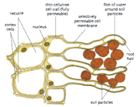
  • Roots contain root hair that provide large surface area: A plant may contain millions of root hair, which together provide large surface area for the absorption of water.
  • The epidermis of root hair is permeable to water: The root hair has very thin walls that are freely permeable to water. The thin walls help in easy movement of water and minerals in and out of cells.Absorption-by-Roots-Root-hair-in-a-freshly-germinated-seed
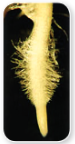
  • Most of the absorption of water and minerals occurs near root tips. The soil particles, which are usually coated with water and dissolved minerals, adhere tightly to the root hair. The soil solution flows in and out of the root hair cells.
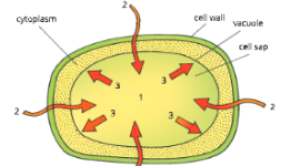
  • Root hair contains cell sap, which is at a higher concentration of minerals than the surrounding water: Root hair grows from the outer layer of the cortex. Between the cells of the cortex are large air spaces (vacuoles) that allow diffusion of gases and movement of water. The spaces allow water to get into the root by capillary action. The large vacuoles in the plant cells contain a solution called cell sap, which contains dissolved salts and is therefore of a higher concentration than the surrounding water. This facilitates osmosis, as a result of which water from outside is drawn inside the root hair.Absorption-by-Roots-Fully-grown-root-hair
    Absorption-by-Roots-Cross-section-of-root-showing-association-of-soil-particles-with-root-hair.

How Do Absorption And Conduction Of Water And Minerals Occur?

The absorption of water and minerals from the soil by the root hair, their movement through the thickness of the root and then upward conduction through the stem to the leaves of the plant takes place through the following processes.

  • Diffusion
  • Osmosis
  •  Imbibition
  • Active transport
  • Turgidity and flaccidity
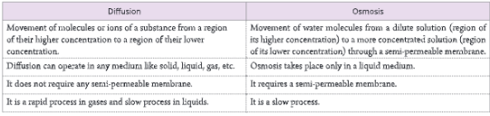
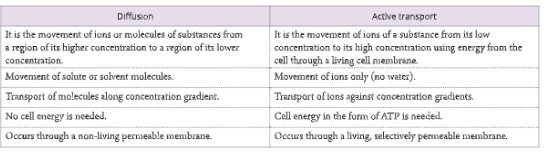
Diffusion

The movement of molecules of a substance from a region of their higher concentration to a region of their lower concentration is called diffusion.
Large molecules move much more strongly than small molecules. If you add a small drop of dye (like ink) to one end of a tub of water without disturbing it, the dye starts dissolving. It would take a long time for the ink molecules to diffuse throughout the tub and reach a state of equilibrium.

Absorption-by-Roots-The-process-of-diffusion
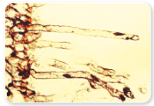
A-barrier-separates-two-kinds-of-molecules

Absorption-by-Roots-The-process-of-diffusion

When the barrier is removed, random movement of molecules results in both kinds of molecules moving from a region of higher concentration to a region of lower concentration.

Absorption-by-Roots-The-process-of-diffusion.

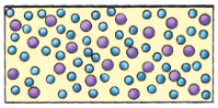
Eventually, an equilibrium (even distribution) is reached. The diffusion gradually slows down as equilibrium is approached.

Absorption by Roots Activity 1

To study the diffusion of a soluble dye in water. Take a beaker containing water. Put a small crystal of potassium permanganate in one corner of the beaker. Observe for some time. You will observe that the potassium permanganate crystal slowly starts dissolving. After some time, the molecules of the dye distribute uniformly throughout the water.

Absorption-by-Roots-Experimental-set-up-to-study-diffusion-of-potassium-permanganate-dye-in-water.

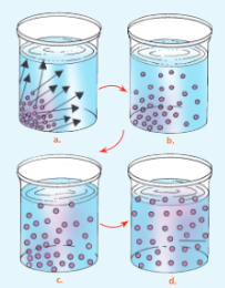
  • The molecules of dye are more concentrated in crystal form. When added to water they begin to dissolve.
  • The molecules move away from the region where they were added (region of higher concentration) to a region where they are less in number (region of lower concentration).
  • Finally, the molecules of dye have been uniformly distributed (state of equilibrium).

Importance of diffusion

  • Diffusion of water keeps the wall of the internal plant tissue moist.
  • It helps in the distribution of ions and molecules throughout the protoplast.
  • Loss of water vapour from the stomata during transpiration is through diffusion.
  • Aroma (smell) of flowers is due to diffusion of aromatic compounds from flowers to attract pollinators.

Osmosis

  • The movement of water molecules from a region of their higher concentration (more dilute solution) to their lower concentration (less diluted solution) through a semi-permeable membrane is called osmosis.
  • In other words, osmosis is the movement of only water from its pure state or solvent from a dilute solution into a stronger or concentrated solution through a semi-permeable membrane.

Absorption-by-Roots-Movement-of-water-molecules-through-a-semi-permeable-membrane

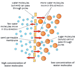
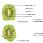
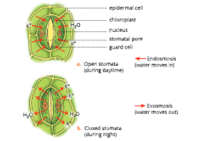
Endosmosis and Exosmosis

  • The inward movement of water molecules through a semi-permeable membrane when the surrounding solution is less concentrated is called endosmosis (endo: inward). Endosmosis leads to swelling up of cells.
  • The outward movement of water molecules through a semi-permeable membrane when the surrounding solution is more concentrated is called exosmosis (Exo: out.vard). Exosmosis leads to the shrinking of the cells.

Absorption By Roots Activity- 2

To demonstrate osmosis through a thistle funnel.

  • Take a thistle funnel and fill it with concentrated sugar solution. Cover the mouth of the thistle funnel with cellophane paper. Tie the cellophane paper as shown in the Figure.
  • Now take a beaker filled with water. Invert the thistle funnel in the beaker and suspend it with a stand as shown in Figure 4.7. Mark the level of sugar solution on the stem end of funnel and level of water in the beaker. Leave the set-up for about two hours.
  • Set the same experiment but without taking sugar solution in the thistle funnel. Instead, take water in the thistle funnel and mark the reading. This serves as a control set-up.
  • After about two hours you will observe that the level of sugar solution in the thistle rises only in the experimental set-up and not in the control.

Absorption-by-Roots-Experimental-set-up-to-demonstrate-osmosis.

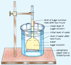
  • The level of water in the beaker in the experimental set-up drops slightly while it remained unchanged in the control.

Conclusion

This shows that:

  1. the water molecules are able to move from a dilute solution (water in a beaker) into the concentrated sugar solution (in the thistle funnel) through the cellophane paper.
  2. Sugar solution from the funnel did not pass into the beaker.
  3. Cellophane paper acts as a semi-permeable membrane. Only water molecules could pass through it.

What will happen if you use a rubber sheet instead of cellophane as a barrier?

There will be no change in the level of sugar solution as the rubber sheet is an impermeable membrane and would not allow the water molecules from the beaker to cross over to the other side.

What will happen if you use muslin cloth instead of cellophane as a barrier?

Since the pores in the muslin cloth are very large, they will not hold back even the sugar molecules and all the sugar solution will flow out to a common level due to gravity.

Absorption By Roots Activity-3

To demonstrate osmosis through a Viking bag.

Repeat the earlier mentioned activity (no. 2) by using a Viking bag. The Viking bag acts as a semi-permeable membrane.

  • Take a Viking bag and tie a knot at one end. Fill this bag with sugar solution from the other end. Insert a long glass capillary tube into it. You will find that the sugar solution rises in the capillary.
    Absorption-by-Roots-Experimental-set-up-to-show-osmosis-through-Viking-bag-containing-sugar-solution.
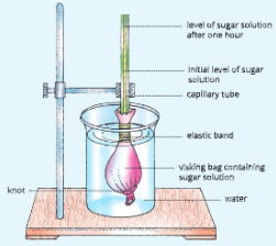
  • Now immerse the visking bag in a beaker containing water and clamp the capillary tube vertically. Leave the experimental set-up for about one hour.
  • After about 1 hour you will find that the level of sugar solution in the capillary tube rises. This happens because the water from the beaker diffuses inside through the walls of the visking bag into the more concentrated sugar solution.

Osmotic pressure

  • The results of the above activities show that the pressure builds up in the sugar solution and forces the solution up the capillary tube. The pressure results from the rapid diffusion of water molecules from the dilute to the more concentrated solution.
  • The osmotic pressure is the minimum pressure that needs to be applied to a solution to prevent the inward flow of pure solvent (water) into the solution when separated by a semi-permeable membrane.
  • Osmotic pressure can also be defined as the pressure required to completely stop the entry of water into a solution across a semi-permeable membrane. It can also be defined as the measure of the tendency of a solution to take in water by osmosis.

Osmotic potential

  • The osmotic potential of a solution is a measure of the tendency of water molecules to diffuse out of it. A concentrated solution that has relatively few water molecules has a low osmotic potential. On the other hand, a dilute solution with a larger proportion of water molecules has a high osmotic potential.
  • Pure water has the highest possible osmotic potential.
  • Movement of water in a plant occurs from a dilute solution of a high osmotic potential to a concentrated solution of a low osmotic potential.

Importance of osmosis

  • Water absorbed by roots moves to the upper parts of a plant from cell to cell through osmosis.
  • Osmosis plays a key role in the growth of radicles and plumules during seed germination.
  • Cell-to-cell movement of water occurs through osmosis.
  • Living cells remain turgid or distended due to osmosis.
  • The stomata are open and close in response to increase or decrease in osmotic pressure of the guard cell.
  • The differences between diffusion and osmosis are given in the Table.

Absorption-by-Roots-differences-between-diffusion-and-osmosis

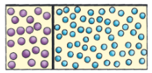
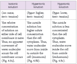
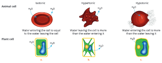
Tonicity: Isotonic, Hypotonic And Hypertonic Solutions

  • The relative concentration of a solution which determines the direction and extent of diffusion is called tonicity. Based on tonicity, solutions can be classified into three types – isotonic, hypotonic and hypertonic.
  • If you take a plant cell and place it in solutions that are of different concentrations you will find that the cell shrinks in a hypertonic solution, swells in a hypotonic solution and remains unchanged in an isotonic solution.

Absorption-by-Roots-Differences-between-isotonic,-hypertonic-and-hypotonic-solutions
Absorption-by-Roots-Behaviour-of-animal-and-plant-cell-placed-in-solutions-of-different-concentration

Imbibition

  • Osmosis is not the only force involved in the absorption of water by plants. Substances such as cellulose and starch are hydrophilic, i.e. they have a strong affinity for water. They can absorb water and swell up.
  • Imbibition is the process by which plant cells (living or dead) absorb water by surface attraction. Imbibition results in swelling of tissues and rupturing of seed coat for germination of seeds. Swelling of wooden doors during rainy season is also due to imbibition.

Passive transport

  • Diffusion, osmosis and imbibition are examples of passive transport in plants. Passive transport always takes place along the concentration gradient and requires no energy from the cell.

Absorption-by-Roots-Black-eyed-pea-seeds-before-and-after-imbibition

Active Transport

The passage of salt or ions of a substance from its lower concentration to a higher concentration utilizing the energy from the cell through a living membrane is known as active transport.

Active transport is just opposite to diffusion. The ions of certain elements such as nitrates, sulphates, manganese, etc., cannot easily pass through the cell membranes of root cells. This is because their concentration is higher inside the root cells than in the surrounding soil water.

Thus, the ions of such elements move inside root cells (the region. of their higher concentration) from outside (the region of their lower concentration) using the energy (ATP). The differences between diffusion and active transport are given in Table.

Absorption-by-Roots-Differences-between-diffusion-and-active-transport

Turgidity And Flaccidity

Turgidity

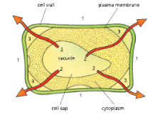
The root hair of plants are permeable to water. The cell sap inside the vacuole contains salts and sugars and is highly concentrated. If this cell is surrounded by water, osmosis will cause water to enter the cell sap.

As a result, the vacuole would expand, pushing the cell cytoplasm against the cell wall. Eventually, a condition will arise when no more water can enter the cell. Such a cell cannot accommodate any more water and is called turgid and the condition is called turgidity.

Turgor pressure

The pressure of the cell contents against the cell wall due to movement of water into the cell during osmosis is called turgor pressure. The pressure exerted by the cell wall on the cell contents is called wall pressure.

Turgor pressure and wall pressure counterbalance each other. As a result, even if the concentration of solute inside the cell is greater than that outside of a cell, further absorption of water does not take place.

Absorption by Roots Turgor pressure in a plant cell

1. Salts and sugars in cell sap make it concentrate inside
2. water enters the cell by osmosis
3. The cell sap volume increases, pushing the cell wall outward making it turgid.

Uses Of Turgor Pressure

  • Turgor pressure keeps the cells and their organelles stretched, which is essential for the proper functioning of a cell.
  • It is necessary for the enlargement of cells.
  • It provides support to living tissues like parenchyma.
  • It keeps the leaves fully expanded and oriented towards the light. In case of loss of turgidity, the shoots droop down and leave wilt. Rapid drooping of the leaves of the sensitive plant, Mimosa pudica in response to touch is due to turgor movement.
  • Flowers, soft stems and other soft parts of a plant are able to maintain their shape due to turgidity or turgor pressure.
  • It keeps a check on the excessive entry of water into the cells.
  • A plant cell may burst when turgor pressure exceeds wall pressure.

Turgor pressure in root cells builds up root pressure

Root pressure is the pressure developed in the roots due to the continued inflow of water by cell to cell osmosis. This helps in ascent of sap upwards through the stem. If you cut off the shoot of a plant, the water

Absorption By Roots Activity- 4

To study turgor pressure in root cells-Root pressure

  • Take a well-watered, potted plant such as balsam and cut it a few centimetres above the soil level.Absorption-by-Roots-An-activity-demonstrating-root-pressure
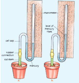
  • Immediately fix a glass tubing over the cut portion with the help of a rubber connection. The other end of this tubing is connected to a manometer.
  • You will observe that water starts coming out of the cut end of the plant and exerts pressure and raises the level of mercury in the connected manometer. This upward movement of water is due to the heavy root pressure.
  • immediately comes out from the cut portion. This is due to root pressure. To understand it better let us perform an activity (activity 4).

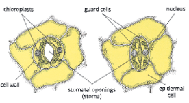
Turgor pressure helps in the opening and closing of stomata

  • During photosynthesis, glucose is synthesized from CO₂ and H₂O. This causes an increase in the osmotic pressure of the contents of guard cells.

Absorption-by-Roots-Cross-section-through-stomata

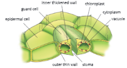
  • As a result, guard cells absorb more water from the neighbouring cells, thus becoming turgid. The high turgor pressure causes the guard cells to bulge out and the stoma opens. At night, since no photosynthesis takes place, there is a shortage of water in the leaf and the guard cells become flaccid, their inner walls become straight and the stomata are closed.

Turgor movement

  • In the sensitive plant, Mimosa pudica, the stimulus of touch leads to the loss of turgor pressure at the base of leaflets and the leaves droop down (fold) within 2-3 seconds of touching. This is an example of the turgor movement.
  • The bending of certain flowers (e.g. sunflower) towards sun is due to the turgor movement.

Flaccidity

Flaccidity reverse of turgidity. If a fully-distended (turgid) cell is placed in a hypertonic solution, the water moves out of the cell due to exosmosis and the cell loses turgidity. The cell is called flaccid and the phenomenon is known as flaccidity. Cells remain flaccid when placed in isotonic solution.

A condition in which the cell contents shrink and the cell loses its turgidity is called flaccidity.

Absorption by Roots Flaccidity in a plant cell
1. Solution outside is more concentrated than the cell sap.
2. Water passes out of the vacuole by osmosis.
3. The vacuole shrinks, pulling the cytoplasm away from the cell wall and leaving the cell flaccid

Plasmolysis And Deplasmolysis

Plasmolysis

  • Shrinkage or contraction of the cytoplasm (cell content) of a cell from its cell wall when placed in a hypertonic solution is called plasmolysis.
  • If we place a living turgid cell in a hypertonic solution, outward movement of water (exosmosis) occurs from the central region of the cell. As a result, the size of the cytoplasm reduces, and the plasma membrane is withdrawn from the cell wall. This is called plasmolysis and the cell is called a plasmolysed cell.

Absorption-by-Roots-portion-of-leaf-of-water-weed-showing-plasmolysis

Absorption-by-Roots-Diagrammatic-representation-of-plasmolysis-in-a-cell
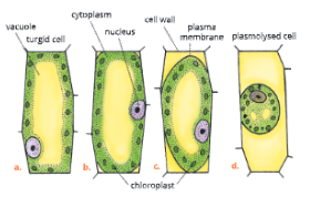
A cell in normal turgid condition;
Successive stages in shrinkage of cytoplasm from the cell wall after being placed in a hypertonic solution

Deplasmolysis

  • If a plasmolysed cell is placed in water, its shrunk cytoplasm swells up and presses against the cell wall.

Absorption-by-Roots-Differences-between-plasmolysis -nd-deplasmolysis

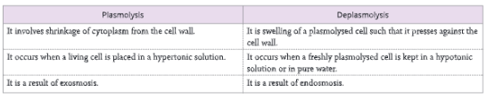
  • This happens due to endosmosis. The swelling up of a plasmolysed cell under the influence
    of hypotonic solution or water is called deplasmolysis. Deplasmolysis is possible only if the cell is alive and its cytoplasm is not dead or damaged. Differences between plasmolysis and deplasmolysis are given in Table.

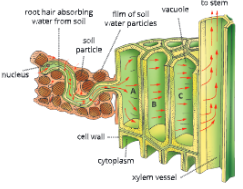
Absorption Of Water And Minerals By The Root

Absorption of water

The absorption of water occurs through root hair. Root hair are thin-walled extensions from the cells. of the outer layer of a root. They grow out pushing between the soil particles. There is a film of water that surrounds the soil particles and in turn root hair also.

  1. The root hair contains cell sap which has a higher concentration of salts than the outside soil water. This causes osmosis and the water from outside diffuses into the cells of root hair (let us take it as cell A). This is due to root pressure.
  2. As water enters the vacuole of cell A, it dilutes the concentration of sugar and salts in its cell sap.
  3. Another cell (assume cell B) next to cell A has a higher concentration of cell sap (salts and sugars). As a result water from cell A moves to cell B.
  4. The water entering cell B makes its cell sap dilute and then moves to cell C. This way water moves from one cell to another by cell-to-cell osmosis.
  5. The water ultimately passes into the xylem vessels at the centre of root and is conducted up the root and stem into the leaves.
  6. The water and minerals absorbed by the roots are transported through xylem tissue through the ascent of sap.

Absorption of minerals

Absorption of mineral elements by the root from the soil takes place by active transport. The water film around the soil particles also contains a low concentration of mineral elements. These mineral elements move from soil into the root cells against the concentration gradient. Energy in the form of ATP is required by the cell for the absorption of minerals.
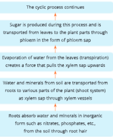
Absorption-by-Roots-Diagram-to-show-cell-to-cell-passage-of-water-from-soil-to-xylem-vessels-in-a-root

Absorption-by-Roots-An-overview-of-transport-of-water-and-nutrients-in-plants

Ascent Of Sap

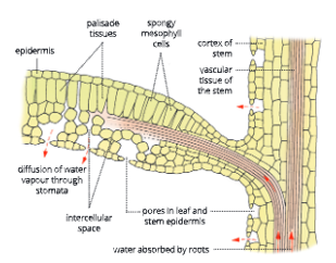
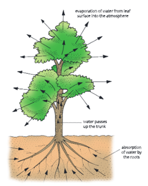
The upward movement of water and mineral salts from roots to the aerial parts (leaves, flowers, branches, etc.) of the plant, against the gravitational force, is called ascent of sap.

The elongated, lignified tracheids and xylem vessels, are placed end-to-end without any cross wall. They form the pipeline for conducting water and minerals from the roots to the leaves.

Water enters the root hair cells by imbibition and then by the process of osmosis. This water from the root hair cells passes into the xylem vessels through the cells of the cortex, endodermis and pericycle.

Ascent of sap takes place from the root, into the stem and finally the leaf veins through the xylem vessels and tracheids by means of a pull exerted by the leaf cells at the top of the sap column.

The cohesive force between the water molecules also helps to maintain the continuity of the water column.

The cohesive force is the force adhesion which keeps molecules of the same substance together, for example, water molecules.

Absorption-by-Roots-Ascent-of-sap-in-plants

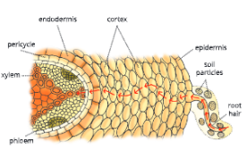
a. Transverse section through a dicot root showing absorption of water by root hair

Absorption-by-Roots-Ascent-of-sap-in-plants
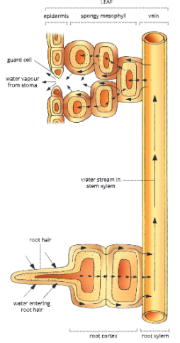
Pathway of water through the plant

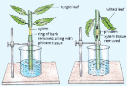
Absorption By Roots Activity 5

To show that xylem is the path of ascent of sap.

  • Cut two leaf shoots of the balsam plant underwater. Keep their lower ends dipped in water. In one shoot remove 2-4 cm long ring of bark (phloem) roughly from the middle region of the shoot.
  • Remove the xylem from the central part of the second shoot (Fig. 4.19b). Fix the shoots with the bola of the help of a stand and leave the apparatus as such for 1-2 days.
  • Result: In the first case (Set-up a), the leaves remain turgid even after 24 hours which shows that water continues to rise even if the phloem is removed. Leaves of the second shoot (Set-up b) get wilted. This shows that when xylem is removed, water cannot rise up.

The experiment proves that water rises through the xylem vessels.

Absorption-by-Roots-Experimental-set-up

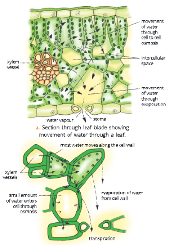
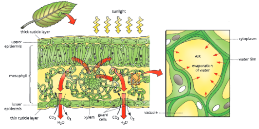
In the leaf blade, water passes from the xylem into the cells of the mesophyll and epidermis by the process of cell-to-cell osmosis.

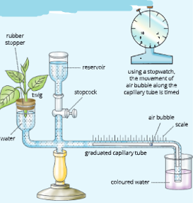
Causative Forces For Ascent Of Sap

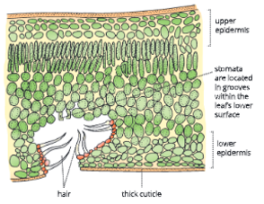
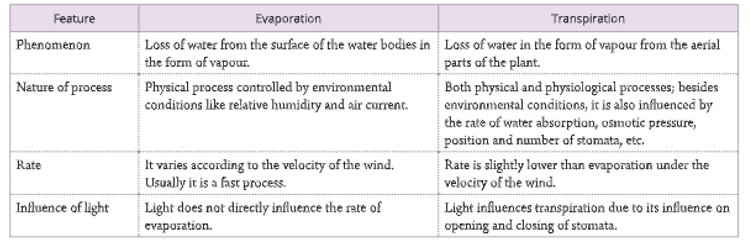
  • As the water moves upward from roots to the leaves, a lot of it evaporates through the stomata present on the leaf epidermis. This process of evaporation of water from leaves and other aerial parts of the plant is called transpiration.
  • The xylem sap (water containing minerals) rises against the gravity without the help of any mechanical pump. The xylem sap is largely pulled upward by transpiration – cohesion-tension mechanism, also called transpiration pull.
  1. Root pressure (pushing xylem sap): Root pressure is a pressure created due to the continuous influx of water in the xylem vessels from the root hair and root cortex. Root pressure causes guttation, the exudation of water droplets that can be seen on leaf surface. In most plants, root pressure is not the major mechanism during the ascent of sap. At the most, root pressure pushes the sap in the xylem vessels up to a certain height. Later on, the sap moves with cohesion, adhesion and transpiration pull.
  2. Capillarity nature of xylem vessels: Xylem vessels are very narrow. This causes the water from a lower level to rise by capillary action in order to fill up the vacuum created at the leaves due to the loss of water by transpiration.
  3. Pulling xylem sap: Transpiration – Cohesion- tension mechanism or transpiration pull: Stomata are the sites of exchange of CO₂ and O₂ between photosynthetic tissues and atmosphere. They are also the sites for transpiration. The air in these stomata is saturated with water vapour since it is in contact with most walls of the mesophyll cells (Fig. 4.20). On most of the days, the air is drier outside the leaf, i.e. it has a lower water concentration outside than inside the leaf. Thus, the gaseous water diffuses outside the leaf through the stomata and there is a loss of water during transpiration.
  4. This leads to a generation of tension (negative pressure) in the leaf due to the unique physical property of water. The thin film of water vapour present in the mesophyll cells replaces the water vapour which is lost from the leaf stomata by transpiration.
  5. During this process, water is pulled on by the adhesive and cohesive forces between the molecules of two different substances.
  6. This tension is the pulling force or suction force which draws water from the leaf xylem through the mesophyll cells toward stomata. The water lost via transpiration is replaced by the water that is pulled out of the leaf xylem.

Absorption-by-Roots-Transpiration-pull

  • Cohesion and adhesion of water: The transpiration pull on water extends from the leaves up to the root tip and even into the soil solution.
  • This cohesion of water is due to hydrogen bonding between water molecules. Each water molecule is attached (adhered) to an adjacent water molecule and this pull is relayed from molecule to molecule down the entire column of water in the xylem.

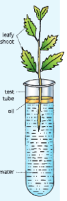
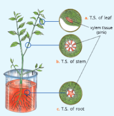
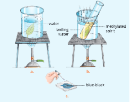
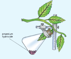
Absorption By Roots Activity 6

To show that water is conducted through xylem tissues.

  1. Take a young, medium-sized balsam plant. Remove this plant from soil, wash it and place it in a beaker. Half fill this beaker with water containing eosin stain solution (pink colour).
  2. Ensure that the roots are completely submerged in the solution. Leave this set-up for about 3-4 hours. Now remove this plant from beaker and wash it in running tap water.
  3. Cut transverse section passing through roots, stem and leaves with the help of a sharp razor or a blade. Mount the sections on the slides and observe under a microscope.
  4. You will observe that in the centre xylem vessels appear red due to conduction of eosin stain dye. This shows that water is conducted through xylem tissue.

Absorption By Roots Summary

  • Plants need water and minerals for many purposes such as growth, photosynthesis, transpiration, mechanical strength and transportation of nutrients.
  • Roots bear root hair that provides enormous surface area. The epidermis of root hair is permeable to water.
  • The movement of molecules or ions of a substance from a region of their higher concentration to a region of their lower concentration is called diffusion. Diffusion of water molecules keeps the cell wall of the internal plant tissues moist and helps in the transpiration of water vapour from stomata.
  • Osmosis is the movement of water from its pure state or diluted solution into a concentrated solution through a semi-permeable membrane. The inward movement of water is called endosmosis and outward movement of water is called exosmosis.
  • The osmotic pressure is the maximum pressure which can develop in an osmotically active solution when it
    Is separated by a semi-permeable membrane to stop further endosmosis of water from a region of low concentration to the region of higher concentration of solute.
  • Imbibition is the process by which plant cells absorb water by surface attraction.
  • The passage of salt or lons of a substance from their lower concentration to higher concentration through a living cell membrane using the energy from the cell is called active transport. The loss of elements moves into roots through active transport.
  • The condition, when a cell reaches a state where it cannot accommodate any more water is called turgidity.
  • The pressure of the cell contents against the cell wall is called turgor pressure. Flaccidity is reverse of turgidity.
  • Shrinkage of cytoplasm of a cell from its cell wall under the influence of a hypertonic solution is called plasmolysis. The condition opposite to it is deplasmolysis.
  • Absorption of water occurs through root hair by the process of osmosis. Minerals move from soil into root hair through active transport.
  • The water and mineral salts enter the root by moving between the cells before entering xylem. Water also enters root hair; then passes through the cells of the cortex and endodermis to reach xylem vessels.
  • The water and minerals absorbed by roots are conducted up through the xylem tissue.
  • The upward movement of water and minerals from the roots to the aerial parts of plants against the gravitational force is called ascent of sap.
  • Xylem sap is pulled upward by root pressure, capillary action of xylem vessels, transpiration-cohesion-tension mechanisms, and cohesion and adhesion of water.

UP Board Notes for Class 10 Science Chapter 11 Sense Organs – The Eye and The Sense of Sight

UP Board Notes for Class 10 Science Chapter 11 Sense Organs The Eye And The Sense Of Sight

Sense Organs Learning Objectives

After completing this chapter, you will be able to:

  • Define a receptor and name various kinds of receptors;
  • Describe the structure and functions of the eye;
  • Trace the pathway of light through the eye to the retina and explain how light is focused for distant and near vision;
  • Explain the causes and consequences of various defects of the eye and methods of their correction;
  • Describe the structure and general functions of the outer, middle, and Inner ears;
  • Explain how the ear serves as an organ for dynamic and static equilibrium.

The eyes, ears, tongue, nose, and skin are the major sense organs in our body, which are sensitive to the sense of light, sound, taste, smell, and touch, respectively. Each of these sense organs is directly connected to the brain. These sense organs have special cells called receptors which receive stimuli from the environment.

UP Board Notes for Class 10 Science Chapter 11 Sense Organs The Eye And The Sense Of Sight Sense Organs Some Key Terms

Stimulus:

A physical event that affects an organism by activating its receptors to bring about a change in its activity. For example, mechanical stimuli (pressure and touch), chemical stimuli (taste and smell), and thermal stimuli (heat and cold).

Sensation:

A general state of awareness of the stimulus.

Receptors:

Sensory cells in tissues that receive stimulus from the environment. Some common specific receptors are:

  1. Photoreceptors: respond to light rods and cone cells in the retina of the eye.
  2. Chemoreceptors: respond to chemicals – taste buds on the tongue, smell receptors of the nose.
  3. Mechanoreceptors: respond to touch – nerve fibers around the hair on the skin.
  4. Thermoreceptors: respond to changes in temperature – receptors on the skin.
  5. Photoreceptors: respond to sound/hearing – receptors in the ear.
  6. Exteroceptors: specialized to detect sensory information from the external environment.

We all respond to the light stimulus. Human beings have two eyes situated in deep sockets or orbits on the front side of the head. Each eye is in the form of a ball called an eyeball that measures about 2.5 cm (1 inch) in diameter. Each eyeball can be rotated with the help of six distinct sets of muscles.

UP Board Notes for Class 10 Science Chapter 11 Sense Organs – The Eye And The Sense Of Sight Accessory Structures Of The Eye

Eyebrows, eyelids, eyelashes, and the tear glands (the lacrimal apparatus) are the accessory structures of the eye.

Sense Organs The lacrimal apparatus of the human eye eye image 1
The lacrimal apparatus of the humane eye


  • The eyebrows protect the eyeball from foreign objects, perspiration, and direct rays of sunlight.
    Eyelids are folds of skin and muscles. The upper and lower eyelids protect the eyes from excessive light and foreign particles, cover the eye during sleep and spread lubricating secretions over the eyeballs. The upper eyelid is more movable than the lower one. Projecting from the border of each eyelid is a row of thick, short hairs called eyelashes. At the base of the hair follicles of eyelashes, sebaceous glands are found. These glands secrete a lubricating fluid into the hair follicles called sebum.
  • Tear glands or lacrimal glands (collectively known as lacrimal apparatus) are a group of glands that manufacture and pour tears (L. lacrima: tears). A lacrimal gland is a compound gland located at the upper sideward portion of each eyelid
    Each lacrimal gland gives rise to 6 to 12 excretory lacrimal ducts that empty their secretion (tears) onto the surface of the conjunctiva of the upper eyelid. From here it passes to the lacrimal canals and then into the lacrimal sac. From here a
  • Nasolacrimal duct conducts the secretion into the nasal cavity in the nose. This duct is responsible for the passage of medicine dropped in the eye to the nose or sometimes into the throat.
  • Tear or lacrimal secretion is a watery fluid containing salts, some mucus, and a bacteriocidal enzyme called lysozyme.

Functions of tears:

  1. After being secreted, it spreads over the surface of the eyeball and serves as a lubricant.
  2. It cleans the front surface of the eyeball by washing away the dust particles.
  3. It moistens the eyeball.
  4. The enzyme lysozyme contained in the tears kills the germs.
  5. Tears help in communicating emotions.

UP Board Notes for Class 10 Science Chapter 11 Sense Organs – The Eye And The Sense Of Sight Structure Of The Eyeball

  • The eyeball is a spherical structure, measuring about 2.5 cm in diameter. Structurally, the eyeball is composed of three layers – the fibrous tunic (anterior cornea + posterior sclera), the vascular tunic also known as uvea (middle layer of the eyeball) (choroid + ciliary body + iris), and the inner retina.
Sense Organs Sclerotic layer or sclera image 2
The structure of a human eye


  1. Sclerotic layer or sclera
  • The sclerotic layer or sclera is the outer tough coat of the eyeball made up of mainly collagen fibers. It can be divided into two regions – the posterior region called the sclera, and the anterior region called the cornea.
  • The sclera is a white coat of fibrous tissue visible through the conjunctiva, which covers all the eyeballs except the cornea. It is also known as the white of the eye. The sclera gives shape to the eyeball and also protects its inner parts.
  • The cornea is a transparent, fibrous coat through which the iris can be seen. The outer surface of the cornea is covered by an epithelial layer which is continuous with the epithelial layer of the conjunctiva. The cornea receives nourishment from tears and aqueous humor.
  • Sometimes, the cornea turns white and opaque and can be replaced by a donated eye.
  1. Uvea
  • The choroid layer is the middle layer of the eyeball and is composed of three parts – choroid, ciliary body, and iris.
  • The choroid is a thin, dark brown membrane that lines most of the inner surface of the sclera. It contains several blood vessels and a large amount of pigment melanin. The choroid absorbs light rays so that they are not reflected within the eyeball. The numerous blood vessels nourish the retina.
  • The choroid expands to form the ciliary body. It consists of circular muscles. The smooth muscles of the ciliary body help in changing the shape of the lens.
  • The iris (arid: rainbow) is a clouded part of the choroid around the pupil. It is a clouded part seen through the cornea. There is a hole (round window) in the canter of the iris, known as the pupil. The light enters the eyeball through the pupil. The iris contains radial and circular muscles.

Sense Organs image 3 effect of light intensity on the size of pupil


The contraction of these muscles constricts the pupil which regulates the amount of light entering the eyeball through the iris

  • In the case of bright light, the circular eye muscles contract, and the size of the pupil is decreased (constriction).
  • In case of dim light, the radial muscles contract, and the size of the pupil is increased (dilation).
  1. Sense Organs Retina – part of the eye where rod and cone cells are located
  • The retina is the third and the inner layer of the eye. It is located only in the posterior part of the eye. It is the light-sensitive layer. It contains light-sensitive cells called rods and cones.
  • The rod cells are sensitive to dim light. They do not respond to color. Rods contain the pigment rhodopsin or visual purple. The rod cells are distributed throughout the retina.
  • The cone cells are sensitive to bright light. The cones are responsible for color vision. Cones contain the pigment rodeos in. Cone cells are mostly confined to the yellow spot or fovea centralis or macula.The differences between rod cells and cone cells are given in Table                                           

Sense Organs differences between rod cells and cone cells image 4

Yellow spot – the area of the best vision

The yellow spot or macula lutea (Fovea centralis) is a spot located inside the eyeball near the Centre of the retina. It contains a maximum number of light-sensitive cells, especially cone cells. The rest of the retina has lesser cone cells and more rod cells. The yellow spot is the region of color vision and also the brightest, clearest and best vision.

Blind spot – the area of no vision

A blind spot is the region of the retina just below the yellow spot. Since there are no light receptors here, no image is formed here, i.e. blind spot is the area of no vision.

senso organs to demonsrate the billnd stop turn the book image 6
To demonstrate the blind spot, turn the book.


clockwise through 90° and hold it at an arm’s length with the two symbols straight in front of your eyes. Close your left eye and concentrate on the cross with your right eye. Bring the book slowly towards you. Keep looking at the cross on the left: eventually, as the dot falls on the blind spot of your right retina, the image of the dot on the right will disappear.

UP Board Notes for Class 10 Science Chapter 11 Sense Organs – The Eye And The Sense Of Sight Sense Organs Lens

  • The main body of the eye is divided into two parts by a biconvex lens which is a transparent crystalline body. The lens lies just behind the pupil and iris. It is flatter at the front than at the back and is soft and slightly yellow in color. It contains transparent lens fibers and an elastic lens capsule made of glycoprotein. There is no blood vessel in the lens.
  • The lens is held in position by the suspensory ligaments which attach it to the ciliary body.

The eyeball is divided into two chambers:

  • the anterior chamber (aqueous chamber) and
  • posterior chamber (vitreous chamber).
  • The anterior chamber (between the lens and cornea) is filled with a fluid called aqueous humor. It is a thin and watery fluid. It keeps the lens moist and protects it from physical shock.
  • The posterior chamber is called the vitreous chamber. It is a large cavity of the eyeball. It lies between the lens and the retina. It contains a jelly-like substance called vitreous humor. The vitreous humor maintains the shape of the eyeball by preventing the eyeball from collapsing and supporting the retina.

UP Board Notes for Class 10 Science Chapter 11 Sense Organs – The Eye And The Sense Of Sight How Do We See – Working Of The Eye

  1. Light rays enter the eye

The light rays enter the eye through the cornea and then pass through the pupil, aqueous humor, the lens, and the vitreous humor (all transparent structures), before reaching the retina.
The pupil adjusts to different intensities of light. In bright light, the pupil constricts (becomes narrow) and in dim light, it dilates (becomes wider) to allow more light.

  1. Image formation on the retina

The formation of an image on the retina requires four steps – a. refraction of light rays, b. constriction of the pupil, c. accommodation of the lens, and d. convergence of the rays.

  1. Refraction of light rays: The eye focuses an image by refracting, or bending the light rays using the cornea and the lens. An upside-down or inverted image is formed at the yellow spot on the retina. Most of the refraction of light occurs in the cornea due to its curved surface.
  2. Constriction of the pupil: The iris contains radial and circular muscles. Radial muscle widens and circular muscle constricts the pupil. This adjustment of the size of the pupil regulates the amount of light entering the eye.
  3. Accommodation of the lens: The ability of the eye to focus on different objects placed at a distance by adjusting the curvature of the elastic eye lens is called accommodation. This is brought about by ciliary muscles which change the focal length of the eye lens to focus on near or distant objects on the retina.

(1) In the distant vision (more than 6 meters), the ciliary muscles are relaxed and the lens becomes flatter or thinner due to the stretching of the suspensory ligaments.


senso organs image formation on the retina in near vision 6
Image formation on the retina in near vision


(2) In the near vision (less than 6 meters), the ciliary muscles contract pulling the choroid forward. towards the lens and tension is released on the suspensory ligaments. The lens becomes shortened, more convex, and thickened due to its elastic nature

In normal conditions, the ciliary muscles are relaxed and the lens remains stretched by the suspensory ligaments. Then, it is less convex and best suited for distant vision.

4. Convergence of the rays: We have two eyes


sense organs eye cpmpared with camera 7
Eye compared with the camera


but we see only one image of an object. This is known as binocular vision or stereoscopic vision. While viewing objects, both eyeballs move toward the object. Thus, both images fall on the corresponding points (fovea centralis) of both retinas at the same time and overlap with each other. This is known as the convergence of the rays.

The eye can be compared with a camera. The major similarities and differences between the eye and the camera are given. A summary of various parts and functions of the eyes is given in Table


senso organs parts and functions of the eye
Parts and functions of the eye
  1. Photoreception by brain

The image formed at the retina stimulates photoreceptors. The light energy of the image formed at the retina produces chemical changes in the rods and cones. This generates nerve impulses that travel through the optic nerve from the retina to the visual area of the brain. At the cerebrum (brain), the sensation of sight is interpreted.

How are the eyes adapted to bright light and dark?

  • When we are in dark or dim light for a long period, then the rate of photopigment (rhodopsin) formation by rod cells in the retina is much faster than the rate at which they are broken down. The pupil dilates to allow more light to enter through the retina to allow viewing of objects in dim light. This is called dark adaptation.
  • When we are in a very bright light for a long period of time, the light-sensitive pigments of the rods are broken down, and the visual purple of the rods gets bleached, reducing their sensitivity. The cone cells in the retina are activated and it forms iodopsin pigment. The further pupil constricts and the size of the pupil is reduced to reduce the amount of light entering the eyes. This is called light adaptation.

Colour vision

  • color vision is possible because of cone cells. When all the cone cells are stimulated, we see white color. When very few cone cells are stimulated, we see black color. Stimulation of separate types of cone cells produces red, green, or blue and their combinations.

Why cannot we distinguish color on a moonlit night?

  • In dim light, like that during a moonlit night, colors cannot be distinguished because cone cells that detect colors do not work in dim light.

UP Board Notes for Class 10 Science Chapter 11 Sense Organs – The Eye And The Sense Of Sight Common Defects Of The Eye And Their Correction

Short-sightedness or Myopia

  • In this condition, light is focused in front of the retina, and a blurred image is formed for distant objects. In myopia, the near objects are seen clearly while the distant objects appear blurred. This happens because either the eyeball is elongated or the lens has become too thickened or curved
sense organs short sightedness an image formed in front of the retina
Short-sightedness -an image formed in front of the retina (myopia)


Long-sightedness or Hyperopia

  • In this condition, the light is focused behind the retina (the image is formed behind the retina). As a result, the distant objects are seen clearly, while the near objects appear blurred. Long-sightedness results due to the shortening of the eyeball or the lens have become. too thin
sense organs long sightedness an image formed behind the retina 10
Long-sightedness -an image formed behind the retina (hypermetropia)


Hyperopia (Hypermetropia) can be corrected by using a convex (converging) lens.

Astigmatism

  • This is a more complicated defect in vision. In this, the surface of the cornea becomes irregular and therefore some of the light rays are focused while others are not. As a result, some parts of the object appear blurred while other parts appear clear
sense organs Astigmatism can result from an irregular cornea or irregular lens.
Astigmatism can result from an irregular cornea or irregular lens.


Astigmatism can be corrected by using a cylindrical lens that bends light rays in one direction only.

Presbyopia

  • Astigmatism can be corrected by using a cylindrical lens that bends light rays in one direction only. Presbyopia In this condition, the lens loses its flexibility causing long-sightedness (the near objects cannot be seen clearly). This defect normally occurs in older people.

Presbyopia can be corrected by using a convex lens.

Night-blindness

  • In this condition, there is difficulty in seeing in dim light (hence the name night blindness). This is because of the non-production of rhodopsin pigment in the rod cells, which function in dim light. In the absence of rhodopsin, these cells cannot function. Thus, there is a lack of normal night vision. It is most often caused due to deficiency of vitamin A.

Night blindness can be cured by having Vitamin A rich diet.

Colour blindness

  • In this condition, a person is unable to discriminate between red and green colors. This is a genetic defect which you have already studied in the chapter on heredity and genetics (X-linked inheritance).

Colour blindness cannot be treated during the lifetime of an individual.

Cataract

  • In this condition, the lens of the eye becomes opaque and as a result, the vision is cut down. Cataracts can be treated by surgical removal of lenses and using convex lenses in the spectacles which compensate for the removed lens. Nowadays, a small plastic lens is implanted behind the iris to correct the defect.

Squint

  • In this condition, either the two eyes somewhat converge (known as cross-eye) or diverge (known as wide-eye). In both these conditions, a person may have double vision. Squint in the eyes can be treated by surgery or by suitable exercises.

UP Board Notes for Class 10 Science Chapter 11 Sense Organs – The Eye And The Sense Of Sight The Ear – Senses Of Hearing And Balance

  • Humans have two ears, one on each side of the head. Human ears are organs for the senses of hearing and balance. The ear is a miniature receiver, amplifier, and signal processing system.

Structure of ear

The human ear is divided into three parts

  • External ear
  • Middle ear
  • Inner ear
  1. The external or outer ear
    It consists of the externally visible part of the ear called the pinna (or by a thin membrane. auricle) and the internal part, the auditory canal. The auditory canal. the passage leading to the eardrum (also known as the tympanic membrane).

Hair and earwax in the ears

The auditory canal contains fine hairs, which filter the air. In addition, the upper wall of the canal contains modified sebaceous glands which secrete earwax. Earwax prevents the entry of foreign material into the ear.

  1. Middle ear

The middle ear is a small air-filled cavity. It is separated from the external ear by the eardrum (tympanic membrane) and from the internal ear by thin bony

sense organs various parts of human ear and mechanism of hearing. 11
various parts of the human ear and mechanism of hearing.


  • The partition which contains two small openings namely, an oval window and a round window.
    The middle ear contains three tiny bones called ear ossicles. These bones are named malleus (or hammer), incus (or anvil), and stapes (or stirrup). The handle of the malleus is attached to the internal surface of the eardrum (tympanum). Its opposite end is connected with the incus. The incus is the intermediate bone and it articulates with the head of the stapes.
  • The flat part of the stapes fits into the oval window. Directly below the oval window is another opening called a round window. The round window is covered by a thin membrane.

Ear ossicles (malleus, incus, and stapes) transmit sound vibrations to the internal ear.

  • The anterior wall of the middle ear contains an opening that leads directly into the auditory tube (also known as the Eustachian tube). This connects the middle ear with the throat. The Eustachian tube also helps in equalizing air pressure on both sides of the eardrum helping it for free vibration.
  •  Throat infection can lead to an ear infection.
    This is because the Eustachian tube connects the middle ear with the throat. Thus, any infection in the throat can pass to the ear through the Eustachian tube.
  1. Internal ear

The internal or inner ear is also called the membranous labyrinth. It has two main parts – cochlea and semicircular canals.

sense organs  Internal structure of the human ear and course of perception of sound waves 12
The internal structure of the human ear and course of perception of sound waves


Cochlea: It is a hollow, spiral-shaped (coiled), chamber. It resembles a snail’s shell. It consists of a bony spiral canal that makes about 23%4 turns around a central bony core. Its inner spiral cavity contains three separate channels or canals that run parallel.

These canals are separated by membranes. The median (cochlear) canal is filled with endolymph and the outer two canals are filled with a fluid called perilymph.

  • The middle canal contains a spiral organ called the organ of Corti. The organ of Corti is the organ of hearing. It contains a series of nerve cells and hair cells that join the auditory nerve and help in hearing.
  • The hair cells of the organ of Corti are very sensitive to sound. They can be damaged by exposure to high-intensity noises such as noise produced by engines of jet planes, loud music, etc.(b) Semicircular canals: The inner ear also contains three semicircular canals. These canals are arranged at right angles to each other in three different planes (one horizontal, two vertical) and are filled with endolymph. One end of each canal is swollen to form an ampulla. The ampulla contains sensory cells which help in the balance of the body
  • while moving. The nerve fibers arise from these cells and join the auditory nerve.
  • There is a short stem (vestibule) that joins the semicircular canals and cochlea. It contains two small sacs – the utriculus and sacculus. These also contain tiny hair-like sensory cells which help in the static balance of the body while at rest (i.e. position of the head when not in motion like standing).
  • The external ear, middle ear, and cochlea help in hearing. The sacculus, utriculus, and semicircular canals help in the sense of balance. A summary of various parts of the human ear and their functions is given in the table.


sense organs Summary of various parts of ear and their functions 13
Summary of various parts of the ear and their functions

UP Board Notes for Class 10 Science Chapter 11 Sense Organs – The Eye And The Sense Of Sight Mechanism Of Hearing And Balance

Hearing

Pinna collects and amplifies sound waves which then pass along the auditory canal to the eardrum.

  • Sound waves strike the eardrum (tympanum) and cause vibrations in its thin stretched membrane. The Eustachian tube equalizes air pressure on either side of the eardrum which allows a free vibration.
  • The vibrations reach ear ossicles in the middle ear. Ear ossicles transmit vibrations from the eardrum to the denser fluid in the inner ear.
  • The lever-like action of malleus and incus magnifies the vibrations of the stapes. The vibrating stapes transmit vibrations to the membrane of the oval window.
  • Vibrations from oval window get transmitted to cochlea. This leads to vibration in the fluid of cochlear canals. Vibration of fluid in cochlear canals triggers movement of sensory hair cells of organ of Corti in cochlea.
  • Movement of sensory hair cells is converted to a nerve impulse.
  • Nerve signal or impulse is transmitted to brain via auditory nerve and this results in hearing. Balancing

The following parts of ear are involved with balance.

  1. Static balance with respect to center of gravity: Sensory cells in vestibule
  2. Dynamic balance (while the body is in motion): Sensory cells in semi-circular canals

Movement of fluid inside semicircular canals triggers sensory hair cells of ampulla. This sensation passes to nerve cells and then to brain. The three canals are at right angles to each other so that the brain can detect even slightest tilting of the body in any direction.

UP Board Notes for Class 10 Science Chapter 11 Sense Organs – The Eye And The Sense Of Sight Summary

Receptors are the special cells that receive stimuli from the environment. Some common receptors are photoreceptors, chemoreceptors, thermoreceptors, and mechanoreceptors.

  • Receptors are contained in sense organs. There are five major sense organs in humans eyes (vision), ears (sound), nose (smell), tongue (taste), and skin (pressure and touch). Each of these sense organs is directly connected to the brain.
  • Each eye is in the form of a ball called eyeball that can be rotated by six distinct sets of muscles.
  • Eyebrows, eyelids, eyelashes, and tear glands are the accessory structures of the eye.
  • Lacrimal glands are a group of glands that manufacture tears. Tears serve as a lubricant and also kill the germs that enter the eye.
  • The eyeball has three layers – the sclerotic layer, the choroid, and the retina.
  • The sclerotic layer is the outer tough coat of eyeball that is divided into sclera (white coat of dense fibrous tissue) and cornea (transparent fibrous coat through which the iris can be seen).
  • The choroid layer is the middle layer of the eyeball and is composed of three parts – choroid, ciliary body, and the iris. There is a hole in the center of iris called the pupil.
  • The retina is the third and inner coat of the eye. It contains light-sensitive cells called rods (sensitive to dim light) and cones (sensitive to bright light).
  • The lens focuses light rays from outside and a real, inverted image is formed at yellow spot. The yellow spot is the area of best vision, while blind spot is the area of no vision.
  • In myopia, the image is formed in front of retina.It can be corrected by using a concave lens.
  •  Ear is concerned with two functions – hearing and body balance.
  • Set eardrum into vibration. It sets malleus, incus and stapes (ear ossicles) into motion. From stapes, vibrations move to oval windows, then to cochlear canal and sensory hairs in cochlea. From here the nerve impulse reaches the brain through the auditory nerve

UP Board Notes for Class 10 Science Chapter 11 Sense Organs – The Eye And The Sense Of Sight Sense Organs Structured/Application/Skill Type Questions

1. The diagram given below depicts a defect of the human eye. Study the same and then answer the following questions.
Sense organs defect of the human eye 14

1. Name the defect shown in the diagram.
2. Give two possible reasons for this defect.
3. Name the parts labeled I to iv.
4. Name the type of lens used to correct this eye defect.
5. Draw a labeled diagram to show how the above-mentioned defect is rectified using the lens named
above

2. The following diagram represents a part of the human ear. Study the same and then answer the following questions.

sense organs a part of the human 15

  • Give the biological terms for malleus, incus, and stapes.
  • Name the parts labeled I, ii, and iii in the diagram.
  • State the functions of the parts labeled I and ii.
  • Name the audio receptor region present in the part labeled i.

3. The diagram given below represents the vertical section of the human eye.

sense organs vertical section of the human eye 16

  • Name the parts labeled I to xii.
  • What is the function of the part labeled x?
  • What would happen if part v is damaged or cut?

4. The diagram of the human ear is given below. Study the same and then answer the questions that follow.

sense organs human ear 17

  • What role does the eardrum play in hearing?
  • What common term is given to the parts labeled i, ii, and v?
  • Would there be any difference if these three parts mentioned in Q(2) above were replaced by one big one? Why?
  • Give the biological terms for the parts labeled iii and iv.
  • Name the fluid which fills the parts mentioned in Q(4) above.
  • State the functions of the ear.

5. The following diagram represents a defect of vision of the human eye.

sense organs defect of vision of the human eye 18

  • Name the defect.
  • What is the effect of this defect on man?
  • Mention two causes for this defect.
  • How can this defect be rectified?
  • Draw a neat labeled diagram to show how this defect can be rectified.
  • What is the nature of the image that falls on the retina of a normal eye?

6. The diagram given below refers to the vertical section of the eye of a mammal. Label the parts i to x to which the guidelines point.

sense organs vertical section of the eye 19

7. The following diagram given below refers to the ear of mammals.

sense organs ear of mammals 20

  1. Label the parts i to x to which the guidelines point.
  2. Name the structure which
  • converts sound waves into mechanical vibrations.
  • converts vibrations into nerve impulses.
  • Responds to changes in position.
  • Transmits impulses to the brain.
  • Equalizes pressure in the ear.

8. The diagram given below represents the parts of the human ear.

sense organs represent the parts of the human ete 21

  • Name the parts numbered i to viii.
  • What is the function of the parts labeled ii and vii?
  • Why is it harmful to use a part or any sharp object to remove the wax from the ear?

9. The diagram given below depicts a defect of the human eye. Study the same and then answer the following questions:

sense organs defect of the human eye 22

  1. Name the defect shown in the diagram.
  2. What are the two possible reasons that cause this defect?
  3. Name the type of lens used to correct this defect.
  4. With the help of a diagram showing how the defect shown above is rectified using a suitable lens.

10. Draw a diagram of the human eye as seen in a vertical section and label the part which suits the following functions/descriptions:

  • The layer which prevents reflection of light.
  • The structure alters the focal length of the lens.
  • The region of distinct vision.
  • The part which transmits the impulse to the brain.
  • The outermost transparent layer in front of the eye lens.
  • The fluid present in the anterior part of the eye in front of the eye lens.
  • The structure is responsible for holding the eye lens in its position.
  • The structure maintains the shape of the eyeball and the area of no vision.

UP Board Notes for Class 10 Science Chapter 6 Photosynthesis

UP Board Notes for Class 10 Science Chapter 6 Photosynthesis Learning Objectives

After completing this chapter, you will be able to:

  • Define the term photosynthesis;
  • Describe the importance of photosynthesis for living beings;
  • Construct the generalized equation of photosynthesis;
  • List the raw materials required for photosynthesis and describe the role of chlorophyll;
  • Describe the structure of chloroplast and the functions of its various parts;
  • Explain what happens in the light-dependent reactions and light-independent reactions of photosynthesis and list the end products of the two reactions;
  • Describe various factors affecting photosynthesis;
  • Give suitable experiments to prove the raw materials required and products formed as a result of photosynthesis;
  • Describe carbon cycle occurring in nature.

You know that all green plants can synthesize their own food from simple inorganic raw materials through photosynthesis. Hence, green plants are called producers or autotrophs and the mode of nutrition in green plants is called autotrophic nutrition. In this chapter, you will study about photosynthesis and its importance for life.

UP Board Notes for Class 10 Science Chapter 6 Photosynthesis What Is Photosynthesis?

Photosynthesis

Photosynthesis is a biochemical process by which living cells of plants containing chlorophyll manufacture their own food (glucose) using carbon dioxide and water as raw materials in the presence of light energy. Oxygen is released as a by-product of photosynthesis. Photosynthesis is an important activity that occurs in all green plants.


Significance-of-photosynthesis

Photosynthesis is the only process by which solar energy is converted into chemical energy.

All living beings depend on photosynthesis for two reasons.

  • For food prepared by plants, and
  • For oxygen is released as a by-product.

Significance of photosynthesis

  1. Photosynthesis provides food for all. The process of photosynthesis occurs in green plants which are the primary producers in a food chain.
  2. Photosynthesis is the ultimate biological source of oxygen and energy for all living beings on earth. It is essential for sustaining life.
  3. It is necessary for the synthesis of organic compounds from inorganic compounds.
  4. It takes in atmospheric carbon dioxide (given out during respiration and other activities) and releases oxygen.

UP Board Notes for Class 10 Science Chapter 6 Photosynthesis Raw Materials For Photosynthesis

To perform photosynthesis, plants require carbon dioxide (CO₂), water (H₂O), light energy, and chlorophyll. CO₂ and H₂O serve as raw materials and sunlight serves as a source of energy. The process of photosynthesis takes place in chloroplasts.

Carbon dioxide

The main source of CO₂ for land plants is the atmosphere, which contains 0.03-0.04 percent of CO2. Aquatic plants use CO₂ dissolved in water. Two main processes, photosynthesis, and respiration take place side by side but photosynthesis does not take place in the absence of light whereas respiration continues throughout the day and night.

CO2 Compensation Point

During the day when the light intensity is high, the rate of CO₂ consumption for photosynthesis is higher than that of CO₂ liberation by respiration in plants, hence, CO₂ is continuously absorbed from the atmosphere through stomata.

During morning and evening hours, the intensity of light is usually low. At this time, a stage may come when CO₂ liberated during respiration in plants is equal to CO₂ used in photosynthesis. At this stage, no exchange of CO₂ takes place between the environment and plants. This stage is known as the compensation point.

Water

Plants absorb water from the soil by their root hair. This water is then transported up to the stem and leaves through the xylem vessels. Aquatic plants absorb water through their general body surface because they have poor root system.

Water rarely serves as a limiting factor in photosynthesis because less than 1 percent of the water absorbed by a plant is used in photosynthesis.

Light

Light is very important for photosynthesis. In photosynthesis, light energy of the sun is converted into chemical energy. The sun is the main source of light energy. Artificial light is also effective in photosynthesis but this light should be of a required intensity.

The rate of photosynthesis is affected both by the quality as well as the quantity (intensity and duration) of light. Too much light intensity may destroy chlorophyll. In red-colored light, the rate of photosynthesis is maximum, whereas in green-colored light, photosynthesis does not occur.

UP Board Notes for Class 10 Science Chapter 6 Photosynthesis Chlorophyll – The Photoreceptor to Trap Solar Energy

Leaves contain chlorophyll which is a photoreceptor molecule. Chlorophyll absorbs photons unit of sunlight. Chlorophyll is present in the chloroplast. Chloroplasts are green-colored plastids containing chlorophyll pigments and are mostly present in leaves  That is why leaves are called photosynthetic organs.

Chloroplasts are also present in young stems and fruits. A cell may contain about 40-50 chloroplasts and a leaf may contain more than 500000 chloroplasts per sq mm of leaf surface. The green color of the plants is due to the chlorophyll.


Photosynthesis The internal struture of a chloroplast as seen through an electron microscope
The internal structure of a chloroplast as seen through an electron microscope

UP Board Notes for Class 10 Science Chapter 6 Photosynthesis Structure Of Chloroplasts

A Chloroplast has three parts:

A double membrane envelope covers each chloroplast. There is a minute space available between the membranes. The membranes are selectively permeable.

  1. Stroma: On the inside, the lumen of chloroplast is filled with a colorless ground substance or matrix called the stroma.
  2. Thylakoids: Thylakoids are flattened membranous sac-like structures present in stroma. These thylakoids are stacked over one another like a stack of coins. A pile of thylakoids is called grana (singular: granum).

The closely packed thylakoids arranged in piles are called grana.

The grana are connected to each other by interconnecting bars or lamellae called fret or stroma lamellae.

The chlorophyll pigment is contained in the membranes of the thylakoids. Chloroplasts are mainly located in the mesophyll cells between upper and lower epidermis (palisade and spongy cells) of leaves. Guard cells of stomata also contain chloroplasts.

Chlorophyll is mainly of nine types. Of these, chlorophyll a and chlorophyll b are most important as they receive solar energy to bring about splitting of water. Chlorophyll is composed of carbon, oxygen, hydrogen, nitrogen, and magnesium. Water passes into the palisade cells of mesophyll tissue by osmosis from the xylem and carbon dioxide diffuses in from the atmosphere.

Location and functions of some structures

table

Sunlight is absorbed by the chlorophyll. By using this energy, carbon dioxide and water are utilized in the chloroplast with the help of a number of enzymes to yield sugar which is readily converted into a storable form of food, starch. The oxygen formed in the reaction diffuses out of the cells and is released into the atmosphere through the stomata.

UP Board Notes for Class 10 Science Chapter 6 Photosynthesis Opening And Closing Of Stomata

Stomata (singular: stoma) are minute pores present either on the lower or both the surfaces of the leaf to facilitate the exchange of gases (CO₂ and O₂) between the leaf and the atmosphere.

Each stoma consists of a stomatal aperture and two surrounding guard cells. The guard cells are kidney-shaped and contain chloroplasts. The inner wall of each guard cell is thick and outer wall is thin.
There are two theories for opening and closing of stomata, namely

  1. sugar concentration theory and
  2. K+ ion concentration theory.

Sugar concentration theory

This is an old theory. During the daytime, the cell-sap concentration becomes high due to the accumulation s increased and of sugar. As a result, osmotic pressure water is drawn inside the guard cells from adjoining cells due to endosmosis.

This makes guard cells turgid so that their thin outer walls get stretched and bulge outside. This widens the stomatal pore causing the opening of stomata. The pressure developed in guard cells is turgor pressure.

As the stomata open, the diffusion of gases takes place and CO₂ is let in and O₂ is let out.

During the night, when there is no photosynthesis, carbon dioxide gets accumulated in guard cells.

Stomatal apparatus opening and closing of stomatal pore

Stomatal apparatus opening and closing of stomatal pore.

This carbon dioxide then combines with water to form carbonic acid which has a pH of 5.0. It then converts the sugar into starch which is insoluble in water. As a result, exosmosis takes place and guard cells become flaccid or lose turgidity. Thus, the slit-like stomatal pore narrows down. The inner thick walls straighten and stomata close.

Potassium ion (K*) concentration theory

Recently a new theory of opening and closing of stomata has come into existence. According to this theory, opening and closing of stomata depend on the generation of potassium ion (K+) gradient.

Opening of stomata: During daytime, photosynthesis takes place inside chloroplasts in the guard cells as a result of which ATP is produced. This ATP helps in the pumping of potassium ions (K+) of the adjoining cells into the guard cells. As a result, the concentration of K+ions increases in the guard cells making them hypertonic. Thus, more water from the adjacent cells move into the guard cells making them turgid. The guard cells open out leading to the opening of stomata.

Closing of stomata: During night, just opposite happens. The ATP formation stops during night as no photosynthesis is taking place. Thus K ions move out of guard cells leading to hypotonic condition. The water moves out of the guard cells and they become flaccid, leading to closing of the stomatal pore.

Structure-of-a-Chloroplants

How are the leaves adapted for photosynthesis?

  1. Large surface area: Leaves have large surface area for maximum absorption of light.
  2. More number of stomata: Leaves have more number of stomata to allow rapid exchange of O₂ and CO₂ gases.
  3. Arrangement of leaves at right angles: Leaves are arranged at right angles to light source so as to trap maximum light.
  4. Concentration of chloroplasts on upper epidermis: Chloroplasts are more concentrated on the upper epidermis of the leaf so as to obtain maximum light energy.
  5. Extensive vein system: The vein system is extensively developed for rapid transport of water to and from mesophyll cells.
  6. Thinning of leaves: This reduces distance between cells for faster transport of gases and water.

UP Board Notes for Class 10 Science Chapter 6 Photosynthesis The Mechanism Of Photosynthesis

As already discussed in the previous section, mesophyll tissue in a leaf is the principal site of photosynthesis. Mesophyll tissue is differentiated into palisade and spongy tissue. Water is transported into palisade cells by cell-to-cell osmosis from xylem tissues and carbon dioxide diffuses in from the atmosphere. Sunlight is absorbed by the chlorophyll of the upper layers of mesophyll cells.

By using the light energy from sunlight, carbon. dioxide and water are combined in the chloroplast with the help of a number of enzymes to yield sugar (glucose). It is readily converted into a storable form, starch. The oxygen evolved in this process is given out in the atmosphere through stomata.
The overall chemical equation of photosynthesis is as follows:

6CO₂ +12H₂O ————- > C6H12O6 + 6H₂O +60₂

The six molecules of H₂O liberated at the end of the reaction are those that have been re-formed during chain of reactions in this process.

Phases of photosynthesis
There are two phases of photosynthesis:

  1. the light-dependent phase and
  2. the light-independent phase (dark phase).

Light-dependent phase or reaction – Hill’s reaction (Photochemical phase)

This is the photochemical phase of photosynthesis. As the name suggests, this is a light-dependent reaction, that is, light plays a key role in this reaction. This reaction takes place in the thylakoids of the chloroplasts.

A series of chemical reactions occur in quick succession, initiated by light. The main steps of this reaction are as follows.

Step 1: Excitation or activation of chlorophyll

  • The photosynthetic pigments (chlorophyll) absorb light energy in the form of photons (smallest unit of light energy).
  • After being exposed to photons, the chlorophyll molecule gets activated and emits electrons, which travel through electron transport chain in chloroplasts.

Step 2: Splitting of water (Photolysis)

The splitting of water, also known as photolysis (Gk. photo: light, lysis: breaking), takes place in a light reaction during which water is broken down into highly reactive hydrogen (H) ions and oxygen and electrons (e) are emitted. These electrons travel through the electron transport chain in chloroplasts.

2H₂O energy of 4 photons———–> 4H+ + O₂ + 4e¯

The free oxygen is the oxygen gas given off during photosynthesis.

Photolysis

The splitting of water (H₂O) molecules into hydrogen (HT) ions and oxygen in the presence of light energy is called photolysis. It takes place inside grana.

Step 3: Formation of ATP from ADP (Photophosphorylation)

The above-mentioned reactions in step 1 is mediated by electron acceptors, and adenosine triphosphate (ATP) is synthesized from adenosine diphosphate (ADP) and inorganic phosphate (Pi). In other words, electrons are used to convert ADP into ATP by adding one inorganic phosphate, Pi. This is known as photophosphorylation [addition of phosphate in the presence of light (photons)]. This ATP is used during dark reactions.

ADP + Pi + e + energy → ATP

End products of photolysis

  1. NADP (Nicotinamide Adenine dinucleotide phosphate) is reduced to NADPH: The released H+ (hydrogen ions) are picked up by NADP molecule to form NADPH (reduced form of NADP).NADP+ + 2e + H+- enzymes —— > NADPH
  1. The oxygen (O) is given out as molecular oxygen (0₂).
    20 → O2

Light-independent phase – Calvin cycle (Dark reaction or Biosynthetic phase)

This is the biosynthetic phase of photosynthesis. The dark reaction occurs in the stroma of chloroplasts. This reaction does not require light energy, but it does not mean that t occurs during dark only.

This is a light-independent reaction. The dark reaction occurs simultaneously with the light reaction and the time gap between the two is less than one-thousandth of a second. In the dark reaction, the following steps take place:

  1. NADPH molecules and ATP molecules, both

Ratnasagar Class 10 biology

Summary of light and dark reactions. The light reactions occur in thylakoids of chloroplasts where sunlight is captured, water is split and oxygen is given out, and ATP and NADPH are produced.

The dark reactions occur in stroma of chloroplasts, where carbon dioxide is fixed and reduced after being incorporated into the Calvin cycle. Reduction uses the ATP and NADPH from the light reactions. produced during light reaction are utilized to produce sugar (C6H12O6) from carbon dioxide.

  1. Reduction of carbon dioxide occurs in the stroma of the chloroplast by means of a series of reactions known as the Calvin cycle during which fixation and reduction of carbon dioxide take place.

The fixation of CO₂ is catalyzed by the enzyme Rubisco (Ríbulose-1, 5-bisphosphate carboxylase).

The overall process of a sequence of events taking place in a plant cell during photosynthesis are shown in.

Water is transported into palisade cells by cell-to-cell osmosis. CO₂ diffuses in from atmosphere.
Photons (light energy) from sunlight absorbed by chlorophyll in chloroplast.

Chlorophyll absorbs light energy (photons)

Chlorophyll molecule gets activated

Photolysis: Splitting of water molecules into highly reactive
H+ ions and O₂
2H₂O → 4H* + O₂ + 4e¯

ATP formed from ADP
ADP + Pi + energy → ATP

NADP+ reduced to NADPH

Fixation of CO₂ by enzyme Rubisco

Sugar (C6H₁2O6) produced from CO₂. NADPH and ATP catalyze the reaction. (Calvin cycle)

Glucose either immediately used by cells or stored as starch.

 A simple flowchart to show the process of photosynthesis in plants.

UP Board Notes for Class 10 Science Chapter 6 Photosynthesis Photosynthesis Factors Affecting Photosynthesis

There are a number of factors affecting the rate of photosynthesis. These factors are categorized as follows:

external factors:

  1. light intensity
  2. carbon dioxide concentration
  3. temperature
  4. water availability

internal factors:

  1.  chlorophyll
  2.  leaf structure
  3. protoplasm

UP Board Notes for Class 10 Science Chapter 6 Photosynthesis Photosynthesis External Factors

  • Light intensity: The rate of photosynthesis increases linearly with an increase in the intensity of light and then stabilizes at a point of 0.02% of CO₂. However, extremely high intensities of light do not increase the rate of photosynthesis. Optimum light intensity for photosynthesis varieswith the species of the plant. In fact, very strong light intensity may bleach chlorophyll and retard photosynthesis.
  • Carbon dioxide concentration: In normal conditions, carbon dioxide is the major limiting factor in photosynthesis. The rate of photosynthesis increases with an increase in the CO₂ concentration. The concentration of CO₂ in the atmosphere varies from 0.03 to 0.04 percent. A concentration of 0.02 percent of CO₂ for a short duration is optimum for increasing the rate of photosynthesis. However, over long periods, even 0.05 percent CO₂ concentration in the atmosphere can increase the rate of photosynthesis, provided the light intensity is also increased to support it.Photosynthesis Effect of increasing light intensity and co2 o rate of Photosynthesis

Effect of increasing light intensity and CO₂ concentration on rate of photosynthesis.

Stage 1: Limited light intensity and at CO₂ concentration of 0.02% (In a normal condition) the rate of photosynthesis gets stabilized.

Stage 2: Rate of photosynthesis gets further increased with an increase in light intensity and CO₂ concentration of 0.03%.

Stage 3: Rate of photosynthesis is maximum at CO₂ concentration 0.05% along with maximum light intensity. Then at this point, the rate of photosynthesis gets stabilized.

  • Temperature: In general, an increase in temperature results in an increase in the rate of photosynthesis when other factors are not limiting. Photosynthesis is restricted to a temperature range in which the enzymes remain active. Further, a rise of 10 °C up to the optimum temperature (35 °C) doubles the rate of photosynthesis, for example, a rise from 20 °C to 30 °C or 25 °C to 35 °C doubles the rate of photosynthesis. The maximum suitable temperature when photosynthesis occurs best is about 35 °C above which the rate falls. The process of photosynthesis falls and stops above 40 °C as the enzymes get destroyed. Similarly, low temperature also inhibits enzymatic activity and rate of photosynthesis is reduced.

Photosynthesis Effect of temperature on the rate of Photosynthesis

Effect of temperature on the rate of photosynthesis

  •  Water: Less than 1 percent of the total water absorbed by plants is utilized as a raw material in photosynthesis. Water rarely becomes a limiting factor in photosynthesis.

Internal factors

  1. Chlorophyll: Inadequate amount of nutrients like minerals causes loss of chlorophyll in leaves thereby reducing the trapping of solar energy. As a result, the rate of photosynthesis is reduced.
  1. Structure of leaf: The size and thickness of the leaf, and distribution of stomata influence the amount of CO₂ and light entering the leaf.
  1. Protoplasm: Dehydration of protoplasm and accumulation of sugar and starch in the leaves reduce the rate of photosynthesis.

Rate Of Photosynthesis

(Blackman’s principles of limiting factors)
The three main conditions affecting the rate of photosynthesis, also known as Blackman’s principles limiting factors, are as follows:

  • Optimum condition: CO₂ conc. 0.03% -0.05%, more light intensity and optimum temperature (35 °C) – More photosynthesis
  •  More light intensity, optimum CO₂ but low temperature -Less photosynthesis
  • More light intensity, less CO₂ and optimum temperature – Less photosynthesis
  • Less light intensity, optimum CO₂, and optimum temperature – Less photosynthesis

Thus it is necessary that all the conditions must be optimum to achieve the maximum rate of photosynthesis.

UP Board Notes for Class 10 Science Chapter 6 Photosynthesis Carbon Cycle

The cyclic process in which carbon element is circulated continuously through the living and non-living components of the biosphere is called carbon cycle.
In fact, carbon is the most essential constituent of all the organic compounds present in the living organisms. Carbon dioxide present in the atmosphere is the main reservoir of carbon.
The main steps of the carbon cycle in nature are:

  1. Photosynthesis: Carbon is present as carbon dioxide gas in the atmosphere. Green plants use this carbon dioxide and prepare their food (as carbohydrates) by the process of photosynthesis. When animals eat the plant, plant carbohydrate is converted into animal carbohydrate.
  1. Respiration: When plants and animals respire by oxidizing carbohydrates in their cells to release energy, they give out carbon dioxide, which is returned to the atmosphere.Photosynthesis Carbon cycle in nature

 Carbon cycle in nature

When animals and plants die, their bodies are decomposed by decomposers and carbon dioxide is returned to the atmosphere.

  1. Burning of fossil fuels: Some of the dead plants and animals get buried deep under the earth. They changed into fossil fuels like coal and petroleum through slow chemical changes. Petroleum gives us fuel like kerosene, petrol, diesel, petroleum gas, etc. When these fuels burn, they give out carbon dioxide which goes into the atmosphere.
  2. Weathering of rocks: Some carbon dioxide is present in the dissolved state in water. This gets converted into calcium carbonate (CaCO3) in limestone and other carbonate rocks. Weathering of carbonate-containing rocks or treatment of their minerals gives out carbon dioxide. When acid rain falls on these rocks, then carbon dioxide is released.
  3. Volcanic eruptions: Volcanic eruptions and hot springs also release carbon dioxide into the atmosphere. Thus, there is a continuous exchange of carbon dioxide between atmosphere, water bodies, and living beings.

The different processes that help in the removal of carbon dioxide from the atmosphere are:

  •  Photosynthesis by green plants
  • Formation f fossil fuels (like coal, petroleum, natural gas,
  • Formation of carbonate rocks
  • Formation of carbonate shells, skeleton, etc.

The different processes that help in the addition of dioxide to the atmosphere are:

  • Respiration of plants and animals
  •  Decay of dead plants and animals
  •  Burning of fossil fuels
  • Action of acid rain on carbonate rocks, shells, etc.
  • Volcanic eruptions carbon

UP Board Notes for Class 10 Science Chapter 6 Photosynthesis Experiments On Photosynthesis

Destarching (removing starch)

Since the presence of starch is regarded as an evidence of photosynthesis, the experimental plant should not have starch in its leaves, before the experiment is started.
The leaves of a potted plant may be detached (devoid of starch) by leaving them in a dark place for 2-3 days (48 to 72 hours).
To conduct experiments on plants in open, the selected leaves on a plant must be detached by wrapped in aluminum foil for 2 days and then experimented upon.

UP Board Notes for Class 10 Science Chapter 6 Photosynthesis Experiment 1

To test a leaf for the presence of starch (iodine test)

  •  Detach a fresh green leaf and dip it in boiling water for 1 minute. Boiling will kill protoplasm and enzymes In it so that no further chemical change takes place. Boiling will also make the cell more permeable to water.
  • Now boil the leaf in a test tube containing methylated spirit in a water bath till it becomes colorless or pale white due to the removal of chlorophyll.
  • The leaf now becomes brittle and hard. Place it again in boiling water to make it soft again. Spread the leaf flat on a white surface such as a glazed tile or a Petri dish and pour a few drops of iodine solution (iodine 0.3 g: potassium iodide = 1.5 g and water = 100 mL) on the leaf surface. What do you observe?
  • You will observe that the part of the leaf having starch becomes blue-black, while, the part of leaf without starch becomes brown in color.
    Photosynthesis Experiment set-up to test a leaf for the presence of starch
    Experimental set-up to test a leaf for the presence of starch

UP Board Notes for Class 10 Science Chapter 6 Photosynthesis Experiment 2

To show that chlorophyll is necessary for photosynthesis.

  • Take a plant with variegated leaves (which have chlorophyll only in patches). Such leaves can be found in plants like Coleus, Tradescantia, Croton, etc.
  • Distarch the leaf by placing the plant in dark for 2-3 days.
  • Place the plant again in daylight for a few hours. Detach the leaf and test this leaf for presence of starch.
  • Only the parts which were green previously, turn blue-black with iodine solution. This shows that chlorophyll is necessary for photosynthesis.
    Photosynthesis Experimental set-up to show that chlorophyll is necessary for Photosynthesis

Experimental set-up to show that chlorophyll is necessary for photosynthesis

UP Board Notes for Class 10 Science Chapter 6 Photosynthesis Experiment 3

To show that sunlight is necessary for photosynthesis.

  • Take a potted plant and destarch its leaves by keeping it in dark for 2 days.
  • Take black paper and cut simple ‘L’ shape in it making a stencil. Cover one leaf on either side with one such paper by clipping it. Leave the set up in daylight for 4-6 hours.
  • Detach the leaf and test it for the presence of starch. You will observe that only the part of the leaf that could get light through the cut-out design and the other exposed parts of the leaf turn blue-black with iodine solution, showing the presence of starch in it. This proves that sunlight Is necessary for photosynthesis.

Photosynthesis Experimental set-up to show that sunlight for Photosynthesis

Experimental set-up to show that sunlight is necessary for photosynthesis

UP Board Notes for Class 10 Science Chapter 6 Photosynthesis Experiment 4

To show that oxygen is given out during photosynthesis.

  • Take some water plants such as Elodea or Hydrilla and place them in a beaker containing pond water.
  • Cover these plants with a short-stemmed inverted funnel. Slightly raise the level of funnel above the bottom of the beaker to allow free circulation of water. Insert a test tube full of water over the stem of the funnel.
  • Place the apparatus in sunlight or bright light and observe.
  • You will observe that bubbles of gas appear from the cut ends of stems, rise and collect in the test tube. Test the gas in the test tube by introducing a glowing splinter. The splinter bursts into flames showing oxygen is present in the test tube. Alkaline pyrogallol can be also used for testing the presence of oxygen. Thus, it is proved that oxygen is produced during photosynthesis.

Photosynthesis Experimental set-up to show that oxygen is produced during Photosynthesis

Experimental set-up to show that oxygen is produced during photosynthesis

UP Board Notes for Class 10 Science Chapter 6 Photosynthesis Experiment 5

To show that carbon dioxide (CO₂) is necessary for photosynthesis (Moll’s half leaf experiment).

  • Take a potted plant with detached leaves. Take a conical flask and put some potassium hydroxide into it. Potassium hydroxide absorbs the carbon dioxide. Insert one leaf into this conical flask through a split cork.
  • Leave the set-up in sunlight for few hours.
  • After few hours take out this leaf from conical flask. Take one more leaf from the same plant. Test both these leaves for presence of starch.
  • You will observe that leaf in the conical flask does not turn blue-black while the one that was exposed to atmospheric air turns blue-black.

This shows that carbon dioxide is necessary for photosynthesis.

Photosynthesis Experimental set-up to show that carbon dioxide is necessary for Photosynthesis

Experimental set-up to show that carbon dioxide is necessary for photosynthesis

UP Board Notes for Class 10 Science Chapter 6 Photosynthesis Summary

  • All green plants prepare their own food, hence, the Hydrogen ions produced due to splitting of water are called producers or autotrophs. combine with NADP to produce NADPH.
  • The process by which living cells of plants containing chlorophyll manufacture their own food (carbohydrate) using CO₂ and water as raw materials in the presence of sunlight is called photosynthesis. Oxygen is released as a by-product during photosynthesis.
  • Photosynthesis provides food for living beings directly or indirectly and produces oxygen.
  • Chlorophyll o and chlorophyll b are most important as they receive light energy from the sun to bring about splitting of water molecules during photosynthesis.
  • Chlorophyll pigment is present in chloroplast. Chloroplasts contain two main parts-stroma and grana.
  • Photosynthesis occurs in two phases – light-dependent reaction and light-independent (dark) reaction.
  • Light-dependent reaction (also called photochemical phase) occurs in thylakoids of grana of chloroplasts.
  • Light reaction includes trapping of light energy by chlorophyll, splitting of water, and formation of ATP from ADP.
  • Light-independent (dark) reaction is a biosynthetic phase which occurs in the stroma of chloroplasts. This reaction does not require light energy.
  • During the dark reaction, NADPH molecules and ATP molecules both produced during light reaction, are utilized to produce sugar (C6H₁206) from carbon dioxide.
  • Dark reaction occurs simultaneously with light reaction. and the time gap between the two is less than 1/1000 of a second.
  • As a result of photosynthesis, glucose, water and oxygen are produced. Glucose is either immediately used up by cells or is stored in the form of starch, sucrose, and cellulose. Oxygen diffuses out into atmosphere which is used by living beings for respiration.
  • Light intensity, CO₂ concentration, and temperature affect the rate of photosynthesis.
  • Carbon cycle involves a series of chemical reactions through which carbon dioxide is circulated in nature.

UP Board Notes for Class 10 Science Chapter 3 Heredity and Genetics

UP Board Notes for Class 10 Science Chapter 3 Heredity And Genetics Learning Objectives

After completing this chapter, you will be able to:

  • Define heredity and variation;
  • Understand Mendel’s contributions to genetics;
  • Describe Mendel’s laws of inheritance;
  • Explain the chromosomal basis of sex determination in humans:
  • Describe the pattern of inheritance of sex-linked genes;
  • Identify the characteristics that follow X and Y sex-linked inheritance;
  • Give examples of sex-linked inheritance of diseases, namely, haemophilia and colour blindness.

In the earlier chapters, you have already studied about the structure of chromosomes and what happens to them during meiosis. You have also studied that genes are the carriers of heredity located on the chromosomes. In this chapter, you will study about the fundamentals of genetics, which includes Mendel’s laws of inheritance and sex-linked inheritance of diseases.

UP Board Notes for Class 10 Science Chapter 3 Heredity And Genetics Geredity And Variations

‘Like begets like’, which means young ones resemble their parents, is the well-known dogma associated with heredity. Heredity is the cause of similarities between individuals of a species.

Transmission of genetically based characteristics from parents to the successive generation is called heredity or inheritance.

Though offsprings resemble their parents, they are not identical. They usually differ among themselves and also from their parents. Even twins show resemblance in many respects, yet they show differences in certain features. Differences in the genetic characteristics or traits among individuals of a species are called variations.

The science that deals with the study of mechanisms responsible for similarities and differences among closely related species is called genetics.

W. Bateson was the first one to coin the term genetics in 1905. It is derived from the Greek word genesis meaning to grow into or to become. In other words, genetics is the study of heredity and variations. However, Mendel is known as the ‘Father of genetics.

Some important terms used in genetics

  • Chromosomes: Filamentous bodies present in thenucleus of a cell, composed of chromatin material (DNA-protein complex).
  • Variation: Different genetic characteristics or traits produced in individuals of the same species.
  • Gene: It is the basic unit of inheritance. It is a segment of DNA found on the chromosome, which controls the expression of a character. It is passed from the parents to the offsprings.
  • Homologous chromosomes: A pair of corresponding chromosomes of the same size and shape, one from each parent.
  • Alleles: Alleles are alternating molecular forms of a gene or a pair of matching genes occupying same position on a chromosome, affecting the same characteristic but in two different ways.
  • Dominant allele: A super ruling allele that masks any phenotypic effect of a recessive allele paired
    with it.
  • Recessive allele: An allele that cannot express fully or partially in heterozygous condition, in presence of the other allele.
  • Homozygous condition: A condition in which a pair of homologous chromosomes carries identical (similar) alleles of a gene for a particular character.
  • Heterozygous condition: A condition in which a pair of chromosomes carries non-identical (dissimilar) alleles for a particular character (i.e. one dominant and one recessive).
  • Character: Any heritable feature is called a character.
  • Traits: The alternative forms of a character are called traits.
  • Genotype: The genetic constitution of an organism.
  • Phenotype: Externally visible expression of al gene, which is an inherited feature in an
    individual’s appearance. For example, free or attached earlobes.
  • Mutation: A sudden change in one or more genes or in the number and structure of chromosome in the progeny which had not existed in the parents.

UP Board Notes for Class 10 Science Chapter 3 Heredity And Genetics Inheritance Of Traits -Mendel’s Contributions

We have been using the principles of inheritance for thousands of years. But as a field of science, genetics did not begin until 1866, the year Mendel published the conclusions of his famous experiments on the common garden pea plant.

Gregor Mendel was born in 1822 in a peasant family He then became a monk in an Augustinian monastery at Brunn, Austria. He had great interest in plant breeding and hybridization experiments of different plant varieties.

In order to understand the principle of inheritance of features from one generation to the another, Mendel conducted a series of hybridization experiments over a period of eight years on the common garden pea plant, Pisum sativum.

Heredity-and-genetics-Gregor-Mendel-1

He was the first to systematically study and explain the mechanism of transmission of characteristics from the parents to the offsprings, generation after generation. Mendel published his work in the annual proceedings of the Natural History Society of Brunn.

Mendel is considered as the Father of Genetics or Modern Genetics. He was the first to introduce the concept of genes as the basic unit of heredity. Mendel called genes as factors.

Selection of garden pea as the experimental plant was based on Mendel’s meticulous and careful observations about its unique features, which are as follows:

Such an organism should have the following features.

  1. Shorter lifespan so that a large number of generations can be studied and examined.
  2. Presence of contrasting variants features.
  3. Ease of rearing or cultivation.

Since almost all these features were present in the garden pea plant, Mendel selected it for his experiment.

Experimental plant

Mendel conducted his experiments on Pisum sativum, the garden pea plant, for the following reasons:

  • Pea plants have several distinct varieties. All the varieties have sharp contrasting characteristics, such as colour and shape of seeds.
  • A pea plant bears bisexual flowers with each flower having both the male and female parts.
  • The structure of the flower is such that it completely encloses the reproductive organs until fertilization, which ensures self-pollination.
  • In pea plants, due to self-fertilization, it is easy to get pure lines for several generations.
  • As it is an annual plant, it is possible to study several generations within a short span of time.
  • The flowers of pea plants are adequate in size and easy to handle.
  • Each plant can produce large number of seeds in a single generation.

Mendel selected seven pairs of contrasting characters which are listed in Table.

Heredity-and-genetics-Seven-pairs-of-contrasting-characteristic-2

UP Board Notes for Class 10 Science Chapter 3 Heredity And Genetics Mendel’s Experiments

Mendel conducted his experiments in three stages.

Stage 1

Mendel selected seven pairs of contrasting characters. He self-pollinated the pea plant for several generations. He observed that seeds from tall plants produce only tall plants, and those from plants with purple flowers produce plants that always have purple flowers. He could, thus, obtain pure line (plants that bred true for the selected characteristic).

Stage 2

He crossed two plants showing contrasting expressions of a single trait. These were reciprocal crosses (crosses with contrasting features), for example, he crossed a true-breeding tall stem variety plant (which he called tall plant) with a true-breeding short stem variety plant (dwarf plant).

Such a cross between two parents representing contrasting forms of a single character was called monohybrid cross and the offspring was called a hybrid. In other crosses, he took two or more traits into consideration for his experiments.

Thus, the crosses in which two traits were taken into consideration were designated as dihybrid crosses. The crosses in which three traits or four traits were taken into consideration were termed as tri-hybrid and tetrahybrid crosses, respectively.

He performed the experiment by transferring pollen grains from the anther of the tall plant to the stigma of the dwarf plant. Self-pollination was prevented

by removing all the stamens from the dwarf plant. The plants of parental generation were designated as P₁. The seeds from the dwarf plant were then collected and sown.

Heredity-and-genetics -Steps-involved-in-cross-pollination-of-plants-3

He allowed these plants to self fertilize. He found that all plants that grew from these seeds were tall plants. The plants in this generation were called F₁ generation or first filial generation.

Stage 3

The plants of F, generation were allowed to self-pollinate and the seeds were collected. When these seeds were sown, few plants were tall and few were dwarf, in the ratio of 3:1 (tall: dwarf). These plants were called second filial generation (or F₂ generation).

UP Board Notes for Class 10 Science Chapter 3 Heredity And Genetics  Monohybrid Cross

A cross between two parents representing contrasting forms of a single trait or feature is called monohybrid cross.

In a cross between round and wrinkled seeds, Mendel cross-pollinated flowers of plants raised on the respective type of seeds (round and wrinkled). Before making such crosses Mendel ensured that all the plants involved in the crosses are of pure line. For this purpose, seeds of only those plants were used in the parental generation, which produced the desired trait for at least six generations.

Example 1 Monohybrid cross between tall and dwarf plant

UP Board Notes for Class 10 Science Chapter 3 Heredity And Genetics Cross-Pollination Between Tall And Dwarf Plants

For a monohybrid cross between tall and dwarf plants, Mendel performed the experiment by transferring pollen grains from the flower (anther) of a dwarf plant (tt) to the stigma of the previously emasculated flower of a tall plant (TT). Seeds obtained in F₁ generation were carefully observed and the observations were recorded.

In this experiment, Mendel observed that all the plants of F₁ generation were tall.

UP Board Notes for Class 10 Science Chapter 3 Heredity And Genetics Self-Pollination Of F, Generation Plants

Mendel further planted the F₁ seeds and flowers were again allowed to self-pollinate to produce the F₂ generation plants. In the F₂ generation, it was found that tall plants and short plants were obtained in the ratio of 3:1 (3 tall: 1 dwarf).

So, 3:1 is the phenotypic ratio of the monohybrid cross. In other words, three-fourth plants were tall and one-fourth were dwarf in F₂ generation (Fig. 3.4). Thus, the dwarf plant trait that disappeared in first generation reappeared in the second generation.

Mendel called the expressed trait of tallness as the dominant trait and repressed trait of both tallness and dwarfness characters were inherited in F₁ generation plants, but only the tallness trait was expressed.

The offsprings inherit two copies of traits which may be identical or different depending on their percentage. A single copy of the trait (in this case T) is enough to make tall plant while both copies of the trait have to be ‘t’ for the plant to be short. Thus, the traits like ‘T’ (expressed trait) are dominant traits

Heredity-and-genetics-dwarf-plants-4

while traits like ‘t’ (repressed trait) are recessive traits. In the F₂ generation, it was found that there is one plant with genotype “TT”, two plants with genotype “Tt’ and one plant with genotype ‘tt.

So, 1: 2: 1 is the genotypic ratio of the monohybrid cross in F₂ generation. The traits of
tallness or dwarfness are inherited separately and are not mixed together.

Heredity-and genetics-generation-plants-5

Example 2 A cross between plants with terminal flowers and axial flowers.

Heredity-and-genetics cross-plants-flower-6

Monohybrid ratio in above cases is as follows: Phenotypic (visible feature) ratio = 3:1 (three axial, one terminal) Genotype ratio=1:2:1 (one AA, two Aa, one aa)

Dihybrid cross

A dihybrid cross is the one in which two varieties of pea plants having two contrasting characters are crossed to study the inheritance of two pairs of traits simultaneously.

For example, two plants differing in two characters like seed shape (round/wrinkled) and cotyledon colour (yellow/green) were crossed together.

Cross between yellow round and green wrinkled pea plants

Mendel selected a pure line variety of peas for yellow round seeds and another for green wrinkled seeds. He crossed these plants and observed that all F₁ generation seeds had the features of only one type (yellow coloured and round shape seeds). This showed that:

  • Yellow colour of seed was dominant over green which is a recessive trait.
  • Round shape of seed was dominant over wrinkled shape.

Self-pollination of F, generation plants

In the next step, Mendel self-pollinated these hybrids obtained in the F, generation. When these F₁ generation

seeds were cross-bred to raise the F₂ generation, the F₂ progeny showed four different kinds of phenotypes of seeds. It was observed that not only both the parental types (round seeds of yellow colour and wrinkled seeds of green colour) were present, but two new combination of traits (round seeds of green colour and wrinkled seeds of yellow colour) also appeared.

Thus, there were yellow round, yellow wrinkled, green round and green wrinkled seeds in the ratio of 9 : 3:3 : 1, respectively. Of these, two are of the parental P₁ types and two are new combinations or recombinants. The dihybrid ratio is, therefore, 9:3:3:1.

Interpretation of mendel’s observations

On the basis of the analysis of results of the monohybrid and dihybrid crosses, following conclusions can be drawn:

1. In a monohybrid cross, when a cross is made between the contrasting pair of a trait, only one of the traits appears in the F₁ generation.
2. The trait, which was not present in the offspring of a particular cross in F₁ generation, again reappears in the F₂ generation.
3. In a dihybrid cross, when combination of contrasting pairs of two traits were taken together, only one variety of each trait appears in the F₁ generation.
4. The other variety of each trait reappears in the F₂ generation on the same lines of the dihybrid cross.
5. However, two new combinations of the two contrasting pairs of traits in the F₂ generation also appear.

UP Board Notes for Class 10 Science Chapter 3 Heredity And Genetics Mendel’s Laws Of Inheritance

Mendel postulated three laws of inheritance on the basis of his monohybrid and dihybrid experiments.

Law 1: Law of dominance

When two alleles of contrasting character of a hereditary trait are brought together by fertilization, only one is expressed, while the other is suppressed. The characteristic which is expressed, is called dominant or expressive characteristic, and the characteristic which is repressed (not expressed), is called recessive or suppressive characteristic. Recessive character expresses only when the pair consists of both (homozygous) recessive alleles.

This is Mendel’s first law of heredity law of dominance. It states that when two homozygous individuals with one or more sets of contrasting characteristics are crossed, the characteristics which appear in the F₁ hybrids are dominant and those which do not appear in F₁ generation are recessive.

Law 2: Law of segregation

According to Mendel, each organism that reproduces sexually by producing gametes has two factors (genes) for a characteristic. Of these, one is inherited from the male parent and other from the female parent.

These are alleles for a characteristic. If the alleles are similar, they are in homozygous condition. And, if the alleles are different, they are in heterozygous condition. All gametes produced by a homozygous individual will have similar alleles. Gametes of a heterozygous individual will be dissimilar (different/not alike).

On the basis of results obtained from monohybrid cross, Mendel formulated the second law of inheritance – law of segregation. It states that when a pair of allele is brought together in a hybrid, the members of the allelic pair remain together without mixing and separate or segregate from each other when the hybrid forms gametes. Since each gamete is pure for a characteristic, the law is also known as law of purity of gametes.

Law 3: Law of independent assortment

It states that, when a dihybrid organism forms gametes,

  • Each gamete receives one allele from each allelic pair (or each characteristic), and
  • The assortment of alleles of different characteristics during gamete formation is independent of their parental combinations.

UP Board Notes for Class 10 Science Chapter 3 Heredity And Genetics Transmission Of Traits

All living organisms produce their own kind. So, there must be some common thing that makes an offspring similar to its parents. It is called trait or character that is passed from the parents to the offspring during sexual reproduction. These traits are transferred through genes located on their chromosomes.

UP Board Notes for Class 10 Science Chapter 3 Heredity And Genetics What Are Genes?

Genes:

A segment of DNA on a chromosome which codes for the synthesis of a specific protein is called gene. Genes are the units of heredity. They are located in a linear fashion on chromosomes. Thus, chromosomes are the carriers of genes. These are located within the cell nucleus.

In normal condition, each gene has two alternative forms of a character producing different effects. These alternative forms are called alleles.

Heredity-and-genetics-Alternative-forms-of-the-gene-occupying-the-sam-position-on-the-chromosomes.

 In humans, all chromosomes are present in pairs within the cell nucleus. Therefore, all genes are also in pairs, i.e. each character has two alleles, each present on one chromosome of the pair. Two alleles of a pair are always located at the same position on the chromosome pair.

The position is called a gene locus. Both the parents contribute equally to the DNA of progeny during sexual reproduction. That means, both parents contribute a copy of the same gene. During meiosis, a gamete receives only one chromosome of a pair.

Hence, each gamete (germ cell) has only one allele of the pair of alleles for a character. When two germ cells combine, they will restore the normal number of chromosome in the progeny and ensure the stability of the DNA of the species.

The characters thus pass from the parents to the offsprings in the form of genes on chromosomes. They are transmitted physically. Thus genes are the physical basis of heredity.

Dominant and recessive alleles

Out of the two alleles of a gene, the allele that masks any phenotypic effect of any recessive allele is called the dominant allele, while the allele that is masked is called the recessive allele. For example, the allele controlling the rolling of tongue is the dominant allele and the allele which controls the condition in which tongue cannot be rolled is the recessive allele.

Genotype and phenotype

The genetic constitution of an organism in which the genes are present in various combinations is called genotype. On the other hand, the externally visible expression of genes, which is an inherited feature in an individual’s appearance is called phenotype. For every phenotype, there may be two conditions, homozygous and heterozygous.

Homozygous condition: A condition in which a pair of a homologous chromosomes carries identical alleles on a gene locus for a specific trait.

Heterozygous condition: A condition in which a pair of homologous chromosomes carries non-identical alleles on a gene locus for a specific trait.

Pedigree analysis – From parents to children

In the pedigree analysis, a family’s history for a particular trait is collected. Then, this information is assembled into a family tree describing the interrelationship of parents and children across generations.

Thus, a pedigree is a family tree or a chart describing the inheritance of a particular character across generations. A pedigree analysis of a recessive trait (attached earlobes) is described in Figure 3.7.

In this case, the first born daughter in the third generation has attached earlobes, although her parents lack that trait (they had free earlobes). Thus, thepresence of attached earlobe phenotype is due to a recessive allele.

Heredity-and-genetics-Pedigree-analysis-9

UP Board Notes for Class 10 Science Chapter 3 Heredity And Genetics Sex Determination In Human Beings – Son Or Daughter?

Determining the sex of an individual at prenatal (before birth or during pregnancy) stage is called sex determination. In a number of organisms, one specific pair of chromosome plays a significant role in the determination of sex of the organisms. These chromosomes are named as sex chromosomes. In human beings, there are 23 pairs of chromosomes, out of which one pair is sex

Heredity-and-genetics-Sex-chromosomes-in-humans-10

chromosome. There are two types of sex chromosomes- X and Y. A female contains two X chromosomes (i.e. homomorphic), while a male contains one each, i.e. X and Y chromosomes (i.e. heteromorphic). Rest of the 22 pairs of chromosomes are exactly similar and are called the autosomes.

At the time of reproduction, during gamete formation these paired chromosomes separate. Thus, two types of gametes are formed in males (one containing X chromosome while the other containing Y chromosome).

Half of the male gametes or sperms carry the X chromosomes and rest half have Y one X chromosome. 22 pairs of autosomes are equally distributed in both sperm and ovum, and hence they are similar in terms of autosomes. Thus, the female has 44 + XX chromosomes and male has 44+ XY chromosomes.

How is it determined if the child would be male or female?

  • The sex of the offspring will be determined by the type of chromosome (X or Y) inherited from father.
  • At the time of fertilization, when the sperm and the egg unite to form a zygote, each individual inherits one of the two possible combinations of
  • A zygote (XX) with two X chromosomes (one from father and one from mother) develops into a girl while a zygote, (XY) with one X chromosome (from mother) and one Y chromosome (from father) develops into a boy.

Heredity-and-genetics-Sex-determination-in-human-beings-11

UP Board Notes for Class 10 Science Chapter 3 Heredity And Genetics Sex-Linked Inheritance

Most genes of an organism are located on autosomes. Genes of sexual characters are located on the sex chromosomes. However, genes of some characteristics which are not related to the sex of the organism are also located on the sex chromosomes.

These are sex-linked characters because their inheritance is sex-linked. Inheritance of non-sexual characters or traits due to the presence of an allele on sex chromosomes is called sex-linked inheritance. These characteristics may be present either on X chromosome or Y chromosome or both.

X-linked inheritance

X-linked genes are present on that portion of the X chromosome for which there is no homologous region on the Y chromosome. Examples of inheritance of traits determined by X-linked genes include colour blindness and haemophilia.

These diseases are caused by recessive alleles located on the X chromosome. Mostly males suffer from these disorders and females are rarely affected. A person suffering from colour blindness is unable to distinguish between red and green colours.

Haemophilia is a sex linked recessive disorder that slows down the process of blood clotting in an affected individual. It results in prolonged bleeding following an injury. It is rare to have haemophilia in females but they act as the carrier and may transfer the mutated gene their sons.

Heredity-and-genetics-Plates-to-test-for-colour-blindness-12

UP Board Notes for Class 10 Science Chapter 3 Heredity And Genetics Inheritance Of Colour Blindness Or Haemophilia

Let us understand how colour blindness is an X-linked characteristic. X chromosomes having the two alternative alleles of the colour blind characteristic are indicated as:

Alleles
X-normal (dominant allele)
Xº – colour blind allele (recessive allele)
Possible genotypes of females
XX- normal female
XX° – carrier female (does not show
symptoms of the disease)
X’X’ – colour blind female
Possible genotypes of males
XY – normal male
X’Y – colour blind male
Now let us take two situations.
Situation 1: A cross between carrier female vs normal male

Parents
Carrier mother Normal father
XX°
XY
Punnet square
Sperms from
normal father
X
XX
XXX
X Y
XY

50% of the progeny are normal. One daughter (25% of the progeny) is the carrier of the defective allele. One son (25% of the progeny) is colour blind.

Situation 2: A cross between normal female and colour-blind male

Normal mother
XX
Colour blind father
Xº Y
Sperms from
colour blind father
X Y
XXX° XY
XXX° XY

XX° – Daughters-heterozygous dominant, carrier
of colour blindness but with normal vision.
XY – Normal sons

None of the child colour l but daughters are carriers of the defective alleles for colour blindness.

 From the given crosses, it can be concluded that,

  • an X-linked recessive gene affects more males than females because males need only one copy of the defective allele to express the characteristic.
  • an X-linked recessive characteristic can skip generations because males can receive an X-linked recessive only from their mothers and very few females are homozygous for the gene.

UP Board Notes for Class 10 Science Chapter 3 Heredity And Genetics What Would Happen If The X-linked Genes Were Dominant?

If the X-linked genes were dominant then:

The pattern of inheritance of an X-linked domainant gene is different from the X-linked recessive one. In this case, both males and females are equally affected by the presence of the gene. Irrespective of sex, half the male and female offsprings are affected by the gene or trait. Inheritance of defective tooth enamel is an example of X-linked dominant inheritance.

Y-linked gene

The gene for hairy pinna is an example of Y-linked inheritance. Will it be expressed if it were in the recessive form? Of course, yes, the gene shall be expressed irrespective of it being dominant or recessive. Can you guess the reason? Y chromosome is present singly in the male genotype. Will females be carriers of this gene? The obvious answer is no as the human female genotype does not have a Y chromosome. The characteristics of a Y-linked trait are as follows.

  • They are expressed only in males.
  • They are always passed from father to son.

UP Board Notes for Class 10 Science Chapter 3 Heredity And Genetics Mutation

A sudden change in the amount, arrangement or structure of the DNA or chromosomes of an organism is called mutation. The mutation alters the hereditary material of an organism’s cell and may or may not be inherited cells. A mutation may results in a change in the genetic appearance of a characteristic in a population.

  • Mutations occurring in gamete cells are inherited while those occurring in somatic cells (called somatic mutations) are usually not inherited.
  • A mutation resulting from a change in the amount or arrangement of the DNA is known as chromosomal mutation.
  • A mutation resulting from a change in the structure of gene in DNA is known as gene mutation.

Causes of mutation

Mutation can be caused by a variety of chemical substances such as mustard gas, caffeine, gamma rays and X-rays and sometimes even certain pesticides.

Sickle cell anaemia is an example of disease caused due to mutation. It is a blood disorder characterized by red blood cells that assume an abnormal, sickle shape due to the sudden change in DNA structure.

Genetic engineering

A technique in which the genetic constitution of an organism can be altered by introducing new genes or replacing existing genes into its chromosomes is known as genetic engineering. For example, insulin-producing genes are introduced in a bacteria to produce insulin.

Genetic counselling

This is a counselling of the parents/newly married couples in which they are advised to get them screened rany genetic diseases such as haemophilia, thalassemia, Down syndrome, etc.

UP Board Notes for Class 10 Science Chapter 3 Heredity And Genetics Summary

  • The science that deals with the mechanisms responsible for similarities and differences among closely related members of the species is called genetics.
  • Gregor Mendel is considered as the father of genetics. He postulated three laws of heredity on the basis of his experiments on the sweet pea plant.
  • Mendel’s first law of inheritance: Law of dominance states that when two homozygous individuals with one or more sets of contrasting characteristics are crossed, the characteristics which appear in F, hybrids are dominant.
  • Mendel’s second law of inheritance: Law of segregation states that when a pair of alleles is brought together in a hybrid, the members of the allelic pair remain together without mixing and separate or segregate from each other when the hybrid forms gametes.
  • Mendel’s third law of inheritance: Law of independent
  • assortment states that when a dihybrid organism forms gametes, each gamete receives one allele
    from each allelic pair (of each characteristic), and the assortment of alleles of different characteristics during gamete formation is independent of their parental combinations.
  • Pedigree is a family tree or a chart describing the inheritance of a particular characteristic across generations.
  • Sex determination in humans is based on combination of sex chromosomes. Females have two X chromosomes while males have one X and one Y chromosome.
  • Inheritance of non-sexual characteristics through sex chromosomes is called sex-linked inheritance. It may be either X-linked recessive/dominant or Y-linked.
  • Colour blindness and haemophilia are examples of X-linked inheritance mostly in human males.

UP Board Notes for Class 10 Science Chapter 2 Human Chromosomes

UP Board Notes For Class 10 Science Chapter 2 Human Chromosomes Learning Objectives

After completing this chapter, you will be able to:

  • Recognize chromosomes as carriers of heredity,
  • Explain the structure of a human chromosome;
  • Differentiate between sex chromosomes and autosomes;
  • Describe the functions of chromosomes.

You have already studied in the previous chapter about the behaviour of chromosomes during cell division. You have also studied that chromosomes are hereditary vehicles on which genes are located. In this chapter, you will study about the structure and functions of human chromosomes.

UP Board Notes For Class 10 Science Chapter 2 Human Chromosomes The Carriers Of Heredity

Chromosomes:

  • A chromosome (Gk. chroma: colour; soma: body) is a strand of deoxyribonucleic acid (DNA) molecule associated with proteins found in the nucleus of a cell. The chromosomes contain genes, thus, they serve as the carriers of heredity.

Human-Chromosomes-Gene-is-the-basic-unit-of-heredity-found-on-chromosomes.

  • The chromosomes are hereditary vehicles. The characteristics move from parents to the offspring in the form of genes located on the chromosomes.
  • The genes are composed of DNA and proteins. DNA functions as the genetic material and forms the chemical basis of heredity.
  • A gene is a segment of DNA that codes for the synthesis of a specific protein, which controls the expression of a particular characteristic in an individual. A gene is the basic unit of heredity found on a chromosome.

Number of chromosomes in an individual

  • The number of chromosomes is constant for all individuals in a species and an individual has a fixed and equal number of chromosomes. The number of chromosomes in the somatic cells (body cells) of higher plants and animals is in diploid number represented by 2n (both sets of homologous chromosomes present).
  • In gametes (sperm and egg), it is in haploid number and is represented by n.

The chromosome number of some plants and animals is given in Table.

  • A human body cell has 46 chromosomes. These 46 chromosomes are arranged into 23 pairs of homologous chromosomes.
  • We inherit half our chromosomes from our mother and a half from our father. Thus, we inherit 23 unpaired chromosomes from each parent.

Human-Chromosomes-Number-of-chromosomes-in-some-organisms

Structure of chromosomes

  • A chromosome is a diffuse, thread-like structure within the nucleus of a cell.
  • At the time of cell division, chromosomes condense and become visible under the microscope.
  • At the start of cell division, a chromosome consists of two chromatids joined at some point along the length. At the point of joining, a constriction is formed, called a centromere.Human-Chromosomes-A-highly-magnified-view-of-the-structure-of-a-chromosome-b.-The-chromatin-fibre-and-DNA
  •  The spindle fibres are also attached to the centromere at the time of cell division. These spindle fibres contract and help in the separation of two sister chromatids towards the opposite poles in a dividing cell.
  • On the completion of cell division, these chromatids become chromosomes. These chromatids decondense and form very thin thread-like chromatin fibres. Each chromatin fibre is made up of DNA and histone proteins with a small amount of RNA.

Chromatin fibre

  • Chromatin is the complex of DNA and proteins found in the nucleus. The chromatin material largely consists of two strands of DNA and proteins (mainly histones). DNA forms about 40% while histones form about 60% of the overall part of the chromosome.
  • DNA has negative charges along its length while histones are positively-charged basic protein molecules which are bound to it.
  • This DNA-histone (protein) complex is called chromatin. It has been shown that the DNA helix combines with groups of eight histone molecules to form a structure known as a nucleosome. It has the appearance of beads on a string. In a human cell, there are about two million nucleosomes among 46 chromosomes.

Structure of DNA molecule

  • Rosalind Franklin for the first time in 1953 studied the shape of the DNA molecule. Then, Watson and Crick in 1953 worked out the structure of DNA. Watson and Crick showed that DNA is a large molecule (macro-molecule) consisting of two polynucleotide strands, complementary in nature, wound around each other in a double helix.

Human-Chromosomes-Diagrammatic-structure-of-DNA

  • The strands run in opposite directions, i.e. they are antiparallel. Each single DNA strand is composed of repeating nucleotides. Nucleotides are made of three components, a phosphate, a sugar (pentose) arranged lengthwise and a nitrogenous base attached to the sugar inwards.
  • The sugar-phosphate backbone has nitrogenous bases arranged at right angles giving a ladder-like arrangement. There are two purine bases and two pyrimidine bases.
  • The bases are guanine (G), thymine (T), adenine (A) and cytosine (C). Among them, adenine and guanine are purine bases and thymine and cytosine are pyrimidine bases. The guanine is complementary to cytosine and thymine to adenine.

Human-Chromosomes-Diagrammatic-structure-of-straightened-chains

Chromosome

  • A thread-like strand of DNA and protein present in the nucleus of a cell. They carry genes, the carrier of heredity.

Chromatid

  •  One of the two thread-like strands of a chromosome formed as a result of cell division. Each chromatid contains a double helix of DNA,

Chromatin

  • A complex of DNA and protein found in the nucleus of a cell. A chromosome is packaged and organised into chromatin.

How are new DNA strands formed?

  • Formation of a new DNA molecule is called DNA replication. During replication, DNA double helix opens at one end, freeing the strands.
  • For each of these free strands, new complementary strands begin to form in the opposite direction. This process takes place for the whole length of DNA as given in.Human Chromosomes DNA replication.
  • DNA double helix
  • Two strands of DNA double helix open at one day. A new strand is produced against each simultaneously
  • Each new DNA contains one original (old) strand and one new strand

Types of chromosomes

  • The chromosomes may differ in the position of the centromere. Centromere is the point on the chromosome, marked by a constriction where nitrogenous sister chromatids are attached during cell division.
  • base If the centromere is near the middle, the chromosome is metacentric. If the centromere is towards one end (away from the centre), the chromosome is acrocentric. If the centromere is located at the end, the chromosome is telocentric.Human Chromosomes Structural forms of chromosomes
  • In addition to a centromere, there may also be a secondary constriction on a particular chromosome. The part of the chromosome located distal to (far from) a secondary constriction is called a chromosome satellite.

Sex chromosomes and autosomes

  • In human beings, out of the 23 pairs of chromosomes, a specific pair, i.e. the 23rd pair of chromosomes, determine the sex of the individual. These are called sex chromosomes or allosomes. All other 22 pairs of chromosomes are termed autosomal chromosomes or autosomes.
  • The autosomes carry genes that control somatic traits and play no role in sex determination. The two members of each pair of homologous autosomes are similar in size and shape, but this may not be true for the sex chromosomes.
  • In human males, one sex chromosome is smaller than the other. The larger one is known as X chromosome and the smaller one as the Y chromosome. Thus, the condition in the male may be briefly expressed as XY and in female as XX.
  • The sex chromosomes of human females are described as homomorphic and that of human males as heteromorphic. The human females produce only one type of gametes (all with X) and are said to be homogametic. The human males produce two types of gametes (X type and Y type) and are described as heterogametic.
  • Human-Chromosomes-Human-karyotype
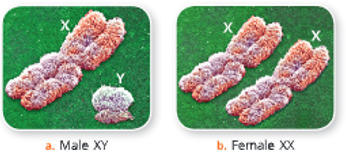
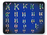
Human karyotype – Human somatic cells have 22 pairs of autosomes and 1 pair of sex chromosomes (XX or XY); Chromosomes are paired by matching their banding pattern and are arranged by size and shape.

Functions of chromosomes

  • Chromosomes are hereditary vehicles that contain genes. All the hereditary information is located on the genes.
  • Chromosomes control the synthesis of structural proteins and thus help in cell division, cell repair and cell growth.
  • By directing the synthesis of enzymatic proteins, chromosomes control cell metabolism.
  • Chromosomes guide development and control cell differentiation.
  • Sex chromosomes (XX and XY chromosomes) determine the sex of individuals.

What is a gene?

Gene:

  • A gene is a basic unit of heredity or inheritance of a character passed from parents to offspring via chromosomes.
  • A gene is a segment of DNA on a chromosome that encodes a particular protein which is expressed in the form of a particular characteristic of the body.
  • Genes are located on specific positions (loci) on the chromosomes.
  • Genes are transmitted from parents to offspring through the gametes.

UP Board Notes For Class 10 Science Chapter 2 Human Chromosomes Summary

  • A chromosome is a thread-like strand of DNA molecule associated with proteins found in nucleus. Chromosomes carry genes and are referred to as hereditary vehicles.
  • Genes are located on chromosomes.
  • The number of chromosomes is constant for a species. Human beings have 46 chromosomes.
  • Depending upon the location of centromere, chromosomes can be metacentric, acrocentric and telocentric.
  • DNA-histone complex in chromosomes is called chromatin.
  • DNA molecule is a double-helical structure. Each single DNA strand is composed of repeating nucleotides.
  • Formation of a new DNA molecule is called DNA replication.
  • There are two kinds of chromosomes in human beings – sex chromosomes (chromosomes that decide the sex of an individual) and autosomes (rest of the chromosomes, somatic chromosomes are same in males and females).

UP Board Notes for Class 10 Science Chapter 5 Transpiration

UP Board Notes For Class 10 Science Chapter 5 Transpiration Learning Objectives

After completing this chapter, you will be able to:

  • Define the terms transpiration, evaporation, guttation and bleeding,
  • Describe transpiration with the help of suitable activities and discuss its significance;
  • Explain the process by which water is made available for transpiration;
  • Explain the mechanism of transpiration;
  • Describe various methods for measuring the rate of transpiration;
  • Describe various factors affecting the rate of transpiration;
  • Explain guttation and bleeding.

Roots of a plant absorb water and minerals from the soil. This water then flows up the root and through xylem vessels in the stem to the leaves. A small amount of this water is used by the plant for photosynthesis and other activities.

Rest of it evaporates from the surface of spongy mesophyll cells and diffuses out of the pores in the leaves called stomata. The loss of water as water vapour from the leaves and aerial parts of the plant is called transpiration.

Transpiration is different from evaporation

Transpiration is different from evaporation. The major differences between transpiration and evaporation are given in Table 5.1.

 Activities to demonstrate transpiration

Given here are certain activities to demonstrate transpiration.

UP Board Notes For Class 10 Science Chapter 5 Transpiration Activity 1

To show that water vapour is given out during transpiration.

  • Take a small-sized, well-watered potted plant. Cover the external surface of the pot and the soil with a cloth.
  • With the help of a transparent polythene bag cover the plant and tie its mouth at the base of the stem. Similarly, take another polythene bag and tie its mouth, this acts as a control. Leave the two in the sunlight observe. You will find that drops of water vapour appear on the inner side of the polythene bag tied around the plant (experimental set-up). This happens due to the saturation of water vapour given out by the leaves. On the other hand, no water vapour appears in the other polythene bag (control).
  • This shows that water vapour is given out during transpiration.

Transpiration-Release-of-water-vapour-by-a-plant-because-of-transpiration.

Transpiration-Differences-between-evaporation-and-transpiration

UP Board Notes For Class 10 Science Chapter 5 Transpiration Activity 2

To demonstrate transpiration in plants.

  • You will require three bell jars and two cobalt chloride papers to perform this experiment.
  • Take two similar-sized, well-watered potted
    plants with broad leaves. Mark plants as A and B.
  • Now cover each plant with a bell jar. In the bell jar containing plant B place strips of dry cobalt chloride paper. Bell jar A Set up A
  • Now you have two set-ups: Set-up A with a potted plant covered with a bell jar, and set-up B with a potted plant having strips of cobalt chloride paper and covered with a bell jar. Take another set-up C having bell jar with cobalt chloride paper but no plant. Seal the edges of each bell jar with vaseline that no air enters the apparatus from outside.Transpiration -experiment-demonstating-transpiration
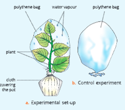
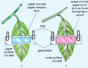
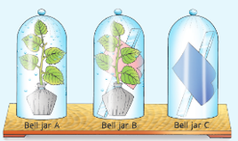
Experiment demonstrating transpiration In plants. Note that bell jars A and B contain water vapour on the inner walls, the cobalt paper turns pink in bell jar B but there is no change in bell jar C

Cobalt chloride paper acts as an indicator of moisture. It is blue when dry but becomes pink when exposed to moisture.

  • Leave the three set-ups undisturbed under the sun for about one hour. Observe after one hour.
  • The bell jar A shows water vapour condensed on its inner walls.
  • The bell jar B shows water vapour condensed on the inner walls. The colour of cobalt paper turns pink in bell jar B.
  • There is no condensation in bell jar C and no
    change in the colour of cobalt chloride paper (i.e. it remains blue). This shows that there was no moisture in the bell jar C.
  • You may conclude that condensation of water vapour occurs and the colour of cobalt paper changes. These are the proofs of transpiration.

UP Board Notes For Class 10 Science Chapter 5 Transpiration How Is Water Made Available For Transpiration?

There is a continuous loss of water from the leaves by transpiration. The water lost from leaves is continuously replaced by more water which flows from leaf xylem vessels. Water flows from xylem vessels to the surface of mesophyll cells of the leaves by two routes.

  • Imbibition: Most of the water flows directly through the porous cellulose walls of the mesophyll cells.
  • Osmosis: A small amount of water flows by osmosis from cell to cell.

Flow of water up the xylem

As a result of transpiration, water pressure of the xylem vessels in leaves and stem becomes lower than that in the root xylem vessels. This pressure difference causes water to be sucked up from the roots into the leaves in a similar manner as we suck cold drink with a straw.

Flow of water from soil to root xylem

Water sucked up from roots to stem and leaves is replaced by water which flows from the soil through the freely permeable cellulose walls of root hair into the root cortex. This flow of water continues as long as a plant transpires.

Types of transpiration

On the basis of occurrence in different regions of a plant, transpiration is of three types:

1. Stomatal transpiration (from the leaves through the stomata)
2. Cuticular transpiration (from the waxy cuticle layer of leaves and stems)
3. Lenticular transpiration (from the lenticels present on the surface of old stems)

The maximum transpiration occurs through stomata on the leaves.

UP Board Notes For Class 10 Science Chapter 5 Transpiration Mechanism Of Stomatal Transpiration

Stomatal transpiration accounts for maximum (about 90%) transpiration in plants. Stomata are the minute openings present on one or both surfaces (epidermal layer) of the leaf. They facilitate exchange of gases between the leaf and the atmosphere. In stomatal transpiration, water vapour is lost through stomata in leaves.

The stomatal transpiration occurs as follows:

  • Water is absorbed by root hair from the soil. This water then moves up through xylem vessels via stem into the spongy mesophyll cells of the leaves through veins.
  • The spongy mesophyll cells in the leaves have large number of intercellular spaces. These cells give out some water as a thin film.
  • The water from this thin film evaporates in the form of water vapour. The water vapour formed then saturates the air in the intercellular spaces.
  • This water vapour then accumulates and reaches the substomatal space through various connecting intercellular spaces by diffusion.
  • From substomatal spaces, the water vapour escapes from the stomata by the process of diffusion. The molecules of water move from the region of their higher concentration to the region of their lower concentration.

Transpiration-from-the-stem,-leaves-and-flowers-of-the-flowering-plant

Transpiration-Process-of-stomatal-transpiration-through-a-leaf.

  • The water from this thin film evaporates in the form of water vapour. The water vapour formed then saturates the air in the intercellular spaces.
  • This water vapour then accumulates and reaches the substomatal space through various connecting intercellular spaces by diffusion.
  • From substomatal spaces, the water vapour escapes from the stomata by the process of diffusion. The molecules of water move from the region of their higher concentration to the region of their lower concentration.

The water lost by cells is replaced by more water from the nearest xylem vessel. Most of the water moves along the cells by imbibition and only a small amount by osmosis.

UP Board Notes For Class 10 Science Chapter 5 Transpiration How Do Stomata Regulate Transpiration?

Stomata Regulate Transpiration As Follows

Stomata are minute openings found in the epidermis of leaves and stem. Each stoma contains a pore surrounded by two guard cells. Both guard cells are firmly joined at both ends but separate in the mid-region of their length.Stomata are present on the lower epidermis of the leaf in dicotyledonous plants.

Transpiration-stomata-with-their-guard-cells

  • The open stomata account for diffusion of water vapour through them. During the day, the cell-sap concentration becomes high due to the accumulation of sugar in the guard cells as a result of photosynthesis.
  • In dicots, the lower surface of a leaf mainly has stomata. Therefore, more transpiration occurs from the undersurface (lower surface) of a leaf.
  • This results in the movement of water into the guard cells from the neighbouring cells. This makes the guard cells turgid and guard cells bulge out and pull apart their inner walls and stomata open. If the availability

UP Board Notes For Class 10 Science Chapter 5 Transpiration Activity 3

To show that more transpiration occurs from the lower surface of a dicot leaf.

(Perform this activity on a relatively dry and non-cloudy day)
This activity should be performed on a dicot leaf. This is because dicot leaf has more stomata on the lower surface and very few on the upper surface.

  • Take a well-watered, potted, dicot plant like balsam or china rose.
  • Take two, 2 x 4 sq cm, pieces of cobalt chloride paper. Place one piece of cobalt chloride paper over a glass slide and hold it on the lower side of the leaf and place other paper on the upper side of the leaf in a similar manner (Fig. 5.6).
  • Fasten both slides (beyond the margins of the leaf) with the help of gem clips. Observe for about1 hour.

Do not detach the leaf from the plant.

  • The piece of cobalt chloride paper on the lower side of the leaf turns pink very fast. while the paper on upper surface remains same or takes a long time to turn pink. This shows that more transpiration occurs from the lower surface of the leaves of dicot plants because of the presence of more stomata.Transpiration-Experiment-to-show-hat-more transpiration occurs-through-the-ower-surface-of-a-dicot-leaf.

of water is reduced, the guard cells lose their turgidity and they become flaccid (lose turgidity) by exosmosis of water from them. This leads to the closing of stomata and transpiration stops.

Cuticular Transpiration

The cuticle is a waxy layer secreted by the epidermis of the leaves and young stems of the plant. Cuticular transpiration accounts for only 3-10% of the total transpiration. It continues throughout day and night.

There is very limited evaporation of water from the cuticle. The cuticular transpiration depends upon the thickness of cuticle. Greater the thickness of the cuticle, lesser is evaporation of water (lower cuticular transpiration) and vice versa.

Can you guess why desert plants have thick cuticle? They have thick cuticle to cut down the loss of water due to transpiration.

Lenticular Transpiration

It occurs in the woody branches of the trees where lenticels are found. Lenticels are special openings on the older stem in place of stomata.The lenticular transpiration accounts for only 0.1% of the total transpiration.

It continues day and night because lenticels do not close. Lenticels remain always open. In lenticular transpiration, water from the cell surface facing the lenticels evaporates directly.

Transpiration-Cross-section-of-old-bark-of-tree-showing-lenticular-transpiration.

Measuring Rrate of Transpiration

There are mainly two methods for measuring the rate of transpiration:

  1. Weighing method
  2. Photometer method

1. Weighing method: In this method, a small lightweight potted plant is weighed before and after a certain period of time in which transpiration has taken place. The loss in weight by the plant during the time indicates the loss of water by transpiration. The following activity shows the measurement of transpiration by weighing method.

UP Board Notes For Class 10 Science Chapter 5 Transpiration Activity 4

To show the loss of water from leaves due to transpiration using weighing method.

1. Take a small potted plant and cover its soil surface with cloth also cover the pot with a polythene to prevent evaporation from the surface of the soil and pot. Weigh the pot at the start of the activity and after 2 hours. The difference in weight is the loss of water by plant due to transpiration.

Transpiration-Measuring-the-rate-of-transpiration-with-the-help-of-a-weighing-balance

  • Take a small plant and fix it in a glass bottle with a graduated side tube as shown in. Fill the bottle with water and note down its reading (water volume). Leave the set-up for 2 hours and observe. Check the level of water in the side tube at leafy the beginning and end of the shoot activity. You will observe that water level has come down. The loss in volume of water can be compared with the loss in weight of the plant with the tube help of a weighing balance.
  • Take a leafy shoot (with no roots) and fix it in a test tube as shown in Figure 5.9. Fill
    the test tube with water and pour some oil on the water surface so as to prevent any water evaporation. Place this test tube in a beaker and weigh the two together. Wait for 2 hours and weigh again. The difference in weight will indicate the loss of water by the shoot due to transpiration.

Transpiration-Experimental-set-up to-measure-the-transpiration

2. Potometer Method: Photometer (Gk. potos: drink, meter: measure) is a device that measures the rate of water intake by a plant (which is almost equal to the water lost by transpiration). There are mainly two types of potometers used in this method – Ganong’s potometer and Darwin’s photometer.

UP Board Notes For Class 10 Science Chapter 5 Transpiration Activity 5

To measure the volume of water uptake by plant during transpiration using potometer method.

Potometer such as Ganong’s potometer is a device made up of glass which is used to measure the rate of transpiration by a leafy shoot. To use the potometer, take a small twig of a plant like Plectranthus and cut the twig obliquely under water to allow larger surface for the water intake.

Fix the twig to one end of the potometer by using a rubber stopper or a cork as aper scopper shown in. Fill the apparatus with water, ensuring there are no air spaces in the set-up.

Introduce an air bubble into the horizontal graduated capillary tube by slightly lifting the tube and then dipping it in a beaker containing coloured water. As the shoot transpires, the water vapour lost is replaced by coloured water drawn in potometer which reaches into the xylem of the stem.

Similarly the air bubble also moves forward in the capillary tube. The reading in the capillary tube gives the volume of water lost in a given time.

Limitations

  • It is very difficult to introduce air bubble in the
  • The twig does not remain alive for a long time.
  • Any change in the external temperature may affect the position of air bubble in the capillary tube.

Transpiration-Ganong's-potometer

UP Board Notes For Class 10 Science Chapter 5 Transpiration Factors Affecting The Rate Of Transpiration

A. External Factors

1. Humidity (Vapour pressure): The humidity of the atmosphere affects the rate of diffusion of water vapour from the stomata. Low humidity in the atmosphere increases the rate of transpiration. On the other hand, high humidity in the air reduces the rate of outward diffusion of water from the sub-stomatal cavity and reduces the rate of transpiration, because the air is already laden with water vapour.

2. Temperature: High temperature increases rate of transpiration by providing the latent heat of vaporization. Rise in temperature increases evaporation of water while low temperature reduces evaporation.

3. Intensity of light: Light affects the rate of transpiration indirectly. More transpiration occurs during the day. Stomata open during light and thus transpiration occurs more in the day. During the night they are closed, thus the rate of transpirationis minimal. Can you guess what happens on a cloudy day?

4. Air movement (Velocity of wind): Moving air sweeps away the water vapour in the air outside the stomata. This speeds up the diffusion of water vapour from stomata. Thus, the rate of transpiration increases with the velocity of wind.

Some plants in dry conditions (xerophytes) have folded leaves or epidermal hair which help to retain moist air around the stomata to prevent transpiration.

5. Atmospheric Pressure: A decrease in atmospheric pressure increases diffusion of water vapour from stomata. The lower the atmospheric pressure, the greater is the rate of evaporation of water from leaves. Thus, the rate of transpiration increases with the decrease in atmospheric pressure.

6. Carbon dioxide concentration: If the increase in the concentration of carbon dioxide is more than the normal (0.03%), the stomata closes. This reduces the rate of transpiration.

7. Water supply to the leaf: Shortage of water in the soil affects the water supply to the leaf and in turn the stomata close and the leaf wilts. This reduces the rate of transpiration.

B. Internal Factor

1. Leaf Structure: The shape of the leaf and the number and position of stomata on it influence water vapour loss. In xerophytes, the cuticle is thick and there is negligible loss of water vapour (less transpiration). On the other hand, in the leaf of the shady plants which has a large surface area and a thin cuticle, the loss of water vapour is more (more transpiration).

UP Board Notes For Class 10 Science Chapter 5 Transpiration How Are Plants Adapted To Reduce Excessive Transpiration?

  • The stomata may be sunken (deep seated) or covered by hair, as in oleander (Fig. 5.11).
  • The number of stomata may be reduced as in xerophytes, like cactus.
  • The leaves may become narrow to reduce leaf surface area, as in Nerium and pine.
  • The leaves may roll over or fold to reduce exposed surface and maintain moisture around the stomata, as in desert grass.
  • There may be very few leaves on a plant to reduce transpiration, such as in Acacia.
  • The leaves may have thick cuticle, as in mango, banyan and most evergreen trees to prevent transpiration.

Transpiration -Sunken-stomata-in-an-oleander-leaf

Significance of Transpiration

Ascent of Sap

Transpiration helps in the ascent of sap due to a suction force or pull created by the loss of water. As water evaporates from leaves, a suction force is developed at the top of the plant, which draws more water from the soil through roots and stems.

Transpiration-helps-in-the-ascent-of-sap

Removal of Excess Water

Plants absorb far more amount of water than is actually required by them. Thus, transpiration helps in removing excess of water.

Cooling Effect

Solar heat during summers increases the temperature of plants, which may be dangerous to the plants. Transpiration helps in reducing the temperature of the leaf surface by evaporation of water vapour from it, which in turn cools the plant.

Distribution of Mineral Salts

The transpiration from leaf surface draws more water from the lower parts and in turn from the soil. This rising column of sap from the soil contains mineral salts. Thus, the mineral salts are distributed to all parts of the plants.

Guttation and Bleeding

Guttation (L. gutta: drop): You must have seen accumulation of water droplets on the surface of leaves in some plants, early in the morning. This is due to guttation. Guttation is exudation of water droplets on the leaf margins of plants such as banana, growing especially in warm humid conditions.

The loss of water in the form of droplets from the margins of the leaves of certain plants during hot and humid conditions is known guttation or exudation It is caused due to root pressure.

When the rate of transpiration is low, the roots of some plants keep accumulating minerals and absorbing water. Thus, more water enters the leaves than is transpired. As a result, the excess water is forced out through specialized structures on the margins of the leaves called hydathodes.

The differences between guttation and transpiration is given in Table .Transpiration-Guttation

Bleeding: During injury to certain plant tissues, the plant sap escapes (bleeds) from the ruptured or cut surface of the plant. This is brought about by the rootpressure generated by the plant.

UP Board Notes For Class 10 Science Chapter 5 Transpiration Summary

  • The loss of water as water vapour from the aerial parts of the plant is called transpiration. Transpiration is different from evaporation.
  • Transpiration produces a suction force which draws water up from the soil to the leaves.
  • There are three types of transpiration – stomatal, cuticular and lenticular transpiration.
  • Stomatal transpiration occurs by diffusion of water vapour through stomata and accounts for maximum transpiration by a plant.
  • Cuticular transpiration involves the loss of water vapour from the surface of leaves and young stems of the plant. It occurs throughout the day and night.
  • Lenticular transpiration occurs through lenticels found in the woody branches of trees. It occurs continuously.
  • Transpiration rate can be measured by weighing method or with the help of a potometer.
  • Rate of transpiration increases with an increase in the intensity of light, temperature, velocity of wind and availability of water. High humidity in the air reduces the rate of transpiration.
  • There are many advantages of transpiration for a plant such as cooling, ascent of sap, distribution of minerals and removal of excess water.
  • Guttation is loss of water from the leaf margins of plants in the form of droplets.

UP Board Notes for Class 10 Science Chapter 1 Cell Cycle and Cell Division

UP Board Notes for Class 10 Science Chapter 1 Cell Cycle And Cell Division Learning Objectives

After completing this chapter, you will be able to:

  • Explain the need for cell division;
  • Describe the cell cycle and list its various phases;
  • Describe two types of cell division and list their phases;
  • Draw diagrams to explain the sequence of events in the two types of cell divisions;
  • Tabulate the differences between mitosis and meiosis;
  • State the significance mitosis and meiosis;
  • Explain how chromosome number is maintained during mitosis while it is halved during meiosis;
  • Recognize homologous chromosomes and crossing over as a means of variations.

All living beings are made up of cells. A cell is the structural and functional unit of life. New cells arise from the pre-existing cells by the process of cell division.

Cell division occurs in all living organisms. In unicellular organisms, cell division directly produces two individuals. In multicellular organisms, life begins from a single cell, the zygote, which divides and redivides into a number of cells to form a complete organism.

In multicellular organisms, there are two types of cells.

  1. The somatic cells or the body cells – They form the body of an organism.
  2. The reproductive cells or sex cells – They are gamete-producing cells. They take part in reproduction of the organism.

UP Board Notes for Class 10 Science Chapter 1 Cell Cycle And Cell Division Why Do Cells Divide?

The cells divide to produce new cells.

The new cells are produced for:

  1. Growth: During cell division, a single cell divides to produce new cells that in turn devide and redivide to form a cluster of similar cells. These cells form tissues and organs. Cell division is essential for the growth of an organism.
  2. Replacement of dead cells: The existing cells in our body a destroyed regularly. These cells should be replaced by new cells for the normal functioning of the body. Cell division of the parent cells in the bone marrow helps in the replacement of dead cells with the new cells.
  3. Repair of tissues: In case of injuries or normal wear and tear of tissues, cells divide and new cells fill up the broken cut ends to heal wounds. Thus, cell division is essential for the repair of the tissues.
  4. Reproduction: The sex cells (sperms and eggs) are formed because of cell division (meiosis). These sex cells contain only half the normal number of chromosomes. During fertilization, these sex cells combine to form zygote. Thus, cell division is essential for reproduction.

UP Board Notes for Class 10 Science Chapter 1 Cell Cycle And Cell Division The Cell Cycle

A cell cycle or a cell division cycle is a series of events in a cell, leading to its division and duplication of its DNA to produce two daughter cells. Mitosis is only one phase of the cell cycle.

A cell cycle starts from the time a new cell is formed and ends when it completes its own division. Then the cell cycle starts again for each new daughter cell formed. A cell cycle has two basic phases:

  •  Interphase
  • M phase or Mitosis

A cell cycle may be defined as a series of events in a cell leading to an increase in the mass and cytoplasmic components of the cell, duplication of DNA, and then division of nucleus and cytoplasm of the cell and finally forming two daughter cells. A cell cycle extends from the time a cell is formed till the time it completes division.

Interphase

During interphase, a cell prepares itself for cell division. The interphase is the longest phase of a cell cycle. It is metabolically the most active phase of the cell cycle. It has three sub-phases:

  1. G₁ or first growth phase: This is the first ‘Gap’ (interval) phase of the cell growth before DNA replication. During this phase:
  •  RNA and proteins are synthesized. Cell size increases due to the increase in the volume of the cytoplasm to almost double.
  • Mitochondria (in plant and animal cells) and chloroplasts (in plant cells) divide.
  • The cell is ready for DNA synthesis.
  • Now the cell enters either the resting phase (Go phase) or a synthesis phase (S phase).
  1. S or synthesis phase: This is the phase of DNA replication. The DNA is synthesized and chromosomes are duplicated during this phase but they remain attached.
  2. G₂ or second growth phase: It is the second ‘Gap’ phase after DNA replication. It is a shorter phase in which RNA and proteins necessary for cell division continue to be synthesized. Now the cell becomes ready for next the cell division, i.e. mitosis.Go phase (Resting phase): This is the phase when the cell has stopped dividing and left the cell cycle. It is the resting state of a cell. The cell remains metabolically active.

M Phase Or Mitosis

  • During M-phase, cell growth stops. Nuclear division (prophase, metaphase, anaphase and telophase) takes place, which is usually followed by cytoplasmic division. The various phases of cell cycle are given in

The cell cycle does not go on endlessly. It stops permanently at some point of time.

cell-cycle-and-cell-division-Eukaryotic-cell-cycle,-generalized.-The-length-of-each-part-differs-among-different-cell-types.1         

  •  Its duration differs from one cell type to another. For example, nerve cells (neurons) in our brain once formed in embryo do not divide further. All blood cells form and replace the worn-out cells at an average rate of 2-3 million each second. Surface skin cells are continuously replaced by underlying cells. Liver cells divide once every 1-2 years to replace damaged cells.

Cell-cycle-and-cell-division-Summary-of-cell-cycle-2

Types Of Cell Division

There are two types of cell division in higher organisms.

  1. Mitosis – Occurs in somatic cells, leading to growth and development.
  2. Meiosis – Occurs in reproductive cells or sex cells, leading to gamete formation.

Karyokinesis and cytokinesis The cell division has two phases. First is the nuclear division or karyokinesis, leading to division of the parent nucleus into daughter nuclei. This is followed by the division of cytoplasm or cytokinesis, leading to the division of the parent cell into daughter cells.

UP Board Notes for Class 10 Science Chapter 1 Cell Cycle And Cell Division Mitosis

  • (Gk. mitos: thread)
    Mitosis or mitotic cell division is an equational division in which one parent cell divides to form
  • Two daughter cells. The daughter cells formed areidentical to each other and also to the parent cell inevery respect. In mitosis, the same chromosome number of the parent cell is maintained at eachstage of the mitotic division of the cell and henceit is referred to as equational division or homotypic division I

Interphase – The Resting Phase

  • Interphase is the growth period between two successive divisions of a cell. Thus, it is a preparatory phase just before the cell starts dividing. This stage is said to be a resting phase because no external change in chromosomes is visible. However, the

Cell-cycle-and-cell-division -stages-of-mitosis-in-an-animal-cell-3


Cell-cycle-and-cell-division       -mitosis-in-a-plant-cell-4                       

  • cell is metabolically most active and prepares itself for the division by synthesizing almost the double amount of DNA (the genetic material) and RNA. The volume of cytoplasm and nucleus increases during this stage. However, chromosomes are not yet distinguished.                                                                                                               

Karyokinesis – Phases of mitosis

  • The nuclear division or karyokinesis during mitosis occurs in four phases – prophase, metaphase, anaphase and telophase.
  • Somatic cells are body cells (like skin cells, brain cells, etc.) that contain normal number of chromosomes (called diploid number, 2n).
  • Gametes are sex cells (sperm and ova) that contain half the normal number of chromosomes (called haploid number, n).

UP Board Notes for Class 10 Science Chapter 1 Cell Cycle And Cell Division Prophase (Gk. pro: first, phasis: stage)

  • This is the first and the longest phase of mitosis.
  • The chromatin material undergoes condensation (becomes short and thick) and changes into thread-like structures called chromosomes.
  • Each chromosome duplicates to form two sister chromatids. The two sister chromatids lie close to each other and remain attached at a point called centromere.
  • As prophase advances, the chromosomes become shorter and thicker (condensed).
  • In animal cells, the centrosome initiates and regulates the cell division. The centrosome splits into two small round bodies called centrioles. The two centrioles migrate to the opposite poles of the cell.
  • Soon two radiating fibres known as Between the separating centrioles, spindle fibres asters are formed around the centriole at each pole. are formed by the aster.
  • Towards the end of the prophase, the nucleolus and the nuclear membrane disappear.
  • The duplicated chromosomes start moving towards the equator.

Spindle formation in plant cell

  • In plant cells, no centrosome (centriole) is there and asters are not formed. However, spindle formation still occurs and spindle fibres are formed by microtubules (cytoplasmic strands).

Cell Cycle and Cell Division Metaphase (Gk. meta: between, phasis: stage)

  • The chromosomes arrange themselves on the equatorial plane.
  • Each chromosome is attached by a spindle fibre at its centromere.
  • Small disc-shaped structures are present at the surface of centromere. These are called kinetochores. Chromosomes lie at the equator with one chromatid connected by its kinetochore to spindle fibres from one pole and another chromatid connected by its kinetochore to spindle fibres of opposite pole.

Cell Cycle and Cell Division Anaphase (Gk. ana: back, phasis: stage)

  • This is the shortest phase of mitosis.
  • The centromere of each chromosome divides into two halves (sister chromatids) so that each chromatid has its own centromere.
  • The sister chromatids separate and begin to move towards the opposite poles due to the contraction of spindle fibres, and due to the repelling force developed between the sister chromatids.
  • Depending on the position of the centromere, the chromosomes appear as U, V or J-shaped.
  • The anaphase ends when all the chromatids (now behaving like chromosomes) reach the opposite poles.

Cell Cycle and Cell Division Telophase (Gk. telo: end, phasis: stage)

  • This is the last phase in karyokinesis (nuclear division). The events of prophase occur in reverse sequence during telophase.
  • The chromatids (daughter chromosomes) uncoil, elongate and change into network of chromatin threads.
  • The nuclear membrane reappears around the chromatin network at each pole.
  • Nucleolus reappears in each daughter nucleus.
  • Spindle fibres disappear.
  • In animal cells, centrosome organizes itself above the nucleus, thus completing karyokinesis.

The karyokinesis (mitosis proper) is followed by the division of the cytoplasm known as cytokinesis.

UP Board Notes for Class 10 Science Chapter 1 Cell Cycle And Cell Division  Cytokinesis (Gk. cyto: cell, kinesis: movement)

  • It is the division of the cytoplasm to form two daughter cells. It begins during late anaphase and is completed soon after telophase. At the end of the telophase, a furrow appears in the middle of the cell membrane which splits the cytoplasm into two halves to form two daughter cells. Cytokinesis is different in animal and plant cells.

How is cytokinesis different in animal and plant cells?

  1. In animal cells, a constriction (or furrow) appears in the cell(or plasma) membrane. This constriction deepens by theend of the telophase, finally completing the division ofcytoplasm.
  2. In plant cells, a cell plate is formed in the centre of thecell at the equator. The cell plate extends on either sideuntil it completely divides the cell into two daughter cellsIn animal cells, cytokinesis starts from the periphery andproceeds towards the centre, but in plant cells, cytokinesisstarts from centre due to the cell plate formation, andextends towards the periphery.

Differences between mitosis in an animal cell and a plant cell is given in

cell cycle and cell division  Eukaryotic cell cycle, generalized. The length of each part differs among different cell types. 7 differences between mitosis in animal plant cells

Significance of mitosis

  1. Mitosis maintains the same number of chromosomes in all the cells of an individual.
    In other words, mitosis is an equational division in which two daughter cells produced are identical to each other and even to their parent cell. This type of cell division usually takes place in the somatic cells such as tips of roots, stems, etc. In animals, zygote develops into mature organism through mitotic cell divisions.
  2. It plays a significant role in the replacement of cells lost during wear and tear, and in wound healing.
  3. It is responsible for the growth of an organism.A fertilized cell develops into an embryo and finally into an adult as a result of mitotic cell division.
  4. Mitosis helps the cells in maintaining the proper size.
  5. It is a method of asexual reproduction in unicellular organisms.
  6. If mitotic cell division becomes uncontrolled, it may cause tumours or cancerous growth.

UP Board Notes for Class 10 Science Chapter 1 Cell Cycle And Cell Division Meiosis-The ReductionDivision (Gk. meioun: to diminish)

Meiosis takes place in the reproductive cells that produce gametes, i.e. sperms and ova. Meiosis is a modified mitosis in which chromosomes divide once and the nucleus divides twice. As a result of this the number of chromosomes is reduced to half. Thus, the four cells resulting from a meiotic division have a haploid number of chromosomes It means that the number of chromosomes of parent cell (diploid) becomes half in each sex cell. This is because when the male and female gametes fuse during fertilization, the diploid (double) number of chromosome pairs is restored. Meiosis is a reductional division.

Meiosis has two nuclear divisions.

  1. First meiotic division (reduction division)
  2. Second meiotic division (mitotic division/ equational division)

Thus, in a meiotic cell division, all the stages, i.e.prophase, metaphase, anaphase and telophase are repeated twice.

During meiosis, the diploid cells are reduced to haploid cells (number of chromosomes is halved).

Diploid → Haploid
(2n)        (n)

In the absence of meiosis, the number of chromosomes will double and the offspring will not be able to survive.

cell cycle and cell division  Eukaryotic cell cycle, generalized. The length of each part differs among different cell types.6 meiosis_ the reduction division

First meiotic division (Meiosis I)

Homologous chromosomes

  • These are chromosome pairs, with each pair containing one chromosome from the father and one from the mother. They are not identical. They are similar in length, gene position and location of centromere. The gene, however, contains different alleles.

Cell-cycle-and-cell-division-Homologous-pair-chromosomes-4

  • Homologous chromosomes come together (associate) and subsequently segregate into daughter cells during meiosis I. Thus, the number of chromosomes is reduced from diploid (double) to the haploid (single) state. That is why it is known as reduction division.

The following events take place during this division.

Pairing of homologous chromosomes

  • During prophase I, the longest part of meiosis I, the homologous chromosomes attract each other and come to lie in pairs. The pairing of homologous chromosomes is known as synapsis and the pair is known as bivalent.

Chiasmata formation or crossing over

  • Chromosome continues to shorten and thicken. Each chromosome splits lengthwise into two chromatids (sister chromatids) so that each homologous pair now has four chromatids and is termed as tetrad.Cell-cycle-and-cell division-Crossing-over-between -maternal and-paternal-chromatids-5
  • The non-sister chromatids of a tetrad break open and rejoin each other. This is known as crossing over or chiasmata (singular: chiasma) formation.
  • Exchange of some genes or portions of chromatids takes place between paternal and maternal chromatids of a pair of homologous chromosomes during meiosis. This is known as crossing over
  • Homologous pairs arrange themselves at equator during metaphase I, the second phase of meiosis I. Nucleolus and nuclear membrane disappear.

UP Board Notes for Class 10 Science Chapter 1 Cell Cycle And Cell Division Separation Of Homologous Pairs

  • The members of homologous chromosomes completely separate from each other and move. towards the opposite poles during anaphase-I. Nuclear membrane reappears, leading to the formation of two daughter nuclei at telophase I.

Second meiotic division (Meiosis 2)

  • It is similar to mitosis. During this division, the two chromatids of each chromosome separate and move to opposite poles. Nuclear membrane reappears and four cells are formed. Finally each cell formed is haploid (n), i.e. it contains half the number of chromosomes of the original cell (diploid, 2n).

Significance of meiosis

  1. The number of chromosomes is reduced to half in the daughter cells.
  2. It results in the formation of haploid sex cells (sperms and ova), which after fertilization restore the diploid number of chromosomes in the zygote.
  3. During crossing over, that occurs in meiosis, parts of chromatids are exchanged between homologous chromosomes which brings about variations in the offspring.
  4. The four chromatids of a homologous pair of chromosomes are passed on to four different daughter cells. This also causes gametic variation.
  5. It prevents the multiplication of chromosomes, and thus maintains the stability of species.Differences between mitosis and meiosisThe major differences between mitosis and meiosis are given in

Cell-cycle-and-cell-division-Differences-between-mitosis-and-meiosis-6

UP Board Notes for Class 10 Science Chapter 1 Cell Cycle And Cell Division Summary

  • New cells arise from pre-existing cells by the process of cell division.
  • Cell division is necessary for growth, replacement of dead cells, healing of wounds, and reproduction.
  • A cell division starts when a new cell forms. It proceeds through interphase and ends when the cell reproduces by mitosis and cytokinesis. The cell prepares itself for division during interphase.
  • There are two types of cell divisions mitosis and meiosis.
  • Mitosis is an equational division required for growth and development. A diploid cell divides to form two genetically identical diploid cells in mitosis.
  • Mitosis maintains the same number of chromosomes in all the cells of an individual.
  • Meiosis is a special type of cell division which produces sex cells or gametes. It is known as reduction division. One diploid cell divides to produce four genetically different haploid cells as a result of melosis.
  • Meiotic division prevents the multiplication of chromosomes, and thus maintains the stability of species
  • The cell cycle has following two basic phases – Interphase [first growth phase (G₁), synthesis phase (S) and second growth phase (G₂)] and mitosis.
  • Mitosis has four phases prophase, metaphase, anaphase, and telophase.
  • Meiosis has two nuclear divisions – first meiotic division and second meiotic division.

UP Board Notes for Class 10 Science

Chapter Wise UP Board Notes for Class 10 Biology Pdf free download was designed by expert teachers from latest edition of UP Board books to get good marks in board exams. UP Board Class 10 Biology Notes contains Textbook Readers and Supplementary Readers of all chapters are part of Revision Notes for grade 10 Biology. Here we have given notes Class X.

UP Board Notes for Class 10 Science

UP Board Notes for Class 10 Science Chapter 9 Excretion-Elimination of Body Wastes

UP Board Notes for Class 10 Science Chapter 9 Excretion-Elimination Of Body Wastes Learning Objectives

After completing this chapter, you will be able to:

  • Define excretion and mention its importance;
  • Describe the nature of excretory wastes produced in our body,
  • Identify different parts of the excretory system and describe their functions; Excretion – Elimination of Body Wastes
  • Describe the gross anatomy of the kidney,
  • Identify the parts of the nephron responsible for urine formation;
  • Explain the physiology of urine formation; describe the mechanism of osmoregulation by the kidney.

Various waste products are formed in our bodies continuously as a result of many metabolic activities taking place inside the body. These waste substances, if accumulated, would poison cells or slow down metabolism. Hence, the body must get rid of these unwanted substances. The metabolic wastes to be excreted include CO₂, H₂O, fats, ammonia, urea, uric acid, etc.

UP Board Notes for Class 10 Science Chapter 9 Excretion-Elimination Of Body Wastes Excretion-Elimination of Body Wastes Excretion

  • The removal of waste products formed in the body as a result of metabolism is termed excretion. Excretion can also be defined as the removal of mainly nitrogenous wastes from the body. The lungs and kidneys are the main organs of excretion in human beings. The process of maintaining the right amount of water and proper ionic balance in the body is called osmoregulation.

UP Board Notes for Class 10 Science Chapter 9 Excretion-Elimination Of Body Wastes Nature Of Excretory Wastes

  • The wastes formed as a result of various metabolic activities are:

1. Respiratory waste formed as a result of oxidation of glucose (food) (through cellular respiration):

  • These mainly constitute carbon dioxide and water.
  • The carbon dioxide is eliminated from the body through the lungs (during expiration).
  • Water becomes a part of the rest of the water contained in the body.

2. Nitrogenous waste formed as a result of deamination of unwanted amino acids, body’s own proteins and nucleic acids: The three main nitrogenous waste products excreted by animals are ammonia, urea and uric acid. Urea is a highly poisonous waste product and its accumulation beyond a certain concentration in the body may cause death. Urea is eliminated through the kidneys.

3. Other waste like salts (NaCl), excess vitamins and water, which we take directly through food: Excess salts are mainly excreted by kidneys.

4. The excess water is removed by kidneys as urine and some of it is excreted as sweat by the sweat glands on our skin.

5. Bile pigments are formed due to the breakdown of haemoglobin in dead RBCs in the liver. A large amount of these pigments is excreted out in faeces while some of it is excreted in the urine.

UP Board Notes for Class 10 Science Chapter 9 Excretion-Elimination Of Body Wastes Human Urinary System

The urinary system in human beings consists of the following parts.

  1. A pair of kidneys
  2. A pair of ureters
  3. Urinary bladder
  4. Urethra

1. Kidneys

  • The kidneys are reddish-brown, paired structures located on either side of the vertebral column (backbone). They are protected by the last two pairs of ribs. Kidneys in human beings are said to be retroperitoneal because they lie behind the peritoneal lining of the abdominal cavity.
  • Each kidney is a bean-shaped organ, about 10 cm long, 6 cm wide and 4 cm thick. The right kidney is positioned slightly at a lower level than the left kidney as the right side of the abdominal cavity is occupied by the liver. They are held in position by a padding. On the inner concave side of the kidney, a notch called the hilum is present. It is the place from where the ureter and the renal vein come out and the renal artery enters the kidney.

2. Ureters

  • The ureters arise from within the renal sinus of the kidney at hilum. The front end of each ureter is extended into a funnel-shaped structure, the renal pelvis. The ureters transport urine from the kidneys to the urinary bladder. Each ureter opens obliquely in the urinary bladder by a slit-like aperture to prevent the backflow of the urine.

3. Urinary bladder

  • The urinary bladder is a muscular reservoir for storage of urine. It lies in the pelvic cavity of the abdomen. The neck of the urinary bladder is surrounded by sphincters. Sphincter acts like a valve which remain closed until the time of micturition (urination) described below.

UP Board Notes for Class 10 Science Chapter 9 Excretion-Elimination Of Body Wastes Role Of The Urinary Bladder In Urination

  • Besides functioning as a temporary reservoir of urine, the bladder also evacuates the urine at suitable intervals. The act of voiding or discharging urine is called micturition.

Excretion-Elimination-of-Body-Wastes-The-human-urinary-system

  • When enough urine gets accumulated in the bladder to raise its pressure sufficiently, a spontaneous nervous activity (reflex) is initiated. This causes the smooth muscles on the bladder wall to contract and the urethral sphincter to relax. Urine flows to the outside. As the ureters enter obliquely through the bladder wall, their openings get shut due to compression by the contracting bladder muscles and backward flow of urine is prevented.

4. Urethra

  • The urethra is a tube that arises from the neck of urinary bladder and extends up to the outside. In males, it serves as a common passage for urine and sperm. The urethra in females serves as a passage for urine only.

UP Board Notes for Class 10 Science Chapter 9 Excretion-Elimination Of Body Wastes Internal Structure Of The Kidney

  • Each kidney in a longitudinal section shows two distinct regions- an outer renal cortex and an inner renal medulla. The medulla is subdivided into 15 or 16 conical masses, the renal pyramids. Due to presence of these pyramids, the medulla has a striped appearance.
  • Each pyramid has a narrow renal papilla toward the pelvis and a broad base toward the cortex. Renal papillae open into a wide funnel-like structure, renal pelvis. The renal pelvis in turn leads into the ureter.

Excretion-Elimination-of-Body-Wastes-The-structure-and-location-of-nephron-in-a-kidney

Nephron – structural and functional unit of kidney

  • Each kidney consists of more than one million nephrons or uriniferous tubules. Nephron is the functional unit of a kidney. Each nephron consists of – a Malpighian capsule, a nephric tubule (secretory part of uriniferous tubule) and a collecting tubule.

UP Board Notes for Class 10 Science Chapter 9 Excretion-Elimination Of Body Wastes Structure Of Malpighian Capsule

  • It has two parts a Bowman’s capsule and a glomerulus.
  1. Bowman’s capsule: It forms the dilated blind end of the nephron. It is a double-walled, cup-shaped structure. It is lined by a thin semipermeable squamous epithelium. The outer concavity of Bowman’s cup contains a knot-like mass of blood capillaries called glomerulus.
  2. Glomerulus: The afferent arteriole enters the Bowman’s capsule and divides into a bunch of about 50 capillaries. This bunch is called glomerulus. Their diameter gets reduced to increase the blood pressure. Bowman’s capsule together with the glomerulus are called renal capsule or Malpighian capsule.

UP Board Notes for Class 10 Science Chapter 9 Excretion-Elimination Of Body Wastes Nephric Or Uriniferous tubule

  • It is a long, coiled tubule and can be divided into three major -regions the proximal convoluted tubule (PCT), the loop of Henle and the distal convoluted tubule (DCT).
  1. Proximal convoluted tubule (PCT): It is the initial (proximal) convoluted region of the nephric tubule nearer to the Bowman’s capsule. It is a highly coiled structure and is about 12-24 mm in length. It is lined by the columnar epithelium and is provided with microvilli. PCT is located in the cortex region of kidney.
  2. Loop of Henle: It is a U-shaped loop formed in the middle of the nephric tubule. It has a thin descending limb and a thick ascending limb. The descending limb is lined with flattened epithelial cells, while the ascending limb is lined with cuboidal epithelium. It is not convoluted. It runs in the medulla and turns back to cortex region.
  3. Distal convoluted tubule (DCT): It is a convoluted structure and is present in the cortex region. It is lined by cuboidal epithelium. It opens into the collecting tubule.

UP Board Notes for Class 10 Science Chapter 9 Excretion-Elimination Of Body Wastes Collecting Ducts And The Ducts Of Bellini

  • Collecting ducts are larger ducts, each receiving collecting tubules from nephrons. These pass into the renal medulla and join with each other forming still larger ducts of Bellini. These ducts drain the urine collected from the nephrons into the renal pelvis which leads to the ureter.

Blood Supply To Kidney Tubules

  • Each kidney receives blood supply from renal arteries branching off from dorsal aorta. These arteries further branch off into arterioles and each arteriole, called afferent arteriole, enters inside Bowman’s capsule.
  • It branches further and forms a mass of capillaries called glomerulus. The fine capillaries of glomerulus reunite and form efferent arteriole. Efferent arteriole comes out of Bowman’s capsule and surrounds renal tubule. They further rejoin and form renal veins which kidney and empty into posterior vena cava.

UP Board Notes for Class 10 Science Chapter 9 Excretion-Elimination Of Body Wastes Physiology Of Urine Formation

  • Urine formation involves three main processes – ultrafiltration, tubular reabsorption and tubular secretion.

1. Ultrafiltration

  • Walls of glomerular capillaries and Bowman’s capsule are very thin and semipermeable in nature. Hence, they act as ultrafilters.
  • The glomerular hydrostatic pressure or the capillary pressure is the main driving force that tends to move fluid out of the glomeruli. It is exerted by the blood while passing through the glomerulus.

Why is hydrostatic pressure more in glomerulus?

Hydrostatic pressure is more in glomerulus because

  • The afferent arteriole entering the Bowman’s capsule is wider than the efferent arteriole leaving it. As a result, more blood enters the glomerulus while less blood moves out. This is the reason behind the high hydrostatic pressure in the glomerulus.
  • Due to this high filtration pressure in the blood of glomerulus, a part of water and dissolved constituents of blood (like nitrogenous wastes, glucose, amino acids, mineral ions, etc.), are filtered out in the Bowman’s capsule. This forms the glomerular filtrate. Filtration under high pressure is called ultrafiltration.

Excretion-Elimination-of-Body-Wastes-Filtration-in-the-glomerulus-(ultrafiltration)

  • During ultrafiltration, nearly all the liquid components of blood come out of glomerulus and pass into the funnel of Bowman’s capsule. The rest of the blood, left behind in the glomerulus after ultrafiltration is very thick (because the liquid part has been removed) and is carried forward by the efferent arteriole.
  • The glomerular filtration rate in adults is 120 mL per minute and the filtrate produced daily amounts to about 172 litres.

2. Tubular reabsorption or selective reabsorption

  • Our body cannot afford to lose several useful substances that are filtered in the glomerular filtrate. As this filtrate flows through the nephric tubule, water and several useful substances (solutes) are reabsorbed through the wall of the renal tubule (nephric tubule) and returned to the blood flowing in peritubular capillaries. This is called selective reabsorption.
  • Water is reabsorbed by osmosis. Selective reabsorption of glucose, amino acids and salts also take place.

Excretion-Elimination-of-Body-Wastes-Role-of-nephron-in-urine-formation

  • In the PCT, 65-80 per cent of the filtrate is reabsorbed. It includes water, glucose, amino acids and minerals like Na+, K+ and Ca++.
  • In the descending limb, 5 per cent water is reabsorbed.
  • In the ascending limb, only minerals (Na+, K+, Cl-) are reabsorbed. The ascending limb is impermeable to water.
  • In the DCT and collecting ducts, Nat is reabsorbed under the influence of hormone aldosterone and water is reabsorbed under the influence of hormone ADH (antidiuretic hormone).

3. Tubular secretion

  • This process is the converse of tubular reabsorption. The cells of the renal tubule also remove wastes from blood and pass into the filtrate by the process of secretion. Tubular secretion removes ammonia, urea, uric acid, creatinine and hippuric acid from the blood. The function of various parts of renal tubule in formation of urine are given in Table.
  1. Blood from renal artery enters kidneys.
  2. Ultrafiltration: Blood enters glomerular capillaries. Water and small solutes are filtered in Bowman’s capsule.
  3. Tubular reabsorption: Water and many solutes are reabsorbed through the wall of nephric tubule and return to the blood in peritubular capillaries.
  4. Tubular secretion: Cells of renal tubule remove wastes from blood and pass them into filtrate.
  5. Hormonal action adjusts the urine. concentration. ADH promotes water reabsorption. Aldosterone influences reabsorption of sodium and potassium.
  6. Urination: Water and solutes flow to renal pelvis, then eliminated out through urinary tract.Excretion-Elimination-of-Body-Wastes-Function-of-various-parts-of-renal-tubule-in-urine-formation

UP Board Notes for Class 10 Science Chapter 9 Excretion-Elimination Of Body Wastes Hormonal Control Of Urine Formation

  • Urine formation is under the control of two hormones – antidiuretic hormone (ADH) and aldosterone.
  1. Antidiuretic hormone (ADH) or vasopressin released from the posterior lobe of pituitary gland, increases the permeability of the renal tubule to water. When the blood plasma is concentrated, more ADH is secreted and more water is reabsorbed in the urinary tubules. Hence, urine passed out from body is concentrated, for example in summer. When the blood plasma is diluted, less ADH is secreted and less water is reabsorbed in the urinary tubule. In this case, a large amount of dilute urine is passed out of the body, for example in winter.
  2.  Aldosterone, a hormone released from the adrenal cortex, influences reabsorption of sodium and potassium. This hormone regulates the amount of sodium and potassium that is to be retained in the blood and is to be excreted.

UP Board Notes for Class 10 Science Chapter 9 Excretion-Elimination Of Body Wastes Urine And Its Composition

  • An adult man normally passes about 1-1.5 litres of urine in a day. Urine is a transparent yellowish fluid. Its yellow colour is due to a pigment urochrome derived from the breakdown of haemoglobin of worn-out RBCs,
  • Its pH range is 5-8. It has an unpleasant odour. If allowed to stand, the urea present in urine is degraded by bacteria to ammonia, which gives a strong awful smell to it.
  • In normal composition, urine consists of 95 per cent water, and rest 5 per cent is the organic and inorganic substances (solid wastes). Each litre of urine contains nitrogenous organic compounds – urea 2.3 g, uric acid 0.7 g, creatinine 1.5 g and a negligible amount of hippuric acid. Of these, urea is the principal nitrogenous waste in human urine. The non-nitrogenous organic compounds include vitamin C, oxalic acid and phenolic substances.
  • The inorganic substances include ammonia (0.6 g/L) and mineral salts, such as phosphates, sulphates and chlorides of Na, K, Ca and Mg. Sodium chloride (9.0 g/L) is the principal mineral salt in the urine.
  • Abnormal components of urine include albumin, bile salts, bile pigments, glucose and ketone bodies, which occur in the urine in various pathological conditions.

Excretion-Elimination-of-Body-Wastes-The-process-of-urine-formation

Common diseases related to abnormal composition of urine

  • Glycosuria: Urine with high glucose due to diabetes mellitus
  • Albuminuria: Urine with albumin due to high blood pressure
  • Ketonuria: Urine with ketone body
  • Jaundice/Hepatitis/Liver cirrhosis: Urine with bile pigment
  • Pyuria: Urine with pus cells
  • Haematuria: Urine with blood cells due to urinary tract infection or kidney stone
  • Diabetes insipidus: Urine with excess of water (frequent passing out of large quantities of urine) due to deficiency of ADH or vasopressin

Role ADH and insulin in urine

  • The hormone vasopressin or antidiuretic hormone (ADH) regulates the output of urine. This hormone is secreted by the posterior lobe of the pituitary gland. When the secretion of this hormone gets reduced, the amount of urine produced is increased, causing diabetes insipidus disorder. Sometimes hyposecretion of the insulin hormone causes diabetes mellitus disease in which excess glucose is passed with urine.

UP Board Notes for Class 10 Science Chapter 9 Excretion-Elimination Of Body Wastes Osmoregulation By The Kidney

  • The water and solute content of body fluids is regulated by the kidney. This function of the regulation of osmotic concentration of the blood by the kidney is called osmoregulation.
  • In human beings, the kidney is extremely flexible in its working. Generally, in summer when most of water is lost from the body by perspiration, the urine passed out is hypertonic.
  • However, in winter as there is no perspiration, the urine passed out is hypotonic. This flexibility in the working of human kidney and renal tubules is brought about by the movement of water and Na+ in and out of nephron under the influence of certain hormones, thus osmolarity is regulated.

When there is excess of water in body fluids

In such a condition, the urine passed out of the body is more dilute (hypotonic) than the body fluids. It is achieved by two processes:

  • Excess of water increases blood volume which increases the hydrostatic pressure in glomerulus, hence, more nephric or glomerular filtrates are formed.
  • The sensation of excess of water in the body fluid is received by osmoregulator cells in the hypothalamus part of the brain.
  • These cells in turn influence the posterior lobe of the pituitary gland to inhibit or reduce the release of antidiuretic hormone (ADH). A deficiency of ADH reduces the permeability of cells in the distal convoluted tubule and collecting duct, thereby decreasing the real notion of water.
  • More filtration and less reabsorption of water produce excessively dilute urine. This brings down the volume of the body fluids to normal.

If the body fluids fall below normal

  • During summer, due to excessive sweating, more water is lost, the body fluids reduce. As a result, more and more water is reabsorbed by kidney tubules back into the blood and urine turns thick and yellow. To maintain water balance of the body, the following changes take place in the body.
  • The glomerular filtration slows down due to decreased filtration pressure in the glomerular capillaries.
  • The osmoreceptor cells of the hypothalamus send impulses to the posterior pituitary lobe to release ADH. ADH increases the reabsorption of water in the distal convoluted tubule and the collecting duct by making them more permeable to water.
  • Less filtration and more reabsorption of water leads to the discharge of small amounts of hypertonic urine. This raises the volume of body fluids to normal.

Urine is slightly thicker in summer than in winter

  • During summer, when the temperature is high, lot of water is lost from the body through sweating (perspiration). To make up for this loss, more and more water is reabsorbed by the kidney tubules and put back into the blood making urine more concentrated (thicker).

Why do we feel thirsty despite drinking water at regular intervals?

  • If enough quantity of ADH is not released, water cannot be retained by the kidney tubule and is lost as urine. This causes increase in thirst. Whatever water we drink is lost again. In severe conditions, it causes dehydration and electrolyte imbalance in the body.

UP Board Notes for Class 10 Science Chapter 9 Excretion-Elimination Of Body Wastes Renal Failure And Artificial Kidney

  • Artificial kidney is used to filter the blood of a patient whose kidneys are damaged. The patient is said to be put on dialysis and the process of purifying blood by an artificial kidney is called haemodialysis.
  • Artificial kidney is a dialysis machine that contains a number of tubes, suspended in dialysing fluid. The patient’s blood is passed through these tubes. During this process, nitrogenous waste products from the blood pass into dialysing fluid. The purified blood is pumped back into the patient’s body.

UP Board Notes for Class 10 Science Chapter 9 Excretion-Elimination Of Body Wastes Summary

  • The removal of waste products formed in the body due to metabolic activities is termed excretion.
  • Kidneys in human beings are said to be retroperitoneal because they lie behind the peritoneal lining of the abdominal cavity.
  • Each kidney In longitudinal section shows two distinct regions an outer renal cortex and an inner renal medulla.
  • Nephron is the functional unit of kidney.
  • Each nephron consists of a Malpighian capsule, anephric tubule and a collecting tubule.
  • Urine formation involves three main processes ultrafiltration, tubular reabsorption and tubular secretion.
  • Yellow colour of the urine is due to the presence of a pigment, urochrome, derived from the breakdown of haemoglobin of worn-out RBCs.
  • The water and osmotic concentration of blood is maintained by the kidney. This phenomenon is known as osmoregulation.
  • The process of purifying blood by the use of an artificial kidney is called dialysis.

UP Board Notes for Class 10 Science Chapter 14 Population Problems And Control

UP Board Notes for Class 10 Science Chapter 14 Population Problems And Control Learning objectives

After completing this chapter, you will be able to:

  • Define the terms-population density, demography, birth rate, death rate, growth rate, etc.;
  • Trace the trends for rise in Indian population over the decades;
  • List the factors responsible for rapid rise of population in India and the world;
  • Describe the disadvantages of enormously increasing population;
  • Explain the need for controlling population growth;
  • List and describe various methods ntraception for population control.

India is the second-most populous country in the world, next to China. India comprises more than 15% of the world’s total population. The ever-increasing human population is putting an ever-increasing load on resources. It is therefore the prime responsibility of each one of us to help educate people and take necessary steps to check our fast-growing population.

Few terms to remember

  • Population: The number of people living in a particular area or a country at a particular time is called the population of that area or country.
  • Demography: The statistical study of human population of a region is called demography. It deals with population growth, ratio of age and sex and population density in a particular region.
  • Population density: The number of persons per unit area at any given time.
  • Birth rate or natality: The number of live births per 1000 persons in a year.
    Population-problems-and-control-Enormous-growth-of-human-population-causes-many-problems-1
  • Death rate or mortality: The number of deaths per 1000 persons in a year.
  • Growth rate of population: The difference between the birth rate and the death rate. If the birth rate is more than death rate, the population grows. If the birth rate is lesser than death rate, the population declines. The population growth rate in 2016 was 1.19%.
  • Census: It is an official enumeration of the population done periodically. The census is the most comprehensive source of demographic, social and economic data.

UP Board Notes for Class 10 Science Chapter 14 Population Problems And Control Population

Population growth in India

The human population is growing. The population of India was about 24 crores in the year 1901. Except for a slight fall in 1911-21, the population of India has been steadily increasing for the last 100 years (Table 14.1). The total population of India has multiplied by about 4 times within last one hundred years (1911-2011). As per the census of 2011, as on March 2011, the population of India was 1,210,193,422,

Population-problems-and-control-The-population-of-India-during-last-100-years-2
i.e. about 121.02 crores. This alarming rate of rise in human population is a cause of concern.

Factors responsible for population explosion in India

  • Advancement in agriculture: This has helped in the availability of more food, and hence, less starvation and malnutrition.
  • Advancement in medicine: This has helped in controlling epidemic diseases, leading to increasing life span. Thus, more and more people are living longer, reaching reproductive age and producing more children.
  • Religious and social customs: Because of the prevailing social customs and religious beliefs, many people do not adopt family planning measures, leading to the rise in population.
  • Industrialization: This has helped in better production, storage and distribution of food, more employment opportunities and more prosperity.
  • Illiteracy: Even though a sizeable number of our population is educated, a large proportion is still illiterate, ignorant and superstitious. They are not aware of the benefits of family planning. This results in overpopulation.
  • Economic reasons: Children are considered to be helping hands to increase the income of the family.
  • Desire of a male child: In most societies, the male child is considered to be essential for keeping up the name of the family from one generation to another. Due to this, the parents want to have at least one son and in this process they give birth to many children.
  • Lack of recreation: Because of poverty and poor standard of living, there are not many recreation facilities available and sex provides recreation to such people.

Factors responsible for increase in human population in the world

  • Better health care is available for all. There are more facilities, doctors and medicines for health care.
  • Better food supplies are available. The food shortage has been minimized due to agricultural revolution. As a result more and better quality of food is produced and made available to all.
  • Fewer deaths as compared to ancient times. This s due to the discoveries of medicines and vaccines which have controlled and even wiped out many diseases. More patients get cured and live longer.
  • Large-scale immunization against epidemic diseases.
  • Better nutrition facilities are available, specially for growing children. As a result, more and more children are healthy and are reaching the reproductive age.

UP Board Notes for Class 10 Science Chapter 14 Population Problems And Control Growth Curves

What are population growth curves?

Population growth curves

Curves obtained by plotting the number of individuals in a population against time are called population growth curves. The factors that influence population growth curves are mainly environmental factors. There are two types of population growth curves,

  • J-shaped growth curve, and
  • S-shaped (sigmoid) growth curve.

J-shaped population growth curve

increasing exponentially and becomes double in size during a constant period of time. The growth is exponential and the curve is known as J-shaped curve as given in.

S-shaped (sigmoid) population growth curve

In this population growth curve, the population first increases slowly and then more rapidly. Finally it slows down and fluctuates within a narrow range due to limitation by environmental factors. Thus, an S-shaped (sigmoid) curve is obtained showing the maximum population size an environment can sustain.

Population-problems-and-control-J-shaped-or-exponential-3

Population-problems-and-control-S-shaped-(sigmoid)-4

UP Board Notes for Class 10 Science Chapter 14 Population Problems And Control Problems Posed By Increasing Population

The problems posed by increasing population are of two types:

1. Problems posed to large families
2. Problems posed to the country

Problems posed to large families

  • Poor health of the mother: Because of frequent pregnancies, the mother may suffer from ill-health.
  • Poor housing: More family members means more space. The family may not afford a good, clean and spacious house.
  • Economic pressure: The large family will need more resources. This will lead to immense economic pressure on the parents affecting the quality of life.
  • Malnutrition: The family members may not get enough food. This will lead to malnutrition and deficiency diseases.
  • Poor medical care: The family members will require constant medical support. However, because of medical facilities being expensive, it may not be possible to provide everyone with adequate medical support.
  • Improper education: Proper educational facilities may not be provided to the children.

Problems posed to the country

A high population growth in India has drastic environmental implications like overcrowding, decrease in per capita income, and depletion of food, land, fuel and consumer resources. Like atomic explosion, population explosion is also equally harmful for our existence.

  • Urbanization and environmental degradation: Due to increasing population, farmlands in the rural areas can no longer support the living expenses of additional people. This has resulted in a continuous migration of a large number of people to urban areas with the hope of finding jobs and a better life. This has led to an increase in the urban population. The growth in urban population puts pressure on the urban environment by increasing the number of squatter settlements, and slum dwellers in slums with no proper sanitation facilities, causing pollution of air, water and soil.
  • Increasing population and transportation: Increase in population results in a corresponding
  • increase in the means of transport in rural areas in general and in urban areas in particular. The total number of vehicles has increased tremendously. Increase in the number of automobiles has increased the pollution load (air pollution, water pollution and solid waste pollution).
  • Increasing population and education: We all know that education is one of the most important aspect for economic and social upliftment. Although the literacy rate is growing, but we still have the largest illiterate population in the world. The increasing population further adds up to the problems of providing education to all.
  • Increasing population, agricultural development and environmental degradation: In order to meet the food requirement of the ever-increasing population, new agricultural techniques have been adopted. The application of modern scientific techniques, agrochemicals (fertilizers and pesticides), expansion in irrigation facilities and the development of high-yielding varieties of seeds have created hazardous environmental problems.
  • Increasing population and deforestation: Due to substantial increase in the agricultural land, lots of forests have been cut down. Due to deforestation, excessive irrigation and natural hazards, such as floods, etc., land is being degraded and wasteland is increasing. The increased use of fertilizers and pesticides to boost agricultural productivity has immense adverse effects on land and water resources of our country. Agricultural land has been extensively polluted due to pollution from fertilizers and pesticides.Population-problems-and-control-Deforestation-5
  • Population, industrial development and environmental pollution: Growth in human population has resulted in increased requirements of food, shelter and day-to-day products. To meet these requirements, industry has expanded and industrial growth has made many Indian cities centres of health hazards, due to environmental pollution.
  • Increasing population and water: The availability of water is limited. Increasing population will need more water. Thus, availability of water will be scarce with the increase in population.
  • Increasing population and depleting energy sources: Energy is needed for almost all our day-to-day activities whether for cooking, transportation, factories or at home. Presently we are largely dependent on fossil fuels such as coal and petroleum for energy. At the current rate of consumption, our fossil fuel reserves will be exhausted within a few hundred years. Increasing population will need even more and more energy. This will further complicate the situation.
  • Increasing population and depletion of mineral reserves: Our mineral reserves are limited, once finished they cannot be replenished (i.e. they are non-renewable). More population means more requirements of minerals, leading to fast depletion of mineral resources.Population-problems-and-control-Industrial-development-causes-environmental-pollution-6 

UP Board Notes for Class 10 Science Chapter 14 Population Problems And Control Population control and Family Planning

It is very necessary to control the overgrowing population and to educate people to accept small family norm and create awareness about population explosion and its impact on the family, society and the nation. The government has taken many measures for providing family planning guidance and support, and family welfare measures.

There are various ways to prevent fertilization and hence to check the increasing population. Some of these are discussed here.

Education

The most effective method is to impart education to the people about various ways of fertility control. Education helps to make people aware of the advantages of a small family and the disadvantages of a large family.

Preventive Methods

These methods prevent pregnancy and obstruct the fusion of the egg and the sperm. Some of the important preventive methods are discussed below.

Surgical Methods

1. Vasectomy (for males): This is a method of sterilization in males. In this method, each sperm duct or vas deferens is cut and tied (ligated) at both ends by a thread. The small piece between the two ligatures is then removed.

By doing so the sperms cannot reach the ejaculatory duct and hence cannot be deposited in the female reproductive tract during intercourse. This procedure is easy and quick and has no harmful effect on manliness of any kind and does not reduce libido.

Population-problems-and-control-Vasectomy-7

2. Tubectomy (for females): This is a method of sterilization in females (Fig. 14.7). It involves cutting of the Fallopian tubes or oviducts and tying the ends to prevent passage of ova down the

Population-problems-and-control-Tubectomy-8

  • Fallopian tubes. In this method, the eggs continue to be released but do not pass through the oviducts and hence, no fertilization takes place. Further when sperms are deposited during intercourse they cannot travel to the oviduct and hence no fertilization can occur. In a new method, tubectomy is performed with the help of a laparoscope. With the help of a laparoscope the abdominal organs can be viewed by a flexible optical fibre lens and a cold light source. This surgery is performed by making a small (1 cm) hole in the abdomen.

Contraception methods

  • These methods involve prevention of fertilization and conception. Contraception results in birth control and is the basis of family planning. Contraception may be natural or mechanical. The main methods of contraception are summarized in Table.

1. Natural methods of contraception: If copulation is avoided for a few days, i.e. at +3 days of the first day of ovulation which is likely to be the time

Population-problems-and-control-Main-methods-of-contraception-in-a-nutshell-9

  • period when the ovum is available in the oviduct, fertilization can be avoided. This is called rhythm method of contraception.
  • Another natural method of contraception is coitus interruptus. In this method, the penis is withdrawn from the vagina prior to ejaculation. All these methods require extreme self-discipline and self-control.

2. Mechanical (Barrier) methods of contraception: Mechanical barriers are used to prevent the passage of semen to the Fallopian tube or to prevent implantation.

  • Condoms or nirodh: It is a thin rubber tube worn over the penis before sexual intercourse. The ejaculate gets collected in this tube and is not discharged into the vagina.
  • Diaphragm or cervical cap: It is a large thin rubber cap fixed on a flexible metal ring. It is fitted over the cervix of uterus in a woman’s body to prevent the passage of sperms into the cervical canal.Population-problems-and-control-Diaphragm-or-cervical-cap-10

Population-problems-and-control -Intra-uterine-device-or-loop-11

3. Intrauterine device (IUD) or loop: It is a very effective method to avoid conception. IUD or loop is made of plastic or stainless steel. It is inserted in the uterus. Its insertion causes certain secretions which prevent the implantation of embryo in the uterine wall.

4. Chemical methods of contraception

  • Spermicides: In this method, strong spermicidal (sperm-killing) chemical creams, jellies, etc., are applied in the vagina before copulation, which kill the sperms and prevent fertilization.5. Hormonal methods of contraception
  • Oral contraceptives or pills: Oral contraceptives prevent ovulation in females. These pills should be taken daily. Birth control pills contain synthetic hormones resembling oestrogen and progesterone which prevent ovulation but allow monthly shedding of the uterine lining through menstrual bleeding.

Corrective methods

  • Medical Termination of Pregnancy (MTP): If conception has taken place, the birth of a child can be avoided by corrective methods. Medical Termination of Pregnancy (MTP) can be done
  • by induced abortion or aspiration by a trained doctor. Abortion is a method by which pregnancy can be terminated by either mechanical method or by using hormones. Aspiration involves a virtual vacuuming of the uterine contents by a suction device.
  • MTP should be considered as a last step that can be taken. It should be taken only in case of an emergency or if there is an evidence of a genetic disease in the foetus and removal of foetus is necessary for the life of the mother. Abortion. can be legally permitted only within 5 months of pregnancy by a trained doctor on the request of the would be mother. Even husband’s consent is necessary for this.

UP Board Notes for Class 10 Science Chapter 14 Population Problems And Control Summary

  • India is the second most populous country in the world only next to China. As per the 2011 census, population of India was 121.02 crore.
  • The statistical study of human population is called demography.
  • Population density means number of individuals per square kilometre at any given time.
  • Advancement in agriculture, medicine, industrialization, religious and social customs, illiteracy, economic reasons and desire of a male child are some factors responsible for the growth of population explosion in India.
  • Increasing population poses problems to the family as well as the nation.
  • The enormous increase of the population can be controlled by education, preventive methods, contraception and corrective methods of population control.
  • Preventive surgical methods include, vasectomy (in males) and tubectomy (in females).
  • Rhythm method and coitus interruptus are the natural methods of contraception.
  • Condoms, diaphragm and intra uterine device are the mechanical contraceptives.
  • Spermicides are the chemical contraceptives and oral contraceptive pills are the hormonal contraceptives.
  • If conception has taken place, the birth of the foetus can be avoided by corrective methods. MTP can be done using aspiration method or induced abortion.