NEET Biology Notes Structural Organization In Animals Introduction To Animal Tissues
A group of cells similar in structure, function, and origin is called tissue. In a tissue, cells may be dissimilar in structure and function, but they are always similar in origin.
- The term “animal tissue” was coined by Bichat.
- Bichat is known as the “father of histology.”
- The term “histology” was coined by Mayer (1819).
- The study of tissues is called histology, Along with cytology (study of cells), histology was placed under the term “microscopic anatomy.”
- Marcello Malpighi is considered to be the founder of microscopic anatomy.
Based On Their Location And Function, Animal Tissues Are Classified Into Four Types:
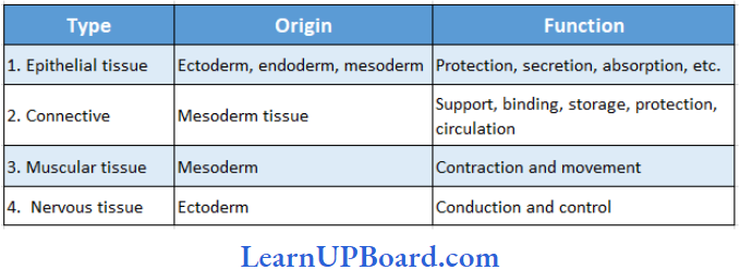
NEET Biology Notes Structural Organization In Animals Epithelial Tissue
Cells of the epithelium are set very close to each other, separated by thin films of extracellular material. Neighboring cells are held together by cell junctions. The epithelial tissue rests on a non-cellular basement membrane, which separates it from the underlying connecting tissue.
” structural organisation in animals bank of biology”
The Basement Membrane Is A Non-Cellular Membrane Made Up Of Two Layers: The upper thin layer is called the basal lamina, made up of glycoproteins and mucopolysaccharides and secreted by epithelial cells and the lower thick fibrous layer is called the reticular lamina made of reticular fibers and collagen fibers, which are a part of underlying connective tissue.
- Blood vessels are absent in the epithelial tissue. Materials are exchanged between epithelial cells and ves¬sels of the connective tissues by diffusion across the basement membrane.
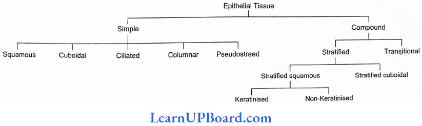
- The epithelial tissue is classified into simple and compound epithelium.
Read and Learn More NEET Biology Notes
Specialized Junctions Between Epithelial Cells: To provide mechanical support to tissues, the plasma membrane of the adjacent epithelial cells is modified to form structures called as intercellular junctions.
Fight Junctions (Zonula Occludens): They help to prevent substances from leaking across the tissue. Plasma membranes in the apical parts become tightly packed together or are even fused.
Interdigitations: These are interesting, finger-like processes of the cell membranes of the adjacent cells.
Intercellular Bridges: These are minute projections that arise from adjacent cell membranes. They make contact with one another.
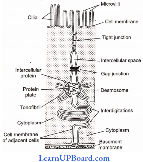
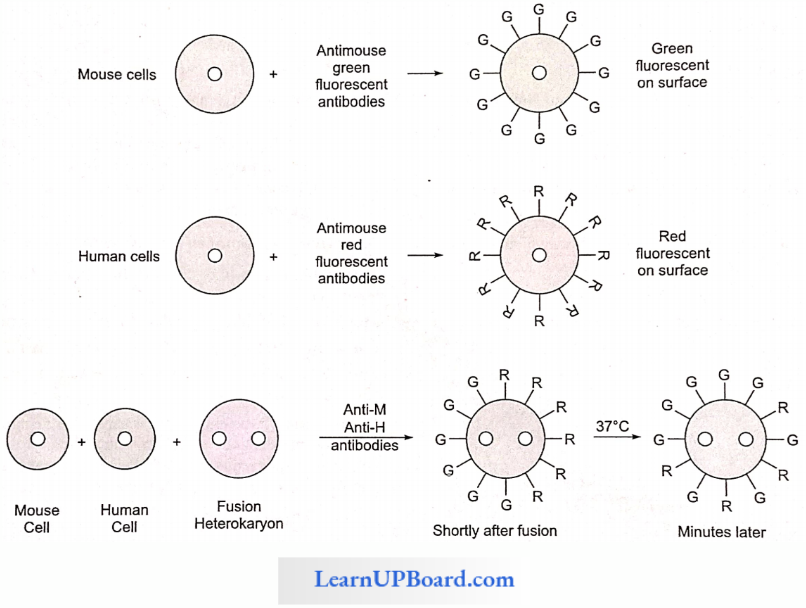
Gap Junctions: they facilitate the cells to communicate with each other by connecting the cytoplasm of adjoining cells, for rapid transfer of ions, small molecules, and sometimes big molecules also.
Intermediate Junctions (Zonula Adherens): These usually occur just below tight junctions. The intercellular space at these places contains a clear, low electron-density fluid. There is a dense plaque-like structure on the cytoplasmic side of each plasma membrane from which fine microfilaments of actin (protein) extend into the cytoplasm. There are no intercellular filaments between the adjacent cell membranes. There is an adhesive material at this point. They probably serve anchoring functions.
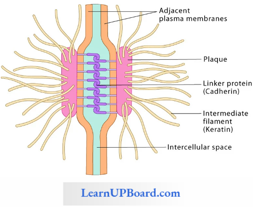
Desmosomes (Macula Adherens): They perform cementing to keep the neighboring cells together. These are like zonula adherens but are thicker and stronger and have disc-like junctions. They have intercellular proteins. The plaque-like structures (protein plates) are much thicker. The microfilaments that extend from microfilaments are called tonofibrils. Desmosomes serve anchoring functions.
Hemidesmosomes (single-sided desmosomes) are similar to desmosomes, but the thickening of the cell membrane is seen only on one side. Hemidesmosomes join epithelial cells to the basal lamina (outer layer of basement membrane).
Simple Epithelium
- Simple epithelium is formed of a single layer of cells.
- The adjacent cells are held together by means of a desmosome, resting on the basement membrane.
- Simple epithelium occurs mainly on secretory and absorptive surfaces.
- It helps in nutrition, excretion, and secretion, but not in protecting the underlying tissue.
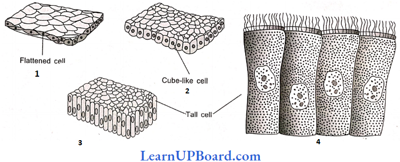
- Squamous Epithelium
- It consists of a layer of thin, flat, scale-like cells with prominent nuclei.
- The cells have irregular boundaries that fit closely into those of neighboring cells.
- It forms the inner lining of lung alveoli and blood vessels (endothelium).
- It is also known as pavement or tessellated epithelium.
- Cuboidal Epithelium
- It has cells that are polygonal in outline, but ap-pear cuboidal in vertical section.
- It lines small salivary and pancreatic ducts and thyroid vesicles.
- The cells participate in secretion, excretion, and absorption.
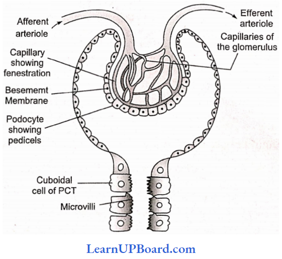
- The cells of cubical epithelium in absorptive surfaces often bear microvilli on their free ends. This gives a brush-like appearance to their free border. They are, therefore, called brush-bordered cubical epithelial cells, for example, in the proximal tubules of kidneys.
- Microvilli greatly increase the area of the free surface of the cell and thereby enhance absorption.
- Columnar Epithelium
- It is characterized by the presence of tall cells that are shaped like polygonal columns.
- The nucleus is usually located at the base of the cell.
- Columnar epithelium covers the inner surface of the intestine, stomach, and gall bladder. It also occurs in gastric and intestinal glands.
- Its function is secretion or absorption.
- The intestinal mucosa is lined by brush-bordered columnar epithelium which is highly absorptive.
- Ciliated Epithelium
- The ciliated epithelium consists of columnar or cubical cells bearing cilia on their free surfaces.
- The function of the cilia is to move particles, free cells, or mucus in a specific direction over the epithelial surface.
- Ciliated columnar epithelium lines the inner surfaces of some hollow organs such as fallopian tubes, bronchioles, and small bronchi.
- The ciliated columnar epithelium lining the ventricles of the brain and spinal canal is called as ependyma.
- Cilia Is Of Two Types: Kinocilium is motile cilium with 9 + 2 organization whereas stereocilium is non-motile and ciliated columnar epithelium and does not contain basal granule. 9 + 2 organization is also absent. Stereocilia are found in some parts of the male reproductive tracts such as the epididymis and vas deferens.
- Pseudostratified Epithelium
- Pseudostratified epithelium covers the inner linings of the trachea and large bronchi.
- Although made up of a single layer of columnar cells, it appears two-layered, because some cells are shorter than the others and have their nuclei at different levels.
- The shorter cells lack cilia and secrete mucus which traps particles on the epithelial surface, whereas the longer cells are ciliated.
- Ciliary movements propel the mucus and the particles toward the larynx.
- Pseudostratified non-ciliated columnar epithelium tissue is found in the urethra of the male and parotid salivary glands.
- Squamous cells show some keratinization.
NEET Biology Notes Structural Organization In Animals Epithelial Tissue Points To Remember
Special Types Of Epithelium
- Neuro Sensory Epithelium: In between pillar-shaped supporting cells, modified sensory cells are present. On the free end, sensory hair is present. The base of these cells is attached to the sensory nerve. For example,
- Gustatory Epithelium: Covers the taste bud of the tongue and receives taste sensation.
- Olfactory Epithelium: The Schneiderman membrane receives smell sensation.
- In Stato-acoustic Organ: Lining of the internal ear.
- In The Retina Of The Eye: Receives optic sensation.
- Myoepithelium: Around mammary and sweat glands
- Pigmented Epithelium (Cuboidal): In the retina of eye
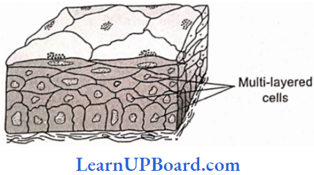
Compound Epithelium
- It consists of more than one layer of cells.
- Only the cells of the deepest layer rest on the basement membrane.
- Being multilayered, compound epithelia have little role in secretion or absorption, but they provide protection to underlying tissues against mechanical, chemical, thermal, and osmotic stresses.
- Compound epithelia may be stratified or transitional.
- Stratified Epithelium
- The stratified epithelium has many layers of epithelial cells.
- The deepest layer is formed by cuboidal cells.
- But the morphology of the superficial layers varies in the different kinds of stratified epithelia.
- In stratified cuboidal epithelium, the superficial cells are cuboidal.
- It lines the inner surfaces of larger salivary and pancreatic duct
- Stratified non-keratinized squamous epithelium covers moist surfaces such as those of the buccal cavity, pharynx, and esophagus.
- It has several superficial layers of living squamous cells and deeper layers of interlinked polygonal cells,
- Stratified keratinized squamous epithelium covers the dry surface of skin. It has many superficial layers of horny, scale-like remains of dead squamous cells, and several deeper layers of living polygonal cells.
- Heavy deposits of the insoluble protein keratin in the dead superficial cells make the epithelium impervious to water and highly resistant to mechanical abrasions.
- In contrast, non-keratinized stratified epithelium cannot prevent water loss and can afford only moderate protection against abrasions.
- Transitional Epithelium
- Transitional epithelium is much thinner and more stretchable than stratified epithelium.
- It has a single layer of cuboidal cells at the base, two to three middle layers of large polygonal or pear-shaped cells, and a superficial layer of large, broad, rectangular, or oval cells.
- It lines the inner surface of the urinary bladder and ureters.
- It allows considerable expansion of these organs to accommodate urine because stretching considerably flattens and broadens the cells of superficial and middle layers.
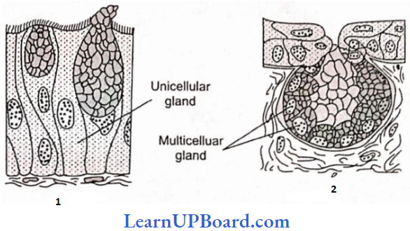
- Glandular Epithelia
- The cells of glandular epithelia are generally columnar or cuboidal.
- The Glandular Epithelium Can Be Classified Into Two Types: unicellular, consisting of isolated glandular cells (for example, goblet cell of the alimentary canal), and multicellular (for example, salivary glands), consisting of a cluster of cells.
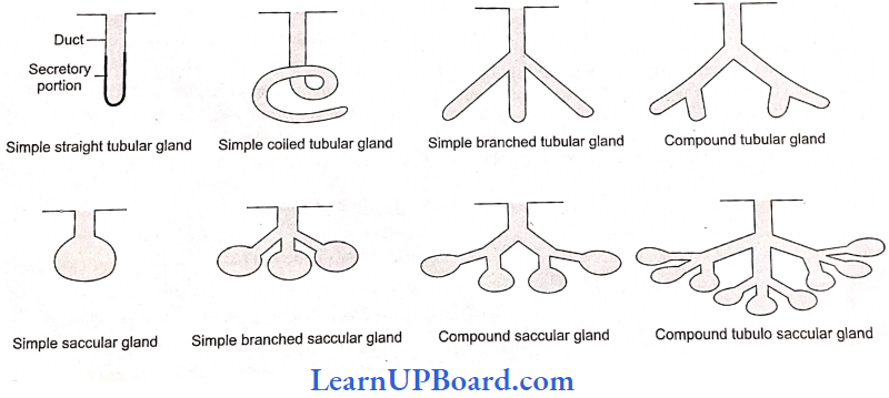
- A gland with a single unbranched duct is called a simple gland.
- The secretory part of the gland consists of epithelial cells arranged in the form of tubes (tubules) sacs (acini, alveoli), or a combination of both.
- The duct is also made up of epithelial cells.
- A gland with a branched system of ducts is called a compound gland.
- In these glands, the secretory tubule or acinus may be coiled or branched and open into the single duct of the gland.
- Compound glands are present in the pancreas and sub-mandibular salivary glands.
Types Of Simple Glands
- Simple Tubular: Simple tubular glands are present in the intestine (for example, crypts of Lieberkuhn).
- Simple Alveolar: The terminal part forms the alveolus, for example, mucous glands in the skin of frogs, and poison glands in toads.
- Simple Coiled Tubular: The terminal part is coiled, for example, sweat glands.
- Branched Tubular: Gastric glands in the stomach.
- Branched Alveolar: Example, sebaceous gland.
- Types Of Compound Glands
- Compound Tubular Gland: Example, mammary glands of prototherians.
- Compound Saccular Or Alveolar Gland: Example, salivary glands, (sub-maxillary and sub-lingual).
- Compound Tubulo Alveolar Or Tubulo Saccular: They are tubular as well as alveolar and are found in mammary glands, pancreas, parotid salivary glands, Cowper’s glands, and Bartholin’s glands.
- Exocrine And Endocrine Glands: Exocrine glands have a secretory portion that contains the cells for secretion of milk, digestive enzymes, mucus, saliva, ear wax, oil, and ducts, which transport their secretions to the respective sites of action, for example, salivary gland, tear gland, gastric gland, and intestinal glands. Endocrine glands are glands of “internal secretion” whose secretions are usually secreted directly into the blood. Examples are follicle-stimulating hormone from the anterior pituitary or thyroxin from the thyroid. When a gland performs both exocrine and endocrine functions, it is called a mixed gland or heterocrine gland (for example, pancreas, testes, ovaries).
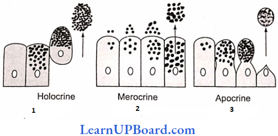
- Classification Of Glands Based On The Mode Of Secretion
- Holocrine Glands: In holocrine glands (for example, the sebaceous gland), the product of secretion is shed with the whole cell leading to its destruction.
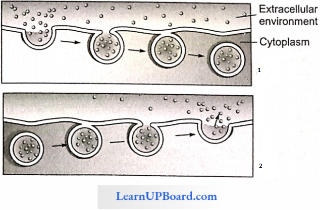
- Merocrine Glands: When the secretory granules leave the cell by exocytosis (simple diffusion) with no loss of other cellular material, the glands are called merocrine glands (for example, pancreas, salivary glands, intestinal glands, and sweat glands)
- Apocrine Glands: In apocrine glands (for example, mammary glands and axillary sweat glands), only the apical portion of the cytoplasm is discharged along with the secretory product.
NEET Biology Notes Structural Organization In Animals Connective Tissue
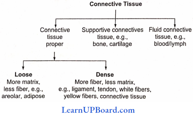
All connective tissues in the body are developed from mesoderm. O. Hartwig called them mesenchyme because they originated from embryonic mesoderm. Only connective tissue constitutes 30% of the total body weight (muscle: 50%, epithelium: 10%, nervous: 10%). On the basis of the matrix type, connective tissue is of three types:
- Connective Tissue Proper: Soft and fibrous matrix.
- Connective Tissue Skeleton: Dense and mineralized matrix. Due to the deposition of minerals, it becomes hard.
- Connective Tissue Vascular: Liquid and fiber-free matrix
Connective Tissue Proper: Connective Tissue Proper is composed of three components:
- Different types of cells,
- Fibers, and
- Matrix.
Cells Of Connective Tissue Proper
- Fibroblast Cells
- The largest cell of connective tissue proper.
- Maximum in number.
- The cell body and nucleus both are oval-shaped.
- A branched cytoplasmic process arises from these cells so they appear irregular in shape.
- Rich in rough ER because their main or primary function is to produce fibers. Fibers are composed of protein.
- They are the chief matrix-producing cells.
- Old fibroblast cells (fibrocytes) are inactive cells.
- These are also considered undifferentiated cells of connective tissue because they can be modified into osteoblast and chondrioblast cells to produce bone and cartilage.
- Fibroblast Cells Function: To produce fibers and to secrete matrix.
- Plasma Cell (Cart Wheel Cell)
- Less in number.
- Amoeboid in shape.
- Chromatin material is arranged like spokes in a wheel, hence is also called cartwheel cells.
- According to research, these cells are formed by the division of lymphocytes. So they are also called clones of lymphocytes.
- Plasma Cell (Cart Wheel Cell) Function: Produce, secrete, and transport antibodies.
- Mast Cells/Mastocytes
- Numerous, amoeboid, and small in size.
- Structurally and functionally similar to basophils.
- Two- to three-lobed S-shaped nucleus.
- The cytoplasm contains basophilic granules which can be stayed with the basic dye methylene blue.
- Mast Cells/Mastocytes Function: These are important cells of connective tissue proper as they perform certain important functions:
- Histamine is a protein, a vasodilator, which increases the permeability of blood capillaries. It takes part in allergy and inflammatory reactions.
- Serotonin, also called 5-hydroxytryptamine, is a protein, a vasoconstrictor, and decreases blood circulation, but increases blood pressure. At the site of cut or injury, serotonin causes a decrease in blood loss.
- Heparin, a mucopolysaccharide, is a natural anti-coagulant that prevents the clotting of blood in blood vessels by preventing the conversion of prothrombin into thrombin.
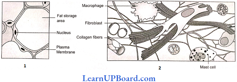
- Adipose Cells/Fat Cells: These arc oval-shaped cells that store fat. Fat is collected in the form of fat globules formed by the fusion of small oil droplets. On the basis of the number of fat globules, adipocytes are of two types:
- Monolocular Adipocytes/White Fat Tissue Cell
- In these cells, a single large and central fat globule is present.
- The nucleus and cytoplasm is peripheral and the cytoplasm is less in amount.
- Due to the compression of the fat globule, the nucleus becomes flattened in shape.
- These adipocytes form white fat.
- Multilocular adipocytes/brown fat tissue cell
- In these cells, two to three fat globules are distributed in the cytoplasm around the nucleus.
- The cytoplasm is more in quantity.
- The nucleus is rounded and found in the center
- These adipocytes form brown fat.
“is earthworm and frog in neet syllabus 2023 “
- Mesenchymal Cells
- Less in number. Small-sized with cytoplasmic process having an irregular shape.
- Oval-shaped nucleus
- These are undifferentiated cells of connective tissue because they can transform into any cell of connective tissue proper (totipotent in nature).
- Mesenchymal Cells Function: To form other cells of connective tissue.
- Macrophages/Histiocyte/Plasmatocytes
- These are the second largest in size and number.
- These are amoeboid in shape with bean or kidney-shaped nuclei.
- Cytoplasm quantity is more agranular, but due to the presence of more number of lysosomes, it appears granular.
- They are phagocytic in nature and destroy bacteria and viruses by phagocytosis. They arise by the fusion of monocytes.
- Also called as the scavenger cells of connective tissue, because they destroy dead or damaged cells to clean connective tissue.
- They are named differently in different organs:
- Lung: Dust cells
- Liver: Kupffer cells
- Blood: Monocytes
- Brain: Microglia cells
- Thymus gland: Hessels granules
- Spleen: Reticular cells
- Lymphocytes
- Less in number and small in size, having an amoeboid shape.
- A large nucleus is present and cytoplasm is present as a peripheral layer. Cytoplasm quantity is less.
- They produce, transport, and secrete antibodies.
- They divide to form the plasma cells of connective tissue properly.
Fibers
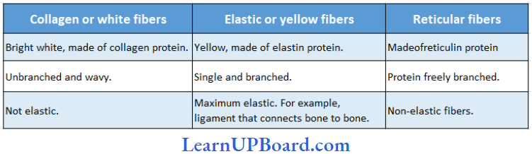
- Collagen Fibers (White Fibers)
- They are shining white fibers composed of collagen protein (tropocollagen).
- It is present in maximum quantity in vertebrates (only collagen fibers constitute one-third part of connective tissue fibers in human beings).
- They are wavy and tough fibers and are always arranged in bundles called fascia.
- On boiling, they convert into gelatin.
- They can be digested by pepsin enzyme.
- Elastic Fibers (Yellow Fibers)
- Precursor in color and composed of elastin protein.
- They are branched fibers but are always arranged singly. Branches of these form a network.
- In these fibers, maximum elasticity is present.
- They are highly resistant to chemicals.
- When boiled they do not dissolve.
- They can be digested by trypsin enzyme.
- Reticular Fibers
- Precursor of collagen fibers, delicate with no elasticity.
- Also known as argyrophilic fibers, since they can be stained with silver salts.
- They are composed of reticulin protein; highly branched fibers that always form dense networks.
- These are mainly distributed in lymphoid organs such as the spleen or lymph nodes.
Differences Between Collagen, Elastic, And Reticular Fibers
Matrix: Matrix is composed of mucopolysaccharide which is present in the form of hyaluronic acid.
Types of Connective Tissue Proper
Loose Connective Tissue: It consists of cells scattered within an amorphous mass of proteins that form a ground substance. The gelatinous material is strengthened by a loose scattering of protein fibers such as collagen, and elastin, which makes tissue elastic, and reticulin, which supports the tissue by forming a meshwork.
- Adipose Tissue
- Adipose tissue is a connective tissue rich beneath the skin, around kidneys in mesentery and bone marrow.
- Besides fibroblasts, macrophages, collagen fibers, and elastic fibers, the adipose tissue also contains large, spherical, or oval cells called fat cells or adipocytes.
- The cytoplasm and organelles in adipocytes are pressed by fat globules into a narrow annular layer just beneath the plasma membrane.
- The adipose tissue synthesizes, stores, and metabolizes fat.
- There are two kinds of fatty tissues. In the white adipose tissue, there is a single large fat droplet in the cells surrounded by a small amount of the cytoplasm.
- The brown adipose tissue cell, on the other hand, has many small droplets of fat, suspended in a considerably larger amount of cytoplasm. Whereas brown fat cells contain many mitochondria, white fat cells have comparatively few.
- The color in the brown fat is due to the high concentration of iron-containing cytochrome pigments.
- Brown fat is particularly found in newborn babies and hibernating mammals.
- It accounts for 5-6% of the body weight of the newborn rabbit and also of man.
- Brown fat has a larger capacity for generating heat.
- It is because of brown fat that new-boni mammals generally do not shiver in spite of lower temperatures outside.
- Brown fat cannot be a substitute for food. Adipose tissue may be examined from the fat bodies of frogs or from the skin of rabbits.
- Adipose Tissue Functions:
- Prevents heat loss by forming a heat-insulating layer beneath the skin.
- Forms shock-absorbing cushions around kidneys and eyeballs.
- Acts as a food reserve.
- Areolar Tissue: Areolar tissue occurs beneath the epithelia of many hollow visceral organs, skin, and in the walls of arteries and veins. It contains different types of cells:
- Fibroblasts: They are the principal cells of this tissue. They are irregularly shaped flat cells with long proto¬plasmic processes. Fibroblasts synthesize two kinds of protein collagen and elastin. Fibroblast secretes the major amount of matrix.
- Macrophages/Histiocytes/Plasmatocytes: They are phagocytic in nature.
- Mast Cells/Mastocytes: They are irregular or ovoid cells and contain basophilic granules which are made of:
- Histamine: An inflammatory substance produced during allergic reactions.
- Heparin: Natural anti-coagulant.
- Serotonin: Vasoconstrictor.
- Plasma cells/cartwheel cells synthesize antibodies. The areolar tissue joins different tissues and forms the packing between them and helps to keep the organs in place and in normal shape.
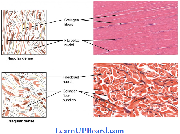
Dense Connective Tissue: Fibers and fibroblasts are compactly packed in dense connective tissues. The orientation of fibers might show a regular or irregular pattern and is called dense regular or dense irregular tissues, respectively.
- In the dense regular connective tissues, the collagen fibers are present in rows between many parallel bundles of fibers, for example, tendons and ligaments.
- Dense irregular connective tissue has fibroblasts and many fibers (mostly collagen) that are oriented in different directions. This tissue is present in the skin, perimysium, perineurium, and around bones as periosteum.
- White Fibrous Tissue: It carries only a few fibroblasts scattered amidst the dense network of thick collagen fiber bundles. It has great tensile strength. The presence of white fibrous tissue at the joints between skull bones makes them immovable.
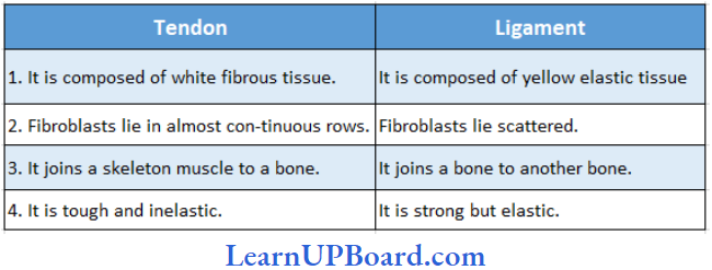
- Tendon: It is a dense, strong, and fibrous connective tissue with thick parallel bundles of collagen fibers. A few flat, elongated tendon cells lie in single rows between the fiber bundles. The tendon forms the strong inextensible attachment of a skeletal muscle to a bone. Colloidal protein gelatin is obtained by boiling collagen.
- Ligament: Ligaments connect the bones at the joints and hold them in position. A sprain is caused by excessive pulling of ligaments. They are made of bundles of elastic fibers and a few collagen fibers. Many-year-old mummies still have their arteries intact due to well-preserved elastic fibers.
Difference Between Tendon And Ligament
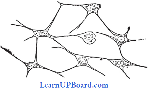
Reticular Tissue: It consists of star-shaped reticular cells whose protoplasmic processes form a network, These cells are phagocytic in function. Matrix and some other types of cells are are also found in the spaces of the network. Reticular tissue is present in the spleen, lymph nodes, bone marrow, etc.
Supportive Connective Tissue
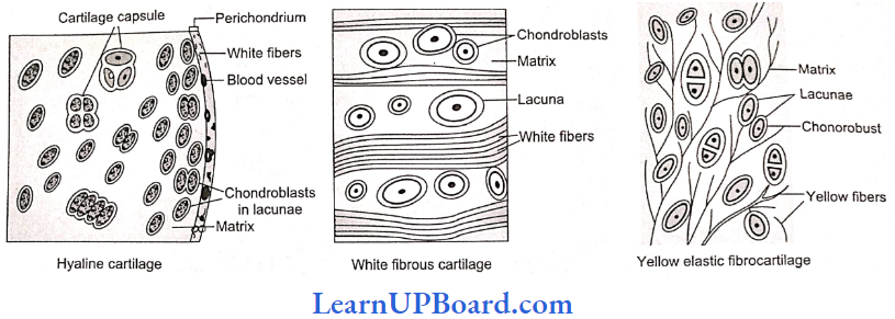
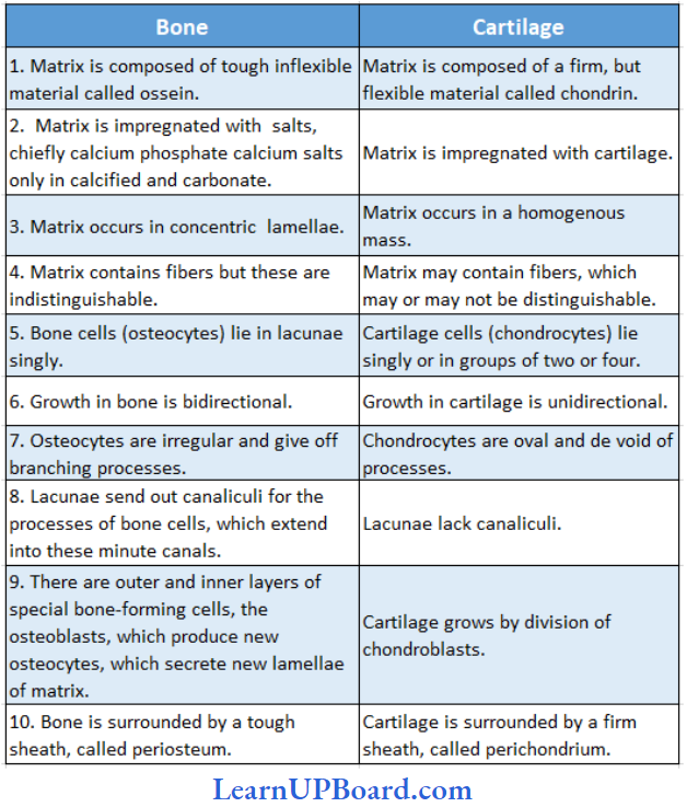
Cartilage: Cartilage is a solid but semi-rigid and flexible connective tis¬sue. Chondrocytes are large, blunt, angular cartilage cells. They occur in clusters of two or three cells in small spaces (lacunae) scattered in the matrix.
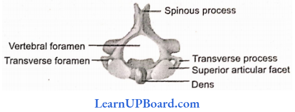
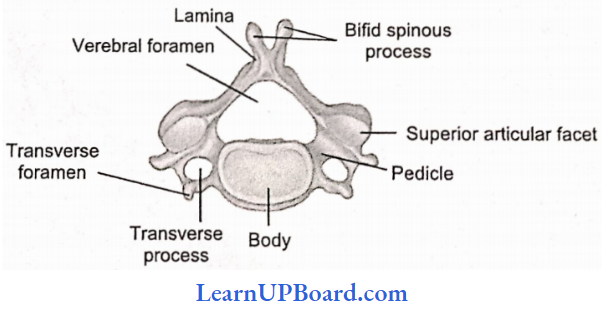
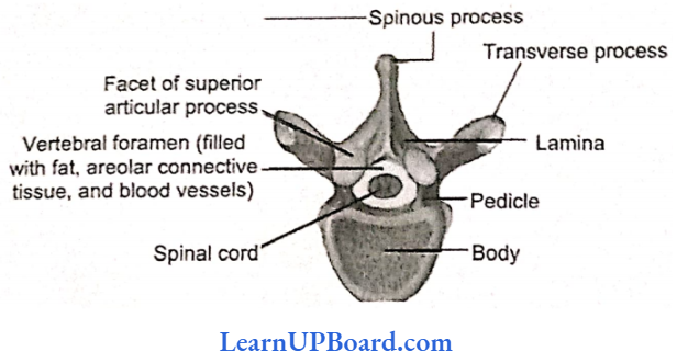
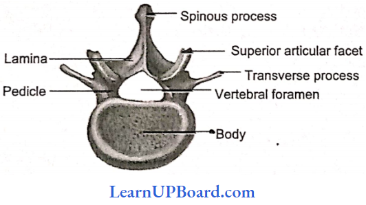
- Hyaline Cartilage: In hyaline cartilage, the matrix is apparently fiberless and glass-like (hyaline), but trans-lucent. It occurs in the larynx, nasal septum, tracheal rings, and costal cartilage. It gives those structures a definite but pliable form. White fibrocartilage carries thick dense bundles of collagen fibers between rows of chondrocytes in lacunae. It occurs in joints between vertebrae. Its collagen fibers make such joints strong but less clastic and only slightly movable.
- Nucleus Pulposus: In the center of the intervertebral disc, a soft area is present called nucleus pulposus which is supposed to be a remnant of notochord.
- Elastic Cartilage: It contains a dense network of elastic fibers between scattered chondrocytes. It forms the Eustachian tube, epiglottis, and pinna of the ear. The elastic fibers make those organs considerably elastic and pliable.
- Calcified Cartilage: Initially, it is like hyaline cartilage but later on it gets hardened like bone due to the dep¬osition of calcium salts, for example, supra scapula of a frog’s pectoral girdle, pubis of the pelvic girdle of the frog.
Bone: It is a solid, rigid connective tissue. The matrix of the bone has the deposition of apatite salts of calcium and phosphorus. For example, hydroxyapatite salts and fluorapatite salts.
- 60-70% of bone is made up of inorganic matter and 30-40% is made up of organic matter.
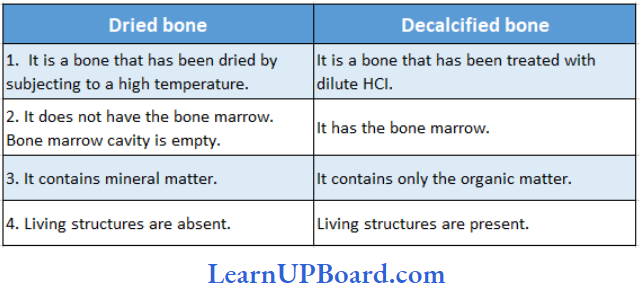
- If the bone is put in dil. HCI will become decalcified, soft, and flexible. Nothing will happen to the bone if we put the bone in KOH.
- Osteoblasts are bone-forming cells that secrete ossein protein.
- Osteocytes are bin cells, they are metabolic inactive cells present in the lacuna.
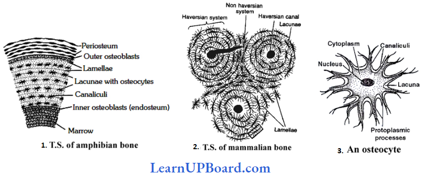
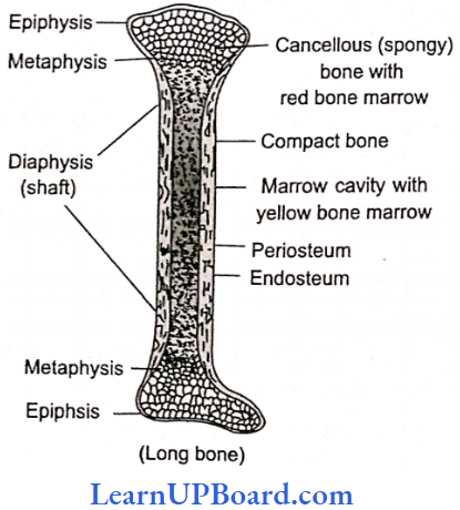
- Bone is a solid, rigid, and strong connective tissue. Its matrix is heavily deposited with apatite salts of calcium and phosphorus. Flat irregular spaces called lacunae occur in the solid matrix. Each lacuna lodges a nathone cell or osteocyte. A bone cell has irregular¬shaped and long cytoplasmic processes. These processes extend into minute canals (canaliculi) radiating from each lacuna.
- Compact bone forms the dense outer layers of all bones. It is composed of many parallel, longitudinal column-like structures called Haversian systems, cemented to each other. Haversian canals are connected to each other by Volkmann’s canals. In each Haversian system, several concentric layers (lamellae) of bony matrix encircle a longitudinal central canal (Haversian canal. This canal carries blood vessels and nerves. Lacunae-containing osteocytes occur in a layer between two lamellae.
“structural organisation in animals neetprep “
Spongy Bone: The ends of long bones are composed of an open lattice of bone called spongy bone. The spaces within contain marrow’, where most blood cells are formed. It carries no concentric organization like the Haversian system. It consists of a network of many fine irregular bony plates or trabeculae. Each trabecula consists of many irregularly arranged lamellae with lacunae between them. It has red bone marrow. Spongy bone is also called as cancellous bone and is found in epiphysis, i.e., the ends of long bones.
Differences Between Bone And Cartilage
Differences Between A Dried Bone And Decalcified Bone
NEET Biology Notes Structural Organization In Animals Connective Tissue Points To Remember
Types Of Bones
- Cartilage Bones/Endochondrial/Replacing Bones: They are formed by the replacement of cartilage by the bone, for example, hu-merus, femur, vertebrae, ribs, and girdle bones, except clavicle. Chondroblasts are cartilage-eater cells.
- Membrane/investing bone/dermah Examples, skull bones, clavicle, etc. The bones are formed in the dermis of the skin and are invested over the already present cartilage.
- Sesamoid Bones: They are formed by the ossification of the tendons, for example, the patella.
- Visceral Bones: They are those bones that get detached from the skeleton and come to lie in visceral organs, for example,
- os Cordis: Present in the interventricular septum of the heart of deer.
- os Falciparum: Palm of mole.
- os Penis: Penis of rats and carnivores.
- os Palbebrae: In the eyelids of a crocodile.
- os Rostralis: Snout of pig.
- Bone China: Porcelain was first made in China during the Tang dynasty. English found a new way of making porcelain with bone ash. Bone china is a form of porcelain made from burned animal bones. Bone ash is mixed with kaolin and white clay. The bone ash increases the porcelain’s translucence.
- Word Roots And Origin Of Periosteum: Greek peri means “around” and osteon means “bone.”
Fluid Connective Tissue (Blood)
- Blood is a fluid connective tissue.
- Its cells are quite distinct from other connective tissue cells both in structure and function.
- The extracellular material in the blood is a fluid devoid of fibers.
- Fluids outside the cells are generally called extracellular fluids (ECF).
- Blood is heavier than water.
- The extracellular material in the blood is a straw-colored, slightly alkaline (pH = 7.4), aqueous fluid called plasma.
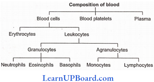
- Constituents, having characteristic forms, float in the plasma. They are collectively called the formed elements of blood. They include the blood cells and blood platelets.
- Blood cells are of two types: erythrocytes and leukocytes.
- Blood circulates within blood vessels in higher animals.
Plasma
- Plasma contains three major classes of plasma proteins, viz., serum albumin, serum globulins, and fibrin¬ogen.
- Plasma proteins serve as a source of proteins for tissue cells.
- Tissue cells may utilize plasma proteins to form their cellular proteins.
- Additionally, albumin and globulins retain water in blood plasma by their osmotic effects.
- A fall in plasma proteins leads to the movement of excessive volumes of water from blood to tissues. That is why hands and feet get swollen with accumulated fluid (edema) in persons suffering from dietary deficiency of proteins.
- Albumins and globulins also transport many substances such as thyroxine and Fe3+ in combination with them.
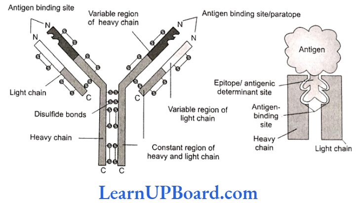
- One class of globulins, called immunoglobulins, acts as antibodies.
- Plasma proteins also maintain the blood pH by neutralizing strong acids and bases. Thus, they act as acid-base buffers.
- It is a slightly alkaline, non-living intercellular substance that constitutes about 60% part of the blood.
- It is a pale yellow but transparent and clear fluid.
Composition of Plasma: Plasma forms 55-60% by the blood volume.
- Water: Water alone forms about 90% to 92% of the plasma. Solids form about 8% of the plasma.
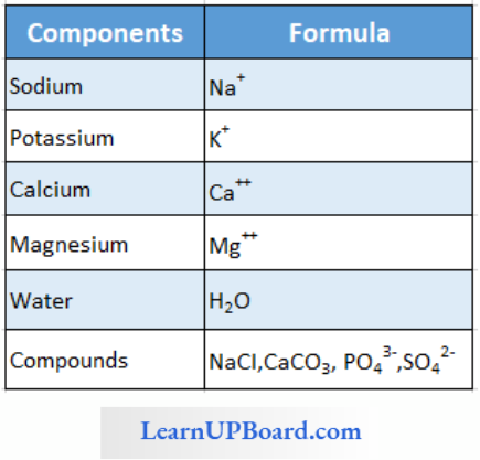
- Mineral Salts: These are chlorides, bicarbonates, sulfates, and phosphates of sodium, potassium, calcium, iron, and magnesium. All salts constitute about 0.9% of plasma. Buffer of the blood is sodium bicarbonate.
- Nutrients: These include glucose, fatty acids, phospholipids, cholesterol, fats, amino acids, nucleosides, etc. Mineral salts have been mentioned above.
- Plasma Proteins: They constitute about 7-8% of plasma. These mainly include albumin 4.4%, globulin 1.5-2%, prothrombin, and fibrinogen both 0.3%.
- Defence Proteins: Immunoglobulins which act as antibodies and some other substances, such as lysozyme and properdin (a large protein) are always found in the plasma. They destroy bacteria, viruses, and toxic substances that may enter into the blood from outside.
- Excretory Substances: These include ammonia, urea, uric acid, creatinine, etc.
- Dissolved Gases: The water of blood plasma contains oxygen, carbon dioxide, and nitrogen in dissolved form.
- Anticoagulant: Blood plasma contains a conjugated polysaccharide, heparin, which prevents the coagulation of blood inside blood vessels.
- Hormones: These are secreted and released in blood by endocrine glands.
- Vitamins And Enzymes: Different kinds of vitamins and enzymes are present in the blood plasma.
“is cockroach in neet syllabus 2023 “
Functions Of Blood Plasma: These can be summarized as under:
- Transport,
- Retention of fluid in the blood,
- Maintenance of blood ph,
- Body immunity,
- Prevention of blood loss,
- Conducting heat to skin for dissipation, and
- Uniform distribution of heat all over the body.
Blood Glucose
- Glucose is mainly absorbed in the small intestine.
- Glucose is also absorbed in the stomach.
- After absorption, glucose reaches the blood.
- Excess of glucose is converted into glycogen by insulin hormone in the liver and muscles.
- Whenever it is required, glycogen is changed back into glucose by glucagon hormone.
- Usually blood glucose level is about 80-100 mg per 100 mL of blood, 12 hours after a normal meal. But its concentration rises soon after a carbohydrate-rich diet.
- If the blood glucose level exceeds 180 mg per 100 mL, it starts appearing in urine. This condition is called glucosuria.
- Fasting glucose is 70-110 mg/dL. Glucose PP is 110 mg/dL (PP: post-prandial or after breakfast).
- If it is higher, it causes diabetes mellitus (hyperglycemia).
- If it is less, it causes hypoglycemia (less amount of glucose in the blood).
Blood Cholesterol
- Usually, cholesterol is considered a harmful substance. But it is quite useful in limited amount.
- Cholesterol is used in the synthesis of biomembranes, vitamin D, bile salts, and steroid hormones.
- Its normal amount is 80-180 mg in 100 mL of blood plasma.
- Cholesterol comes in the blood plasma either by intestinal absorption of fats by the synthesis from the liver or by both.
- Saturated fats such as ghee and butter increase cholesterol level in the blood.
- Increased blood cholesterol may lead to its deposition in the internal wall of the blood vessels such as arteries and veins which cause high blood pressure and heart problems.
Functions Of Plasma Proteins
- Prevention Of Blood Loss: Fibrinogen and prothrombin play a role in blood clotting.
- Retention Of Fluid In The Blood: Albumin helps in osmotic balance.
- Body Immunity: Certain globulins called immuno-globulins (glycoproteins) act as antibodies in blood and tissue fluid. Antibodies belong to a class of proteins called as immunoglobulins.
- Maintenance of pH: Plasma proteins serve as acid-base buffers. It means they maintain pH of the blood by neutralizing acids and bases.
- Transport Of Certain Materials: Thyroxine (hormone) is bound to albumin or specific globulin for transport in the plasma.
- Distribution Of Heat: Plasma proteins help in the uniform distribution of heat all over the body.
- Enzymes: Some proteins acting as enzymes also occur in the plasma.
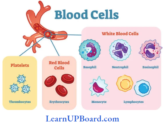
Blood Cells
1. Erythrocytes
- Erythrocytes (red blood corpuscles or RBCs) are the most numerous of the formed elements of blood.
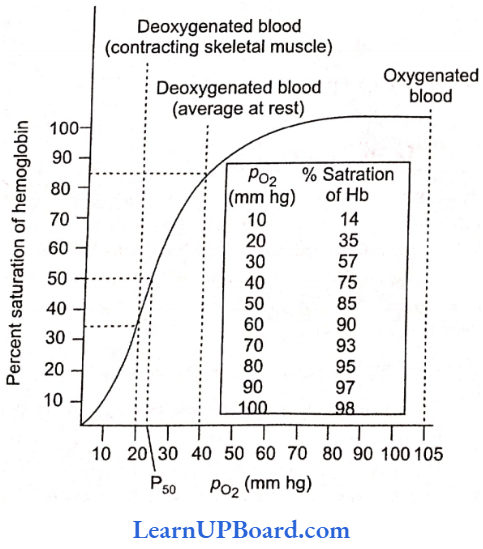
- Their most important characteristic feature is the presence of hemoglobin (Hb), the red oxygen-carrying pigment.
- The total number of erythrocytes per microliter (1μL = 1 mm³ =10-6 L) of blood is known as the total count of RBC.
- It averages 5 million and 4.5 million in adult men and adult women, respectively.
- The total count would be low in anemia and after profuse bleeding.
- On the contrary, the abnormal rise in the total count of RBC is called polycythemia.
- Anemia is caused due to the deficiency of folic acid, vitamin B12, and hemoglobin.
- The size and shape of erythrocytes vary in different classes of animals.
- In fishes, amphibians, reptiles, and birds, erythrocytes are usually nucleated, oval, and biconvex. But in mammals, they are non-nucleated, biconcave, and circular.
- Only camel and llama possess oval red blood corpuscles.
- Human erythrocytes measure 7-8 μm (1 μm = 10-6 m) in diameter and 2μm thickness near the rim.
- Old and damaged erythrocytes are phagocytosed and destroyed by macrophages.
- The pigment part (porphyrin) of hemoglobin is then catabolized to the yellow pigment bilirubin which is excreted in the bile.
- The pale yellow color of plasma is largely due to bilirubin.
- If a sample of blood is rendered non-coagulable by adding potassium or sodium oxalate and then centrifuged at a high speed in a graduated centrifuge tube (hematocrit tube), the centrifugal force rapidly sediments the erythrocytes to the bottom of the tube. They become packed into a solid, red. bottom layer while plasma forms a clear, fluid upper layer. On the upper surface of the erythrocyte layer, leukocytes form a thin, buff-colored layer.
- From the graduations on the tube, the relative volume of erythrocytes may be read as a percentage of the total blood volume. This is called the hematocrit value or packed cell volume.
- It normally forms 45% of the blood volume.
RBCs of mammals are circular, biconcave, and non-nucleated, except those of the family Camelidae (camel, which has non-nucleated and oval RBCs). The largest RBCs are found in amphibia. The smallest RBCs are found in mammals.
- In mammals, the smallest RBCs are found in “musk deer,” Tragiilns jammies (1.5 pm).
- In mammals, the largest RBCs are found in elephants. (9.4 pm).
- The graveyard of RBC is the spleen.
- Life span of RBCs in men is 120 days, in frogs 100 days, and in rabbits 80 days.
- The radioactive chromium method (Cr51) is used for the estimation of the life span of RBC.
Count of RBCs:
In embryo = 8.5 million/mm³
In man = 5-5.5 million/mm³
In woman = 4.5 million/mm³
Daily destruction of RBCs = 1%
ESR (Erythrocyte Sedimentation Rate): It is measured by Wintrobe’s method. It is the rate of the settling down of RBCs. It is also estimated by Westergen’s method.
- ESR is very useful in diagnosing various diseases including tuberculosis.
- ESR in men is 0-5 mm/h and in women 0-7 mm/lt, according to Westergen’s method.
Hemocytometer: It is an instrument for counting the number of both WBCs and RBCs.
Rouleaux: In resting and slow-flowing blood, RBCs aggregate to form rouleaux (RBCs are piled on top of each other). Fibrinogen favors rouleaux formation.
Bone Marrow: It is the main site for the formation of RBC. The volume of bone marrow at the time of birth is 70 mL. In adults, the volume of bone marrow is 4000 mL.
Structure Of RBC Of Man: Biconcave, nonnucleated, and bounded by Donnan’s membrane (plasma membrane of RBC). Hemoglobin is filled in RBC which is a respiratory pigment.
Normal Range Of Hemoglobin
- Infants: 16.5 ± 3.0 g/dL (dL = deciliter)
- Children 3 Months: 11.0 ± 1.5 g/dL
- Childen 10-12 Months: 13.0 ± 1.5 g/dL
- Men: 15.5 ± 2.5 g/dL
- Women: 14.0 ± 2.5 g/dL
Structure Of Hemoglobin: Each molecule of hemoglobin contains four molecules of heme and one molecule of globin. These are attached by coordinate bonds. Heme is a protoporphyrin compound and has four pyrrole groups joined together to form a ring structure. In Hb, Fe is present in (Fe++) ferrous form. Heme is 5% and Globin is 95%. Globin is made of four polypeptide chains.
2. Leukocytes
- Leukocytes (white blood corpuscles or WBC) are devoid of hemoglobin and are consequently colorless
- Leukocytes are nucleated blood cells.
- They are of two major classes: granulocytes (with cytoplasmic granules) and agranulocytes (without granules).
- Granulocytes are of three types, viz., neutrophils, eosinophils, and basophils, each with lobed nuclei.
- Agranulocytes are of two types, viz., lymphocytes and monocytes.
- Neutrophils and monocytes protect the body against microbes by phagocytosing them.
- Lymphocytes secrete antibodies in the blood to destroy microbes and their toxins.
- The number of leukocytes per microliter (1 μL = 1 mm³ = 10-6 L) of blood is called the total count of WBC. It is 6000-8000/mm³ of blood normally.
- It may rise abnormally in acute infections (for example, pneumonia), inflammations (for example, appendicitis), and malignancies (for example, leukemia).
- In some conditions such as folic acid deficiency, the total count falls abnormally (leukopenia).
- The total count of WBCs is also of diagnostic value in many diseases.
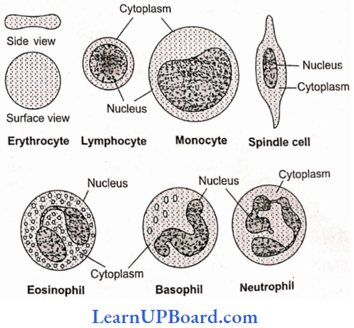
- Monocytes have kidney-shaped nuclei.
- The process by which monocytes and neutrophils squeeze out through thin capillary walls is diapedesis.
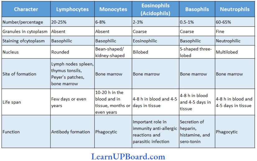
- Neutrophils: They are maximum in number, stain equally with both basic and acidic dyes, and have many lobed nuclei, granules are in abundance in the cytoplasm, and help in phagocytosis.
- Eosinophils: They have a bilobed nucleus. They get stained with acidic stains. Their number increases during allergic reactions (eosinophilia).
- Basophils: They get stained with basic dyes. Their nucleus is S-shaped. Coarse granules are few in the cytoplasm. Basophils release heparin and histamine in the blood and have a function similar to the mast cells.
- Lymphocytes: They have large and rounded nuclei. The cytoplasm forms a thin peripheral film. They have their stem cells in the bone marrow and are differentiated in the bone marrow or in the thymus. Lymphocytes are of two types: B- lymphocytes and T lymphocytes. B-lymphocytes produce antibodies against antigens and they mature in the bone marrow.
- Monocytes: They are the largest leukocytes (12-15 pm). The nucleus is kidney-shaped. They are produced from bone marrow monoblast cells. They help in phagocytosis.

Differences Between Different Types Of Leukocytes
3. Blood Platelets
- Also called thrombocytes, blood platelets are non-nucleated, round, or oval, biconvex disc-like bodies.
- They are 2-3 pm in diameter and their number normally varies from 0.15 to 0.35 million/mm3 or 150,000-350,000 platelets/mm3.
- They bud off from the cytoplasm of very large megakaryocytes of the bone marrow.
- Their normal life span is about a week.
- When a blood vessel is injured, platelets get clumped at the injured spot and release certain chemicals called platelet factors. These promote blood coagulation.
- Thrombocytopenia is a decrease in the platelet count and purpura is a group of bleeding diseases due to thrombocytopenia.
Blood Coagulation
- When blood oozes out of a cut, it sets into gel within a few minutes. This is called coagulation.
- Coagulation is brought about by the hydrolysis of soluble fibrinogen of plasma to insoluble fibrin. This is catalyzed by an enzyme called thrombin.
- Fibrin precipitates as a network of fibers. This network traps many blood cells, particularly RBCs, to form a red solid mass called the blood clot.
- The clot seals the wound in the vessel to stop the bleeding.
- The straw-colored fluid left after clotting of blood is called serum.
- Serum cannot be coagulated as it lacks fibrinogen.
- Thrombin occurs in normal blood as an inactive globulin called prothrombin.
- It must be activated to thrombin before blood coagulation can occur.
- In the case of injury to a blood vessel, coagulation-promoting substances called thromboplastins are released into the blood from clumped platelets and damaged tissues.
- Thromboplastins help in the formation of the enzyme prothrombinase. This enzyme hydrolyzes prothrombin to thrombin to initiate coagulation.
- Ca2+ ions are essential for both activation and action of thrombin.
- Blood normally contains an anticoagulant, heparin, which prevents the activation of prothrombin. Heparin is released from “mast cell” granules.
- Blood also contains antithrombin, which inhibits any thrombin formed accidentally.
- Blood drawn from a blood vessel can be kept coagulated by adding a pinch of oxalate (sodium or potassium oxalate) to it.
- Oxalate precipitates Ca2+ and consequently prevents coagulation.
- Chilling of blood also delays coagulation because lesser temperature depresses the action of courage-location-promoting enzymes.
NEET Biology Notes Structural Organization In Animals Blood Cells Points To Remember
ABO Blood Clotting Factor
- Karl Landsteiner reported for the first time ABO blood groups in human beings.
- A, B, and O blood groups were discovered by Landsteiner (1900), while the AS blood group was found by de Castello and Steini (1902).
- Agglutinogens (antigens) are present on the surface of red blood corpuscles and agglutinins (antibodies) are found in the blood plasma. Both antigens and antibodies are proteins.
- When two different types of blood are mixed, the red blood corpuscles form a clump.
- The clumping of red blood corpuscles is called agglutination
Clotting Factors
- Thirteen factors help in blood clotting.
- These factors are mainly produced in the liver.
- Vitamin K is required in the synthesis of these clotting factors.
- These factors are represented in Roman numbers.
- 1: Fibrinogen
- 2: Prothrombin
- 3: Thromboplastin
- 4: Ca+2 (cofactor in each step of blood clotting)
- 5: Proaccelerin
- 6: Accelerin (rejected)
- 7: Proconvertein
- 8: AHG (anti-hemophilic globin; absent in hemophilia-A)
- 9: Christmas factor
- 10: Stuart factor
- 11: PTA (plasma thromboplastin antecedent)
- 12: Hagman factor
- 13: FSF factors (fibrin-stabilizing factor) (Laki Lorand factor)
- Other natural anticoagulants
- Hirudin: Found in leech.
- Anophelin: Found in female Anopheles
- Lampredin: Found in Petcromyzon (Lamprey)
- Cumerin: Obtained from plants
- Warfarin: Obtained from plants
- To collect blood in a bottle in a blood bank, artificial anticoagulants such as sodium citrate, and sodium oxalate. EDTA (ethylenediaminetetraacetic acid), etc., are used. These chemicals act as calcium-binding units and remove Ca+2 ions from the blood.
Blood Group
- Agglutination is due to the interaction of antigens and antibodies.
- There are two kinds of antigens that are named A and B.
- There are also two kinds of antibodies which are called a and b.
- Antigen A and antibody a are incompatible (antagonistic) cause self-clumping and cannot exist together. The same is the case with antigen B and antibody b. Thus, A and b can exist together and B and a can exist together.
- Corpuscle factors A and B can occur together if their antagonistic plasma factors a and b are not present.
- Plasma factors a and b can occur together if their antagonistic corpuscle factors A and B are absent.
Blood Groups With Corresponding Antigens And Antibodies
Rh Factor
- Another antigen, Rh antigen, similar to one present in Rhesus monkeys (hence Rh), is also observed on the RBC surface of the majority (nearly 80%) of humans.
- In India, the percent ratio of Rh is 97% Rh positive and 3% Rh negative. In the world, it is 80% Rh positive and 20% Rh negative.
- Individuals in which Rh antigen is present are called Rh positive (Rh+) and those in whom this antigen is absent are called Rh negative (Rh–).
- An Rh– person, if exposed to Rh+ blood, will form specific antibodies against the Rh antigens. Therefore, Rh group should also be matched before transfusions.
- A special case of Rh incompatibility (mismatching) has been observed between the Rh– blood of a pregnant mother and with Rh+ blood of the fetus.
- Rh antigens of the fetus do you get exposed to the Rh-blood of the mother in the first pregnancy as the two types of blood are well separated by the placenta. However, during the delivery of the first child, there is a possibility of exposure of the maternal blood to small amounts of the Rh+ blood from the fetus.
- In such cases, the mother starts preparing antibodies against Rh antigen in her blood. In the case of her subsequent pregnancies, the Rh antibodies from the mother (Rh–) can leak into the blood of the fetus (Rh+) and destroy the fetal RBCs. This could be fatal to the fetus or could cause severe anemia and jaundice to the baby. This condition is called erythroblastosis foetalis. This can be avoided by administering anti-Rh antibodies to the mother immediately after the delivery of the first child.
NEET Biology Notes Structural Organization In Animals Muscular Tissue
Muscles cause movements of limbs and internal organs and also locomotion of the organism. Cells of muscle tissue can shorten forcefully and again return to the relaxed state. This specialized property is called contractility.
- It is based on the organized arrangement of some protein filaments in the cytoplasm of a muscle cell.
- The cell shortens or relaxes according to the relative positions of different intracellular filaments.
- Whenever adequately stimulated, muscle cells respond by contracting. This property of the muscle tissue is responsible for various movements in an animal.
- Muscle cells are usually called muscle fibers because they are thin and elongated.
- In higher animals, some muscles remain associated with the skeleton, but many others form walls of visceral organs, blood vessels, and heart.
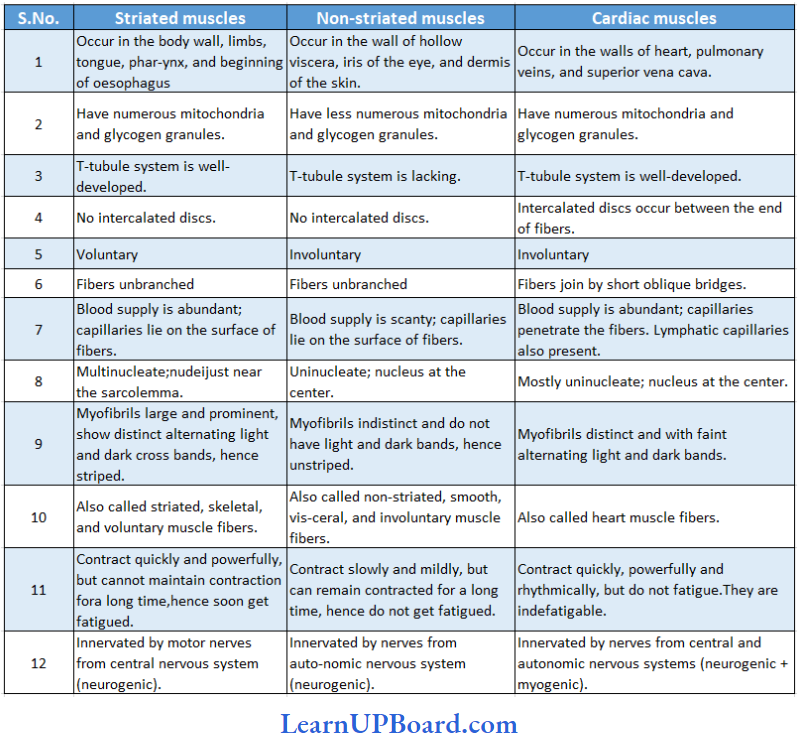
- Muscle tissue may be classified into striated, non-striated, and cardiac muscles, according to their structure, location, and functions.
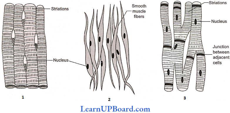
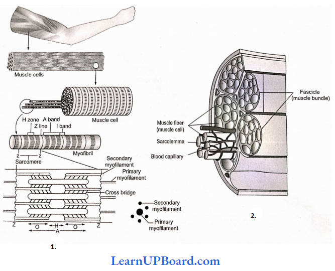
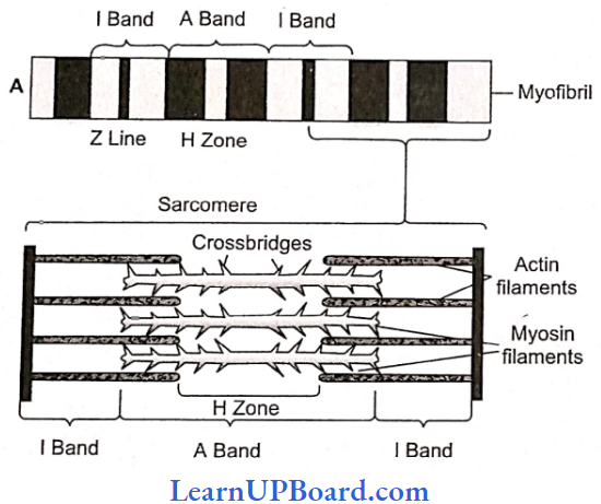
Striated/Skeletal/Voluntary Muscles: Such muscles are attached to bones by tendons. A voluntary muscle is composed of long bundles of striated muscle fib¬ers. Each fiber is a long, unbranched, cylindrical cell. It shows transverse striations in the form of regular alternate dark (A) and light (I) bands.
- At the center of the band is a fine, dense Z band or Z-line (Krause’s membrane). The plasma membrane covering the fiber is called sarcolemma. The cytoplasm inside the fiber is called sarcoplasm.
- The sarcoplasm contains many long, thin, unbranched, cross-striated cylindrical structures called myofibrils. They are arranged along the long axis of the fiber. Dark A bands of neighboring myofibrils are located side by side, and so also are their light I bands. This gives a cross-striated appearance to the entire muscle fiber also.
- A-band has both actin and myosin filaments. The portion of A band, where actin filaments are absent is called H zone. Z line or Krauze’s membrane is a dark membrane that bisects I band or isotropic band.
- Muscle is rich in proteins. Most of these proteins occur as two types of filaments arranged longitudinally in myofibrils. The thick filaments are made up of the protein myosin. Myosin filaments are located inside A bands.
- Thin filaments are more numerous. They are composed of the protein actin. From a fine, dense, dark Z band at the center of each I band, actin filaments extend through I band and encroach between myosin filaments up to a considerable distance into A band.
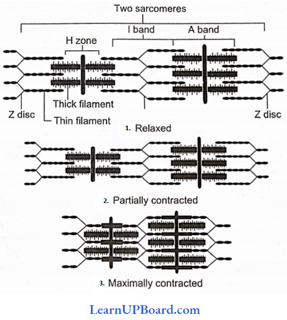
Each segment of the myofibril from one Z band to the next, functions as a contractile unit and is called a sarcomere. Various parts of a sarcomere have a specific arrangement of actin and myosin filaments as given below.
I Band: Has only actin filaments
A Band: Has both actin and myosin filaments
H Band: Has only myosin filaments
Z Line: A membrane to which actin filaments are attached on both sides.
Differences Between Single-Unit And Multi-Unit Smooth Muscles
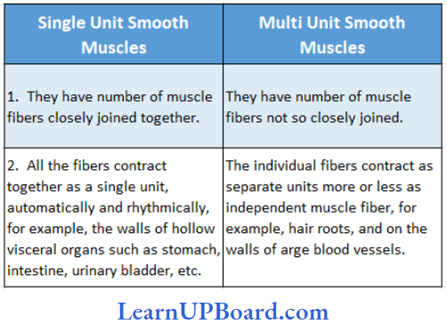
Non-Striated or Smooth Muscles: Non-striated fibers do not show cross-striations; instead, they look smooth. Smooth muscles cannot be moved voluntarily. Hence, they are also called involuntary muscles. Functionally, smooth muscles are of two types: single-unit and multi-unit smooth muscles.
- Single-unit smooth muscles are composed of muscle fibers closely joined together. All its fibers contract together as a single unit. They may contract automatically and rhythmically. Such smooth muscles occur on the walls of hollow visceral organs such as the urinary bladder and gastrointestinal tract.
- Multi-unit smooth muscles are composed of more independent muscle fibers, not so closely joined together. Individual fibers of such smooth muscles contract as separate units. These occur at hair roots and in the walls of large blood vessels, for example, erector pili muscles.
- Smooth muscle fibers are elongated spindle-shaped cells. They are packed parallel to each other in branching bundles. Each fiber contains a single, spindle-shaped nucleus at its thick central part. The smooth muscle fiber is generally shorter than a striated muscle fiber. Mitochondria and other organelles are less extensive and protein filaments are not regularly arranged to give rise to striations.
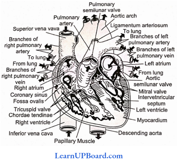
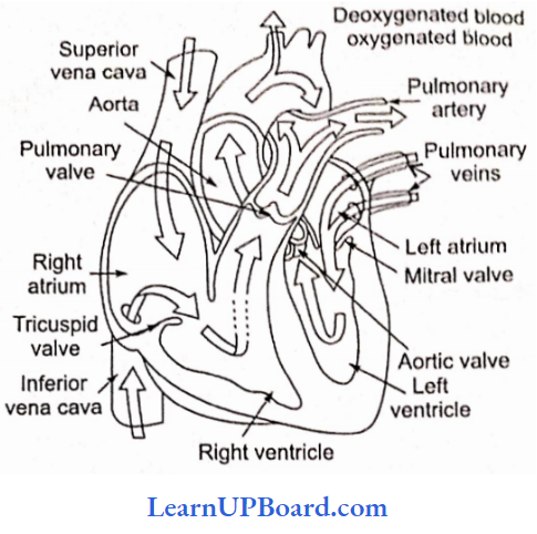
Cardiac Muscle: Cardiac muscle occurs in the heart. It possesses considerable automatic rhythmicity and generates its own wave of excitation. The excitation can also pass directly from one fiber to another in the cardiac muscles. It is not under voluntary control. It shows cross-striations, but striations are much fainter than those of striated muscle.
Between the cardiac muscle fibers, intercalated discs are present. They are specialized regions of the cell membrane of two adjacent fibers. The intercalated discs function as boosters of the contraction wave and permit the wave of contraction to be transmitted from one cardiac fiber to another.
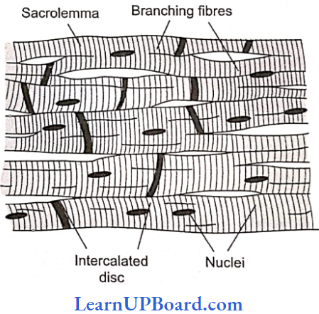
Cardiac muscle cells are short cylindrical cells joined end to end to form rows. They possess abundant cytoplasm with myofibrils (sarcoplasm) and numerous mitochondria and glycogen granules. This is because they need a large amount of energy. Faint but regular, alternately dark and light bands give rise to cross-striations in the cardiac muscle fibers and indicate regular and alternate arrangements of thin and thick filaments in the fiber.
Sarcomeres are also present. Cardiac muscle cells frequently branch to form junctions with neighboring cells. Where two cardiac muscle cells meet end to end, a dense zig-zag junction is formed between them. It is called an intercalated disc. The longest refractory period is present in cardiac muscles.
Differences Between Striated, Non-Striated, And Cardiac Muscles
NEET Biology Notes Structural Organization In Animals Nerve Tissue
Ordinary connective tissue is absent inside the central nervous system. The neurons are held together by supportive cells called neuroglia cells. A nerve tissue is made up of neurons and neuroglia cells. A neuron has a large cell body with two or more, thin protoplasmic processes extending from it.
- One of the processes called the axon is long and conducts nerve impulses away from the cell body. Axon ends in a number of small branches on muscle fibers, gland cells, or other neurons. The remaining one or more processes conduct nerve impulses towards the cell body and are called dendrites or dendrons.
- The axon terminals may form intercommunicating junctions, called synapses, with dendrite terminals, cell bodies, or even axons of other neurons. Nerve impulses pass between neurons through the synapse with the help of chemicals, such as acetylcholine, which are termed neurotransmitters.
- The cell body of a neuron is called the soma. It has various shapes.
- Both soma and the processes are covered by a plasma membrane.
- The soma contains abundant granular cytoplasm and a large nucleus.
- To serve the high-energy needs for impulse conduction, neurons have many mitochondria.
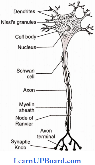
- Light microscopy shows many small conical, angular, or rhomboidal and highly basophilic structures in the cytoplasm of soma and dendrites, called Nissl bodies which are absent in the axon and the axon hillock. Nissl bodies are made up ofribosomes, ER, and m-RNA.
- The processes which arise from neurons are called as neuritis. These are of two types: dendrites and axons.
- Dendrites conduct the nerve impulse toward the nerve cell body and are called as afferent processes.
- Axon is a single, usually long process. The part of cyton from where the axon arises is called as axon hillock. The cell membrane of the axon is called axolemma and its cytoplasm is known as axoplasm. The axon divides to form an axon ending, each with a synaptic knob. The synaptic knobs contain mitochondria and se¬cretory vesicles. The vesicles contain neurotransmitters such as nor-adrenaline, adrenaline, acetylcholine, etc.
Synapse
- Nerve signals travel from neuron to neuron all over the body. These associations are called synapses.
- Synapse is a junction between the axon endings of one nerve fiber and the dendrite of the other.
- At a synapse, the membrane of the axon and dendrites are not in physical contact with each other but there is a narrow intercellular gap, 10-20 nm across, separating the axon tip and target cell. This gap is a synaptic cleft. The neurotransmitter is always released from axon endings and not by dendrites, so there is only one-way transmission of nerve impulses.
Types Of Neurons: Based on the number of nerve processes, neurons are of four types
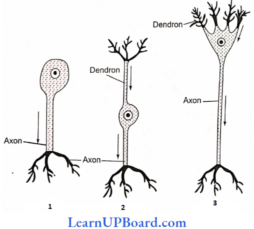
- Unipolar Neurons: These have only axons but no dendrons and are found only in early embryos.
- Bipolar Neurons: These have two processes, one axon, and another dendron, and are found in the olfactory epithelium and retina of eye.
- Multipolar Neurons: These have many processes arising from the cell body; out of them, one (longer) acts as an axon and the remaining as dendrites. Multipolar neurons are most common and are found in the brain and spinal cord.
- Pseudo-Unipolar Neurons: They are actually bipolar but appear like unipolar. A single process arises first which divides to form dendrite and axon. This is found in the dorsal root ganglion of the spinal cord.
Non-Polar Neurons: Each neuron bears several branched processes which are not differentiated into axons or dendrites. On the basis of function, neurons are of three types:
- Sensory (Receptor Or Afferent) Neurons: They connect sense organs with the central nervous system (brain and spinal cord).
- Motor (Effector Or Efferent) Neurons: They connect the central nervous system to the effectors (muscles and glands). They carry motor impulses from the central nervous system to the effectors.
- Interneurons (Connector, Relaying, Or Adjustor Neurons): They are present in the central nervous system and occur between the sensory and motor neurons for distant transmission of impulses. They are neither sensory nor motor.
The extended axon or dendrite of a neuron is called a nerve fiber. It is generally elongated axon. There are two basic types of nerve fibers:
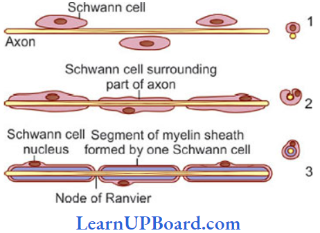
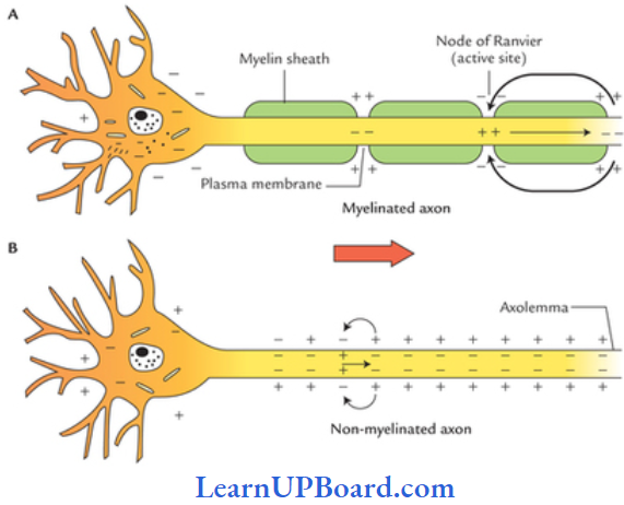
- Myelinated/Medullated Nerve Fibers: These are with the myelin sheath. Myelin sheath is formed by the spiral wrapping of the Schwann cell membrane around the axon. Outside the myelin sheath, a neurilemma is present. Myelin sheath is absent at certain points called nodes of Ranvier. In myelinated nerve fibers, the impulse jumps from one node of Ranvier to the other, which is called saltatory conduction of the impulse. The node of Ranvier is without myelin but with neurilemma. Myelinated nerve fibers are found in cranial and spinal nerves.
- Non-Inyelinated/Non-Medullated Nerve Fibers: These are not covered with myelin sheath and, hence are called non-myelinated or non-medullated nerve fibers. They do not possess nodes of Ranvier but have neurilemma. Myelinated nerve fibers are generally thicker than non-myelinated ones. These fibers are enclosed by Schwann cells that do not form a myelin sheath around these axons and are commonly found in autonomous and somatic neural system.
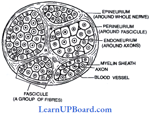
Nerve: A nerve is a collection of nerve fibers surrounded by connective tissue membranes.
- The membrane of the nerve fiber is a neurilemma; outside this, each nerve fiber is surrounded by a layer of connective tissue called the endoneurium.
- A nerve consists of several bundles of nerve fibers called fasciculi.
- Each fasciculus is surrounded by a layer of connective tissue called the perineurium.
- A dense layer of connective tissue that surrounds the entire nerve made of a number of fasciculi is called epineurium.
- A Nerve Can Be Of The Following Types:
- Sensory Nerve: It is made up of only sensory nerve fibers surrounded by connective tissue membrane. It carries the impulse from the receptor to the CNS.
- Motor Nerve: It is made up of motor nerve fibers, which carry the impulse from CNS to the effector organs, i.e., muscles or glands to bring about their movement.
- Mixed Nerve: It has both sensory and motor nerve fibers. All the spinal nerves in our body are mixed.
Neuroglia Cells/Glial Cells: Neuroglia cells are undifferentiated cells with no Nissl granules. They are of the following types:
- Astrocytes/Macrocytes: They are large in size with a number of protoplasmic processes. They form a maximum number of glial cells. They help in the repair of nerve tissue and form the blood-brain barrier.
- Oligodendrocytes: They arc with few protoplasmic processes and form myelin sheath in CNS. There is no neurilemma inside the central nervous system. In the absence of Schwann cells, myelin is formed by the spiral wrapping of nerve fibers by the processes of oligodendrocytes. They are a type of neuroglia cells.
- Microglial Cells: They are mesodermal in origin. They are smallest in size with few feathery processes and help in phagocytosis.
NEET Biology Notes Structural Organization In Animals Nerve Tissue Points To Remember
Cells, tissues, organs, and organ systems split up the work in a way that ensures the survival of the body as a whole and exhibits division of labor. A tissue is defined as a group of cells along with intercellular substances performing one or more functions in the body.
- Epithelial tissues are sheet-like tissues lining the body’s surface and its cavities, ducts, and tubes.
- Epithelia have one free surface facing a body fluid or the outside environment.
- Their cells are structurally and functionally connected at junctions.
- Epithelial tissue is classified into different categories on the basis of the shape and function of cell.
- Diverse types of connective tissues bind together, support, strengthen, protect, and insulate other tissues in the body.
- Soft connective tissues consist of protein fibers as well as a variety of cells arranged in a ground substance.
- Cartilage, bone, blood, and adipose tissues are specialized connective tissues.
- Cartilage and bone are both structural materials.
- Blood is a fluid tissue with transport functions.
- Adipose tissue is a reservoir of stored energy.
- Muscle tissue, which can contract (shorten) in response to stimulation, helps in the movement of the body and specific body parts.
- Skeletal muscle is the muscle tissue attached to bones
- Smooth muscle is a component of internal organs.
- Cardiac muscle makes up the contractile walls of the heart.
- Connective tissue covers all three types of tissues.
- Nervous tissue exerts the greatest control over the response of the body.
- Neurons are the basic units of nervous tissue.
NEET Biology Notes Structural Organization In Animals Earthworm (Pheretima Posthuma)
Phylum: Annelida
Class: Oligochaeta
Genus: Pheretima
Species: Posthuma
There are several types of earthworms. The most common genus of earthworms is Pheretima in India and Lumhricus in Europe. Pheretima has 500 species, 13 of which are found in India.
Habitat: Earthworm is a reddish brown terrestrial animal that inhabits the upper layer of moist soil where it lives inside burrows during the daytime.
- Earthworm inhabits those soils that have abundant organic matter.
- An acre of good moist soil can have up to 50,000 animals. Burrow is made by boring and swallowing the soil.
- The burrows are vertical or oblique.
- They are 30-45 cm deep during moist season but may go as deep as 2 m in summer.
- The burrows are lined by debris or mucus secreted by the animals and are wider at the base.
- During winter, the animal drags organic debris into its burrow and plugs the mouth of the burrow.
- This keeps the burrow warm.
- Even the mouth of the burrow is hidden from view by leaves and small stones.
- The area of the burrow can be recognized by fecal pellets called worm castings.
Habit: Earthworm is nocturnal because it is sensitive to higher light intensities.
- It partly creeps out of burrows during the night for search of food.
- It is only during the rainy season that the earthworm comes out of the burrow even during the daytime.
- After heavy rainfall, they can be seen crawling on the ground in large numbers.
- If the burrow is left, the animal does not re-enter the same.
- It digs a new burrow by pushing the body through the soft soil as well as by eating its way through the soil.
- The worm keeps its skin moist through mucus, coelomic oozing, and from the moisture of the soil. The animal respires through the skin.
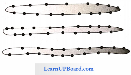
- The body of an earthworm is long and cylindrical has about 100-120 segments (metameres). The first segment is called as buccal segment or peristomium which bears a very small terminal opening the mouth.
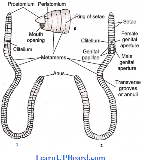
A small projection is also present which hangs over the crescent-shaped mouth and is called prostomium. It serves as a wedge to force open cracks in the soil into which earthworms may crawl. It is sensory in nature.
- The skin of earthworms is brown due to the presence of porphyrin pigment which protects the earthworm from UV radiation.
- In all the body segments, except the first, last, and clitellum, there is a ring of S-shaped setae, embedded in the epidermal pit at the middle of each segment (perichaetine).
- Setae are chitinous structures and are not dissolved in KOH.
- In the intersegmental grooves of 5/6, 6/7, 7/8, and 8/9 segments, four pairs of spermathecal pores are present which are the opening of spermathecae.
- A thick band of glandular tissue clitellum (cingulum) surrounds segments 14-16, forming a thick girdle. Its glands secrete mucus and albumin and also form the cocoon.
- On the ventral surface of the 18th segment, a pair of male genital apertures is present and on the ventral surface of the 14th segment, a median female genital aperture is present.
- On the ventral side of each of the 17th and 19th segments, circular raised pairs of genital papillae are present which help in reproduction.
- The dorsal surface of body is marked by a dark median mid-dorsal line (representing dorsal blood vessels) along the longitudinal axis of the body. The ventral surface is distinguished by the presence of genital openings (pores).
Internal Morphology: The body wall of earthworms is thin, soft, and slimy. From the surface inward, it consists of the cuticle, epidermis, muscular layers, and coelomic epithelium.
Cuticle: It is a thin and clastic non-cellular protective membrane. It is formed of collagen fibers secreted by the underlying epidermis.
Epidermis: This is a single layer of epithelium of tall, columnar cells which arc distinguished into four types as follows:
- Supporting Cells: Unspecialized epithelial cells that form the major part of the epidermis.
- Glandullar Cells: These are of two types more numerous and club-shaped mucussecreting goblet cells and fewer, narrower albumen cells. The mucus, se¬creted by these cells, keeps the body wall moist and slimy. It is also used to lubricate and smoothen the walls of burrows.
- Basal Cells: These are shorter, conical cells wedged in between narrower basal parts of other cells.
- Sensory Cells: These are narrow, columnar cells occurring, here and there, in small groups. Each sensory cell has small sensory hairs at its free end.
Muscular Layer: Beneath the epidermis is the musculature of the body wall. It consists of a thin outer layer of circular and about twice thicker, inner layer of longitudinal muscle fibers. The circular muscle layer is a continuous sheet around the body, but the longitudinal layer is broken into several longitudinal strips or bands, separated from each other by thin connective tissue partitions. These bands appear elliptical or club-shaped in the transverse section. Numerous granules of porphyrin pigment are found scattered in the circular muscle layer.
Coelomic Epithelium: Next to the longitudinal muscle layer is a thin, membrane-like, mesodermal epithelium of flattened, squamous cells. It is the outer envelope of coelomic cavity and hence called the parietal or somatic layer of coelomic epithelium or peritonium.
Coelom Or Body Cavity: Earthworm has schizocoel type of body cavity. It lies between the body wall and the alimentary canal.
- A layer of peritoneum lines both the surfaces, the outer parietal in contact with the body wall and the inner visceral in contact with the alimentary canal.
- The coelom of the first four segments is continuous or undivided.
- The coelom is divided by septa from the fourth and fifth segments onward.
- The septa lying between segments 5/6, 6/7, 7/8, 8/9 9/10, and 10/11 are thick and muscular.
- One of the two septa, either between the eighth and ninth or between the ninth and 10th segments are absent.
- The first six septa are cone-like and run obliquely backward from the body wall to the gut wall.
- The first nine septa, i.e., up to septum 13/14 are without perforations.
- The remaining septa beginning from the septum 14/15 are perforated by numerous apertures.
- Coelomic fluid is milky white and alkaline. It has a fluid matrix of watery plasma containing proteins, salts, and numerous minute nucleated corpuscles. Corpuscles are of the following four types.
- Phagocytes: They move like Amoeba and engulf harmful germs.
- Leucocytes: These are smaller and fewer. Their function is not fully understood.
- Mucocytes: These are elongated, vase-like corpuscles, one end forms an expanded fan-like structure and the other narrow end contains a nucleus. The function of these cells is not known.
- Eleocytes: They are formed by mitosis of the yellow cells of the visceral peritoneum. They contain glycogen and fat and distribute their food to various tissues.
- The coelomic fluid serves as a hydrostatic skeleton to assist the musculature of the body wall in bringing about the locomotion of the body. It oozes out upon the body surface through dorsal pores, keeping the body wall moist to facilitate respiration and to destroy bacteria and other harmful microorganisms.
Locomotion: The earthworm does not have specialized locomotory organs.
- The locomotion is brought about by the circular and longitudinal muscles of the body wall, aided by the chitinous curved setae embedded in the skin.
- Due to the contraction of the circular muscles of the anterior end, the latter becomes thin, elongated, and extends forward.
- At the same time, the setae of the anterior end hold the ground firmly and prevent the animal from slipping backward.
- Now the circular muscles of the anterior end relax and the longitudinal muscles contract.
It causes the shortening and thickening of the anterior segments, and thus, the posterior part of the body is pulled ahead. - The process is repeated and the worm is able to move forward with speed. Earthworms move at the rate of about 15 cm per minute.
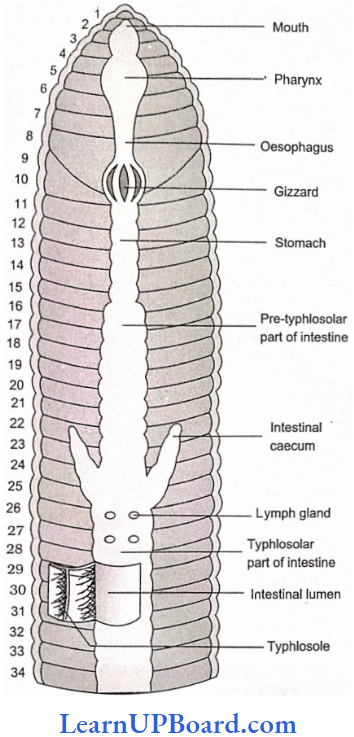
Alimentary Canal: The alimentary canal is a straight tube and runs between the first and last segment of the body.
- In the first to third segments, the buccal cavity is present; the pharynx is present in the fourth segment.
- From the fifth to seventh segment, the oesophagus is present.
- Gizzard is present in the eighth segment.
- The ninth to 1 4th segment is a tubular stomach. Calciferous glands are present in the stomach, which produce CaCO3, to neutralize the humic acid.
- The intestine starts from the 15th segment onward and continues till the last segment.
- A pair of short and conical intestinal caeca projects from the intestine on the 26th segment. They secrete amylolytic enzyme which digests starch. Other enzymes are lipase, cellulase, invertase, etc.
- Surrounding the pharynx there are pharyngeal or salivary glands, made up of masses of chromophil cells, they produce mucin for the lubrication of the food, and also a proteolytic enzyme that can digest some proteins.
- Associated with the intestine arc chloragogen cells which are supposed to be excretory in function.
- The intestine is divided into the pre-typhlosolar region: This runs from the 15th to the 26th segment, it has a small villi.
- Typhlosolar Region: It starts from 27th segment and extends up to 23-25 segments in front of theanus. Typhlosole is a large villus as an internal median fold of the dorsal wall of intestine. This enhances the effective area of absorption after digestion. Post-type solar region: Also known as rectum, it is present in the last 23-25 segments and opens to the outside through a terminal anus.
- Lymph Glands: These are white fluffy bodies that are found arranged on either side of the dorsal vessel from the 26th segment and extend to the successive segments. These glands are believed to produce phagocytes of the coelomic fluid.
Circulatory System
- Earthworms is the first to evolve a closed circulatory system in the evolution of animals.
- The respiratory pigment is hemoglobin which remains dissolved in the plasma, RBC are absent.
- Blood glands are present in the fourth, fifth, and sixth segments. They produce blood cells and hemoglobin.
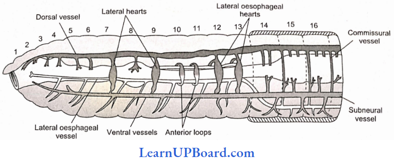
- There are two main blood vessels: the dorsal vessel is the largest vessel and blood flows forward from the posterior to anterior end. The dorsal vessels have a contractile wall; valves are present. Before the 13th segment, it becomes a distributing vessel and behind the 13th segment, it becomes a collecting vessel. The ventral vessel is the main distributing vessel, blood flows backward from the anterior to a posterior end; valves are absent.
- Latero-oesophageal vessels are paired vessels that extend from the first to 13th segment.
- The supra-oesophageal vessel is unpaired and extends between 9th and 13th segments.
Hearts: There are four pairs of hearts with valves.
- Two pairs of lateral hearts, one pair in the seventh segment and one pair in the ninth segment.
- Two pairs of lateral oesophageal hearts in the 12th and 13th segments.
- There are two pairs of lateral loops in which valves are absent. One pair is in the tenth segment and one pair is in the 11th segment. Blood flows in an upward direction in them.
NEET Biology Notes Structural Organization In Animals Earthworm Points To Remember
Earthworm mainly removes nitrogenous waste in the form of urea in soil. But when plenty of water is available, earthworms become ammonotelic. So, earthworm is both ureotelic and ammonotelic.
Excretory System: Excretory organs occur as segmentally arranged coiled tubules called nephridia. They deliver the wastes through a pore to the surface of the body wall or into the digestive tube. Three main types of nephridia
- Pharyngeal Nephridia: These are situated in the segments 4, 5, and 6. They open in the anterior part of the alimentary canal, i.e., the buccal cavity and pharynx. They are without nephrostome and are enteronephrictype.
- Integumentary Nephridia: These are scattered in the body wall. They are the smallest, V-shaped, without nephrostome, and are exophoric type. In clitellar segments, they form forests of nephridia.
- Septal Nephridia: These are the largest, attached to both faces of each intersegmental septum behind the 15th segment. Septal nephridia are the only nephridia with nephrostome or funnel. The terminal duct opens into the septal excretory canal. These canals, in turn, open into two supraintestinal excretory canals.
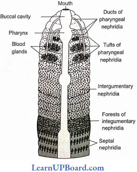
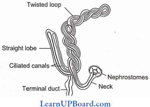
Septal nephridia are enteronephric. Hence, final excretory products are poured into the intestine. Enteronephric condition is an adaptation for the conservation of water or osmoregulation. The excretory products of earthworms are urea (about 50%), ammonia (about 40%), and traces of creatinine. Earthworms are mainly ureotelic.
Nervous System: The nervous system of earthworms consists of central, peripheral, and sympathetic nervous systems.
Central Nervous System
- The central nervous system consists of a brain ring/cricopharyngeal ring and a ganglionated double ventral nerve cord.
- The circumpharyngeal ring occurs in the third and fourth segments. It has a brain consisting of two suprapharyngeal ganglia, two circumpharyngeal connectives, and a pair of subpharyngeal ganglia.
- The ventral nerve cord arises from subpharyngeal ganglia, which is double, solid, and bears paired ganglia in each segment.
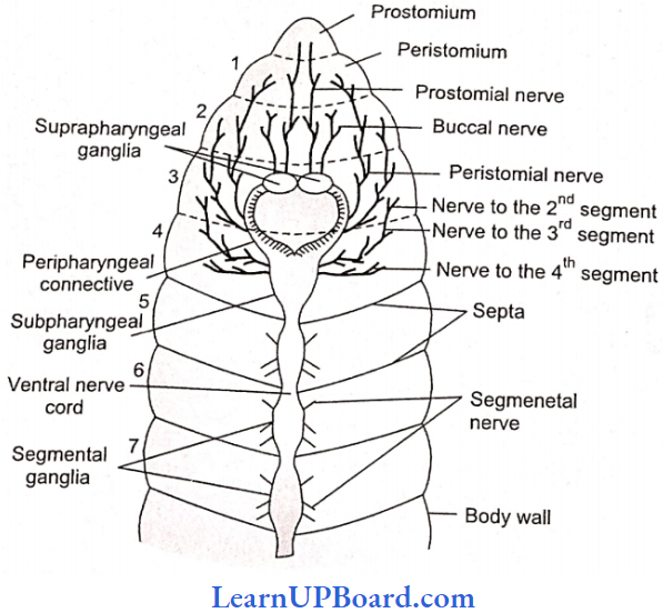
Peripheral Nervous System
- The peripheral nervous system comprises nerves that extend from CNS to supply various parts.
- The nerves are mixed in nature.
- Two pairs of nerves arise from the brain and innervate the prostomium and buccal cavity.
- Nerves from cricopharyngeal connectives supply segments 1 and 2.
- Subpharyngeal ganglia send nerves to the second, third, and fourth segments.
- Each segmental ganglion (actually paired) sends out three pairs of segmental peripheral nerves, one pair from the anterior part and two pairs from posterior part.
- They supply various structures in each segment. All the segmental nerves are mixed in nature, i.e., containing both sensory (afferent) and motor (efferent) nerve fibers.
Sympathetic Nervous System: The sympathetic nervous system consists of various nerve plexes present in the wall of the alimentary canal.
Sense Organs
- A group of specialized cells are found in the skin and lining of the buccal cavity.
- Photoreceptors are located in the prostomium and dorsal epidermis. They perceive light intensity with the help of phagosomes (optic organelle).
- Thigmoreceptors are located in the ventral and lateral epidermis.
- Olfactoreceptors are located in the lining of the buccal cavity.
- Gustatory receptors are located in the lining of the buccal cavity.
Respiratory System
- Earthworm has no special respiratory organs.
- Gaseous exchange takes place simply through the skin, which is thin and highly vascular.
- Effective gaseous exchange takes place only when the skin is moist.
- The skin is kept moist due to damp earth, secretion of the mucus by the epidermal gland cell, and oozing of coelomic fluid through the dorsal pores.
Reproduction
- Earthworms is hermaphrodites.
- There are two pairs of testes present in the 10th and 11th segments. They are surrounded by two testes sacs lying ventrally, one in the 10th and the other in 11th segment.
- There are two pairs of seminal vesicles, one pair in the 11th and the other pair in 12th segment.
- Maturation of sperms occurs in seminal vesicles.
- The testis sac of 10th segment communicates with the seminal vesicles of 11th segment and the testis sac of 11th segment communicates with the seminal vesicles of 12th segment. From each testis sac, the vas deferens carries the sperms up to the 18th segment where they join the prostatic duct from the prostate gland.
- Four pairs of spermathecae are present, one pair in each of the sixth, seventh, eighth, and ninth segments. They receive and store the spermatozoa of another earthworm during copulation.
One pair of ovaries lies in the 13th segment, which opens through a median aperture on the 14th segment. - Accessory glands are present on the ventral surface of the 17th and 19th segments, which open through genital papillae. These are a part of the male reproductive system.
- Testes mature earlier (protandrous).
- Development is direct and there is no larval form.
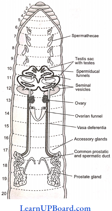
NEET Biology Notes Structural Organization In Animals Cockroach (Periplaneta Americana)
Phylum: Arthropoda
Class: Insecta
Genus: Periplaneta
Species: Americana
Morphology: The body is divided into three distinct regions—head, thorax, and abdomen.
- Their size ranges from 1/4 inch to 3 inches (0.6-7.6 cm).
- The head is hypognathus (facing downward) and is formed by the fusion of six segments.
- Anteriorly, the head bears mouth which is provided with appendages collectively called mouth parts which are used in chewing, cutting, and swallowing.
- The mouth parts consist of a pair of mandibles and maxillae, labium forming the lower lip, and a labrum forming upper lip. Within the cavity enclosed by mouthparts, there is a median flexible lobe called hypopharynx which acts like a tongue.
- Thorax consists of three segments—prothorax, mesothorax, and metathorax.
- A pair ofwings arise from mesothorax which are thick and leathery and are called elytra or tegmina. A pair of membranous wings used in flying arise from metathorax. In houseflies and mosquitoes, the metathoracic wings are reduced to halteres for balancing.
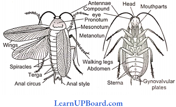
Habit And Habitat: Cockroaches are worldwide and found in such places where darkness, warmth, dampness, and plenty of organic debris is available.
- Cockroaches are nocturnal and omnivorous.
- Three species of cockroaches are commonly found in India: Periplanata americana, Blatta orientalis, and Blatta germanica.
- Periplanata americana is the largest and most common species. It is commonly called American cockroach, Bombay canary, or ship cockroach. In both sexes, wings are present which are larger than the body.
- Blatta orientalis is a black or dark brown mediumsized species. The female has rudimentary wings which are not helpful in flight. The male has wings that are short and are not present up to the end of the body.
- Blatta Germania is the smallest of the domestic species of cockroaches. It is pale yellow-brown in color.
External Features (P. Americana)
- The body is narrow, elongated, bilaterally symmetrical, and flattened, measuring about 3-4.5 cm (34-53 mm) in length and 1.5-2 cm in breadth.
- The color is reddish brown with a pale yellow area around the edge of the pronotum and two dark patches over it.
Exoskeleton
- The entire body of the cockroach is covered with a thick, hard, chitinous cuticle, which is secreted by the epidermis, forming the exoskeleton.
- The exoskeleton of each segment consists of four plate-like pieces called sclerites. The dorsal sclerite is called tergum or tergite, the ventral sclerite is called sternum or sternite, and two lateral sclerites are called pleura or pleurites.
- The sclerites of each segment are joined with each other and with those of the adjacent segments by means of soft and flexible articular or arthrodial membranes. This gives the sclerites some freedom of movement upon each other at their edges.
- The stiff, immovable bristles or spines covering the body and its appendages are in fact the outgrowths of the cuticle while the movable hair-like setae occurring at some places are secreted by special trichogen cells of the hypodermis lying below the cuticle.
Segmentation
- Embryologically, the body of a cockroach is formed of 20 segments: six in the head, three in the thorax, and 11 in the abdomen.
- Due to complete or incomplete fusion between some segments during development, the number of distinct segments is reduced in the adult.
- All segments of the head are fused. The thorax consists of three segments. Only 10 segments are retained in the abdomen of the adult. Of these, only the first seven are distinct in females and the first nine in males. The remaining hinders abdominal segments from becoming small and modified into external genitalia that are hidden under the last distinct segment.
- When wings are removed, the three regions—head, thorax, and abdomen—become distinctly visible. A small, soft, and mobile neck or cervical connects the head with the thorax.
Head
- It is small and triangular, and its narrow end is bent downward in a hypognathous position, i.e., at an angle of 90° with the long axis of the body.
- On each lateral side, it bears a large and blackish compound eye.
- At the base of each antenna, on the inner side, a small rounded and light-colored area called fenestra or ocellar spot representing the simple eye is present. The endoskeleton of head is called the tentorium.
- All sclerites of the head are fused, forming a strong head capsule exhibiting only faint lines of fusion.
- Cephalic Appendages: The head appendages include a pair of antennae, mandibles, first maxillae, and second maxillae (fused as single labium or lower lip), one labrum or upper lip, and one hypopharynx.
- Antennae: These are a pair of long, thread-like appendages, extending forward from an antennal socket located dorsally upon the head capsule near the eye. These are mobile and act as tactile, thermal, and olfactory receptor organs. Each is formed of several small segments called podomeres. The first basal podomere, called scape, is the largest. The second, called pedicel, is narrow and elongated. The remaining long, slender, and many-jointed parts of each antenna is called a flagellum.
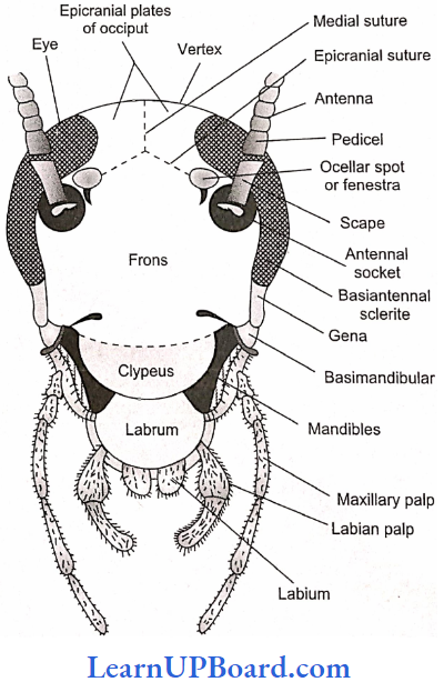
Mouth Parts: The remaining cephalic appendages are small and located around the mouth. Hence, together these appendages comprise the mouth parts of the cockroach. These help in “biting and chewing” its food.
Labrum (Upper Lip): It is the broad, flattened termi¬nal sclerite of the dorsal side of the head capsule, movably articulated to the clypeus that acts as the upper lip. It has an epipharynx (chemoreceptors) on its inner side.
Mandibles: Thick, hard, and triangular appendages beneath the labrum, one on each lateral side of the mouth, which bear pointed, teeth-like processes called denticles.
First maxillae: Located on each side of the mouth next to mandibles. These serve to hold food particles in between the mandibles for cutting and chewing. They also bear olfactory receptors.
Labium (Lower Lip): The second maxillae are fused together forming a single large structure that covers the mouth from the ventral side, hence called the “lower lip” or labium. It bears tactile and gustatory sensory setae.
Hypopharynx: It is a small, cylindrical mouthpart, sandwiched between the first maxillae and covered by labrum and labium on the dorsal and ventral sides, respectively. It bears several sensory setae on its free end and the opening of a common salivary duct upon its basal part.
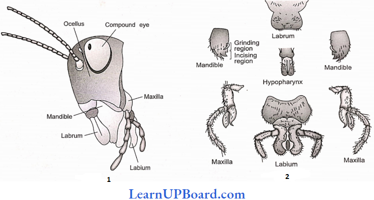
Thorax
- The thorax comprises three segments—prothorax, mesothorax, and metathorax.
- The three thoracic segments are covered by relatively thicker and larger tergites called nota.
- The notum of prothorax called pronotum is very large and covers the neck also. Each of the mesonotum and metanotum bears a pair of wings.
Thoracic Appendages: Each thoracic segment bears a pair of walking legs. Each walking leg consists of segments
- Coxa.
- A triangular short rod-like trochanter, articulated with coxa and femur
- A long, spiny femur.
- A spring-like tibia which represents the longest segment.
- A long tarsus is divided into five tarsomeres or podomeres. The last tarsomere is called pretarsus forming the claws and bearing an adhesive arolium or pulvillus. Similar but smaller adhesive pads called plantulae are located at each joint of the tarsus.
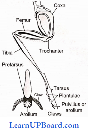
Abdomen: It is the largest and the broadest; relatively more flattened and softer part behind the thorax. There are 10 tergites. In both males and females, the eighth and ninth tergites are mostly covered by the seventh. The 10th tergum is somewhat bowl-shaped and posteriorly bifurcated into two lobes.
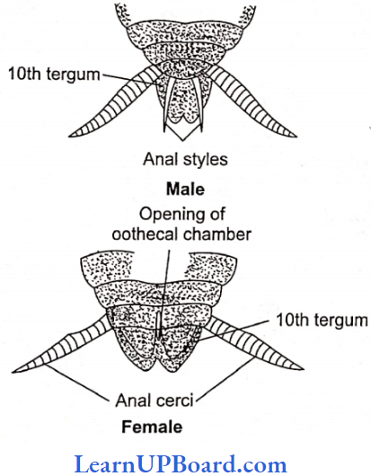
- Ventrally, the abdomen has nine stemites in males and seven in females.
- In females the last stemming (seventh) is larger and boat-shaped and together with indistinct eighth and ninth stemites, it forms a chamber-like structure called gynatrium the posterior part of this chamber is called the oothecal chamber. Behind this chamber, the seventh stemite bifurcates into two prominent oval plates called apical lobes. Female gonopores is located between them.
- In males, the ninth stemite bears a pair of spine-like anal styles.
- In both male and female cockroaches, several small chitinous appendages are located around the gonopore. These help in reproduction and hence are called gonapophyses.
- At several places, certain processes of the exoskeleton extend into the body and form endoskeletal elements that provide attachment to muscles and are hence called apodemes.
Abdominal Appendages
- Abdominal segments lack locomotory appendages. There are certain small structures associated with gonopore, which are different in male and female cockroaches.
- The 10th tergum posteriorly bears a pair of many jointed anal cerci. They bear minute sensory hair sensitive to sound and other vibrations.
- The ninth sternum of males bears, in addition, a pair of small and spine-like, unjointed anal styles.
Digestive System Of Cockroach
The alimentary canal is long and somewhat coiled, di-visible into three main parts, namely, foregut, midgut, and hindgut.
Foregut (stomodeum) is lined by a cuticle and is differentiated into five parts: buccal chamber, pharynx, esophagus, crop, and gizzard. The crop is used for the storage of food.
- The gizzard is muscular and internally provided with six cuticular teeth that crush the food. A stomodeal valve is present between the gizzard and mesenteron.
- The midgut (mesenteron or ventriculus) is short, tubular, and lined with glandular endoderm.
- At the anterior end of the mesenteron, there are six to eight blind glandular hepatic or gastric cecae which secrete digestive enzymes.
- Internally mesenteron is not lined by cuticle but it is covered by a very thin and transparent peritrophic membrane formed of chitin and proteins.
- The peritrophic membrane is secreted by the gizzard which serves to protect the wall of the midgut from abrasion due to the friction of food particles.
- The peritrophic membrane is permeable to digested food and enzymes in the mesenteron.
- The hindgut (proctodeum) is broader than the midgut and comprises the ileum, colon, and rectum.
- The wall of the rectum is provided with six rectal papillae. They help in the absorption of water and salts.
- The cockroach is omnivorous. It feeds on all sorts of organic debris.
- The digestive enzymes of saliva are mainly zymase and amylase.
- Most of the nutrients of food are digested in the crop.
- The absorption of digested food takes place in the mesenteron.
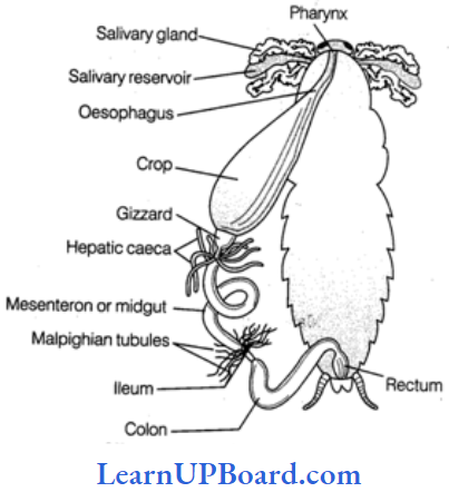
Respiratory System of Cockroach
- The blood of cockroaches is not responsible for the transportation of gases. It serves as a stationary medium for the exchange of gases.
- A complicated system of numerous, shiny, transparent, and branched air tubes or tracheae are found for gaseous exchange in the hemocoel cavity. There are six longitudinal tracheal tubes—two dorsal, two ventral, and two lateral, which are interconnected by transverse commissures. Chitinous rings prevent the collapse of the trachea.
- Atmospheric air enters into and escapes out from this system through 10 pairs of slit-like apertures called stigmata or spiracles located on the lateral sides of the body. Two pairs of these are thoracic and eight pairs are abdominal. The openings of spiracles are regulated by the sphincters.
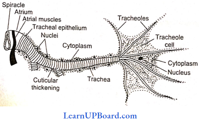
- Thoracic spiracle, arc somewhat larger. One pair of these is between prothorax and mesothorax and the other between mesothorax and metathorax, upon re¬spective pleurites.
- The first pair of abdominal spiracles is dorsolateral upon the tergite of the first abdominal segment, but the remaining seven pairs are present upon the pleurites of the second to eighth segments.
- Each spiracle is surrounded by a ring-like sclerite called peritreme.
Mechanism Of Respiration
- Several tergo-stemal muscles extend vertically between the tergites and stemites of all abdominal segments.
- Harmonious contractions and relaxations of these at regular intervals cause rhythmic expansion and compression of the abdomen leading to inspiration (with relaxation) and expiration (with contraction) of air.
- At rest, the oxygen requirement is less, and tracheolar ends get filled with tissue fluid.
- The movement of O2 is along the pressure gradient as the tracheolar ends are losing oxygen to the cells for performing cellular respiration.
- O2 requirement increases during activity.
- Tracheolar fluid is withdrawn out of Tracheoles.
- Alternate expansion and contraction of the abdominal cavity occurs involving tergo-stemal muscles and abdom¬inal muscles.
- A high level of CO2 in the abdominal cavity makes tergostemal muscles and abdominal muscles to contract pushing out the air from the tracheal system to the outside through spiracles.
- With relaxation, the abdomen expands, i.e., tracheal trunks and tracheae expand, and as a result, air rushes into tracheae and tracheoles via spiracles, which results in inspiration.
Circulatory System of Cockroach
- Blood vascular system is open and lacunar type. Body cavity contains blood (hemolymph) which bathes viscera in it, therefore known as hemocoel.
- The blood vascular system consists of a tubular heart, a blood vessel called the anterior aorta, and a system of ill-defined blood spaces or sinuses.
Heart: It is a long elongated tube situated in the mid-dorsal line of the thorax and the abdomen immediately beneath the terga.
- The heart consists of 13 chambers.
- The last two posterior chambers are very small.
- The chambers are separated from one another by deep constrictions.
- The opening of each chamber into another is guarded by valves that allow blood from behind to forward.
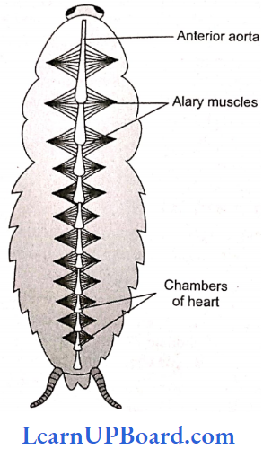
Blood Sinuses: The large body cavity or hemocoel is divided by two membranous horizontal partitions, into three wide and flattened sinuses—the dorsal pericardial sinus containing the heart, the middle perivisceral sinus containing the gut, and the ventral perineural sinus or sternal sinus containing the nerve cord.
- The partition between the pericardial and perivisceral sinuses is called the dorsal diaphragm and between the perivisceral and perineural sinuses is called ventral diaphragm.
- The sinuses intercommunicate by pores in the respective diaphragms.
- A pair of fan-like, triangular alary muscles in the floor of the pericardial sinus in each segment reinforces the dorsal diaphragm by their broad bases and also con¬nects it by their pointed tips with the tergite of the segment.
Circulation Of Hemolymph
- The pumping force that propels the hemolymph is provided by the pulsations of the heart. The respiratory movements of the abdomen and contraction of alary muscles increase this force.
- From the pericardial sinus, the hemolymph enters into heart through the ostia. When the heart is filled, it contracts from behind forwards. This is its systole phase. Soon the heart becomes relaxed in its diastole phase. Then the next systole follows after a short time interval called diastasis. Thus, the heart pulsates about 50 times/ min.
- During systole, the valve-like ostia closes, preventing the backflow of hemolymph into the pericardial sinus. Therefore, some of its hemolymph is pumped into seg-mental vessels while most of it is poured into the head sinus through the terminally opening anterior aorta.
- From the head sinus, the hemolymph flows backward into the thorax and abdomen. While flowing backward from the head sinus, the hemolymph remains in the ventral part due to the presence of esophagus in the dorsal part. So, it fills into the perineural sinus.
- From the perineural sinus, the hemolymph now flows into the perivisceral sinus through the pores of the ventral diaphragm in the abdominal region.
- Then from the perivisceral sinus, it flows into the pericardial sinus through the pores of the dorsal diaphragm. Then, during the heart’s diastole, it fills in the heart through the ostia.
Excretion In Cockroach: Cockroach is uricotelic. In cockroaches, the following structures help in excretion:
- Malpighian tubules
- Fat bodies
- Nephrocytes
- Cuticle
- Uricose glands in some species
- Malpighian Tubules: Malpighian tubules are attached at the junction of the midgut and hindgut. Excretory products, dissolved in hemolymph and absorbed by Malpighian tubes, are discharged into the hindgut.
- Fat Bodies: Some fat bodies are also present in hemocoel which have mycetocytes, urate cells, oenocytes, and trophocytes.
- Nephrocytes: Nephrocytes are present in the lateral wall of the heart and help in the excretion and storage of nitrogenous waste.
- Uricose Glands: In some species, in males, uricase glands are present on the periphery of mushroom glands. These glands synthesize uric acid. Malpighian tubules are analogous to mammalian kidneys, and bodies are analogous to vertebrate liver.
- Nervous System of Cockroach: It consists of a series of fused, segmentally arranged ganglia joined by paired longitudinal connectives on the ventral side.
- Central Nervous System: The central nervous system consists of the brain or supraesophageal ganglion. The brain gives of a pair of short, stout cords, the circumocsophageal connectives, which encircle the esophagus and pass downward and backward over the subesophageal ganglion situated below esophagus.
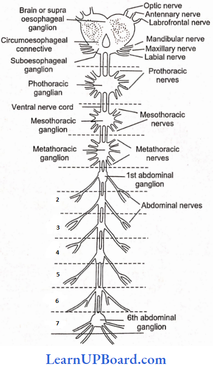
From the suboesophageal ganglion, a double ventral nerve cord passes backward into the thorax, which bears three ganglia in the thorax and six in the abdomen.
Peripheral Nervous System: The peripheral nervous system consists of nerves, which are given off from the ganglia so as to innervate all the parts of the body.
Sympathetic or Somatogastrlc Visceral Nervous System: The stomatogastric nervous system consists of a frontal ganglion which is situated on the dorsal side of the esophagus in the head. From this ganglion, a median unpaired recurrent nerve reaches the visceral ganglion situated on the crop.
Various nerve branches are given off from the visceral ganglion. The frontal ganglion is joined with the central nervous system by nerves that connect it to circumoesophageal commissures.
Sense Organs of Cockroach: Receptor cells are present on the general body surface.
- Proprioreccptors: They are for hearing or receiving sound vibrations. Auditory receptors are present on the antennae and anal cerci.
- Thigmoreceptors: They are the receptors for touch and arc present on antennae, maxillary palps, and legs.
- Olfactory Receptors: They receive various smells and are present on antennae and palps.
- Gustatory Receptors: They are for the sense of taste and are present on maxillae and labial palps.
Eyes: Cockroach has compound eyes. Each compound eye is formed of about 2000 hexagonal ommatidia. Each ommatidium has a biconvex lens or cornea. Below the lens, there are collagen cells that secrete the lens.
- Below the collagen cells is a transparent crystalline cone surrounded by four vitrellae or cone cell.
- The vitrellae secrete the crystalline cone. All this forms the focusing or dioptrical region.
- Below the cone, there is a refractive body, the rhabdome, surrounded by seven retinular cells.
- Each ommatidium is isolated from the other by an iris pigment sheath and retinal pigment sheath.
- The image formed is an apposition or mosaic vision, composed of as many separate but adjacent images as there are ommatidia.
- In mosaic vision, images are sharp but separate and the eye can use only in bright light.
- In cockroach vision is mosaic and an apposition image is formed (although cockroach is nocturnal).
- If pigmented iris sheath is removed from the compound eye of insects, only a superposition image will be formed.
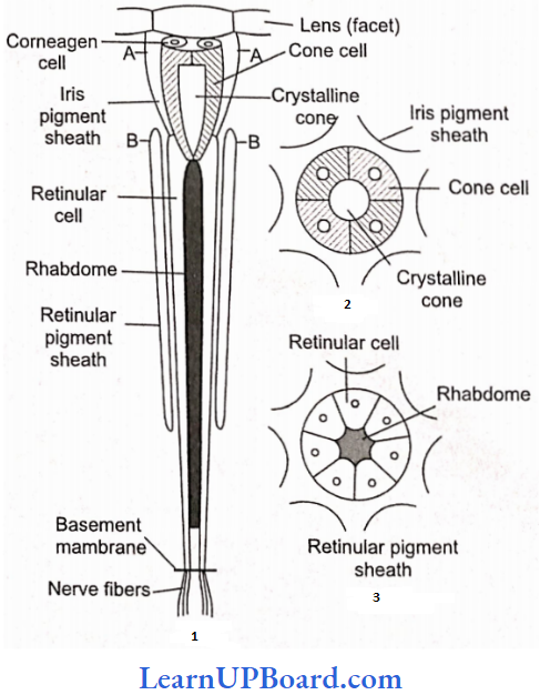
Reproductive System of Cockroach (Male)
- In cockroaches, the sexes are separate, so it is dioecious.
- The testes of cockroaches are located in the abdominal segments 4, 5, and 6.
- The mushroom gland consists of two types of tubules:
- The long slender tubules, utriculi majors or peripheral tubules, and
- Short tubules, utriculi breviores, make up the major part of the gland. It is present in the sixth to seventh abdominal segments which function as an accessory reproductive gland.
- Small seminal vesicles are also found associated with mushroom glands.
- All sperms of a seminal vesicle are glued together into a large bundle called a spermatophore.
- Spermatophore has a three-layered wall: the inner layer is secreted by utriculi majores; the middle layer is secreted by the ejaculatory duct; and the outer layer is secreted by the phallic gland or conglobate gland.
- There are three asymmetrical chitinous structures called male gonapophyses or phallomeres. Right phallomere, left phallomere (largest), and ventral phallomere (smallest).
Reproductive System of Cockroach (Female)
- Female organs consist of ovaries, oviducts, vagina, genital chamber, spermathecae, colleterial glands, and female gonapophysis (ovipositor processes).
- The ovaries of cockroaches are located in the abdominal segments 2 to 6. Each ovary consists of eight ovarioles.
- Two oviducts from each side open into a common oviduct or vagina which opens into the genital chamber by the female genital pore. A pair of spermathecae (left larger pyriform sac) are present near the female genital pore.
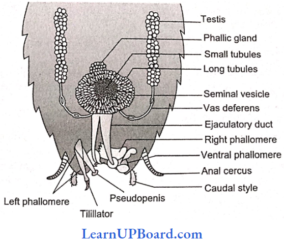
- A pair of colleterial glands also open in the genital chamber.
- The genital pouch or gynatrium is divisible into a genital chamber in front and an oothecal chamber (vestibulum) behind.
- Female genitalia consist of three pairs of chitinous processes hanging from the roof of the oothecal chamber into its cavity.
- The ootheca of cockroaches contains 14 to 16 fertilized eggs. It is formed of a protein secreted by colleterial glands. On average, females produce 9-10 oothecae.
- The nymphs of cockroaches emerge from the ootheca. A nymph resembles an adult in general structure but lacks wings and mature reproductive organs. The next to last nymphal stage has wing pads but only adult cockroaches have wings.
- The instar is a stage in the development of insects (larval instar, nymphal instar). The period between two successive molts in insects is termed stadium.
- In Periplaneta americana, the nymph grows by molting about 13 times to reach the adult form, and in Blatta orientalis, it molts about six times.
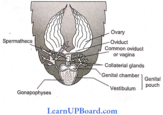
NEET Biology Notes Structural Organization In Animals Frog (Rana Tigrina)
Phylum: Chordata
Class: Amphibia
Order: Anura
Genus: Rana
Species: tigrina
The most common frog found in India is the Indian bullfrog. It is the largest frog and is named as bullfrog because of its large size and loud call.
- Indian bullfrog is found in freshwater marshes, ditches, ponds, and shallow lakes.
- They undergo aestivation (summer sleep) in summer and hibernation (winter sleep) in winter.
- They are carnivorous (feeding upon other animals, insects, etc.), poikilothermic, i.e., the body temperature changes with the environment.
- They develop protective coloration to camouflage, i.e., to hide in surroundings.
- Frogs belong to the phylum Chordata, subphylum Vertebrata or Craniata, superclass
- Gnathostomata, class Amphibia, and Genus Rana.
- The most common species is known as Rana tigrina (Indian bullfrog).
- The scientific name of the common toad is Bufo melanostictus. Frogs exhibit sexual dimorphism.
- Male and female are distinguishable externally only during breeding season when the males develop nuptial pad in the first digit of the forelimbs.
- Vocal sacs are well developed in males so they produce louder sounds as compared to females which are devoid of vocal sacs.
- The vocal sacs help to produce mating calls.
- The total number of bones in a frog is 153.
External Morphology: Skin is made up of the epidermis and dermis. Mucous glands are present in the dermis and their ducts open at the surface.
- Blood capillaries and pigment cells (chromatophores) are present in the dermis.
- Skin is without scales or any other cover or exoskeleton.
- Body is divisible into head and trunk, neck is absent. The trunk is provided with a pair of fore and hind- limbs. The hind limbs are much larger and muscular than the forelimbs. Forelimbs end in four digits and the hind limbs end in five digits. The digital formula of forelimbs is 02233. The digital formula of hind limbs is 22343.
- Shank or crus is associated with hind limbs.
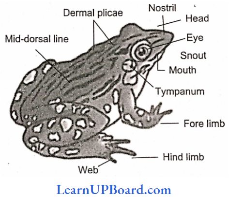
Sexual Dimorphism
- The male and female frogs exhibit certain differences in their external features, which become more pronounced during breeding season.
- Generally, male frogs are larger than females.
- During the breeding season, however, the females become bloated with large ovaries and numerous ova and appear considerably larger.
- Only the males possess a pair of ventrolateral, wrinkled, pouch-like vocal sacs located a little behind the mouth.
- These sacs become especially large and distensible in the breeding season.
- By inflating these repeatedly with air from the lungs, the males produce a loud croaking sound meant to call the females for copulation (amplexus).
- The sound is actually produced by a pair of vocal cords in the larynx; the sacs only increase its pitch, such as resonators.
- The females produce a low-pitched sound by their vocal cords alone.
- The forelimbs in both male and female frogs bear small particular pads dorsally at the joints of digits, but the males also possess a special nuptial, copulatory, or amplcxusary pad on the ventral side of the first finger of each forelimb.
- Normally, these pads appear merely as rough patches but during breeding season, these become thick and sticky.
- In amplexus, the male strongly grips a female under her armpits by means of these pads.
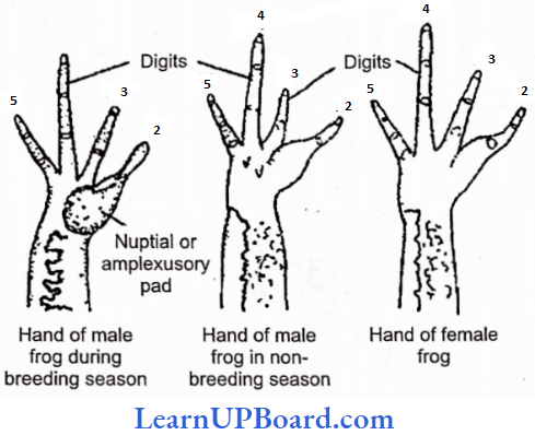
Internal Morphology: Digestive System
- Since the frogs are carnivorous, their alimentary canal is short in length.
- Tadpole larva is herbivorous, so the alimentary canal is very long and coiled in the form of offspring.
- The mouth is present as a terminal, wide opening.
- It opens into the bucco-pharyngeal cavity, which contains numerous maxillary teeth arranged along the margin of the upper jaw. Vomerine teeth are present under the roofof the buccopharyngeal cavity.
- The lower jaw is toothless.
- Opening of the Eustachian tube, vocal sacs (only in males), gullet, and glottis can be seen clearly in the buccopharyngeal cavity.
- The muscular tongue is bilobed at the tip and free from behind. It is used for capturing the prey.
- The gullet opens into a narrow and short tube-like esophagus, which continues in a large and distended stomach.
- It contains a thick muscular layer, which helps in verting food into chyme.
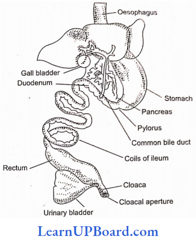
- It secretes gastric juice containing HCl and proteolytic enzymes. The stomach is followed by a coiled small intestine.
- The intestinal wall has numerous finger-like folds called villi and microvilli, projecting into its lumen to enhance the surface area for absorption of the digested food.
- The first part of the small intestine lying parallel to the stomach is called the duodenum. The intestine continues into a wider rectum, opening into the cloaca.
- The urinary bladder opens into the cloacal chamber through the ureter.
- The gastric and intestinal glands occur in the walls of the stomach and intestine, respectively.
- The other important digestive glands associated with the alimentary canal are the liver and pancreas.
- The liver secretes bile which is temporarily stored in the gall bladder before being released into the duodenum.
- Bile helps in the digestion of food by changing its pH from acidic to alkaline and by emulsifying the fats.
- The liver does not secrete any digestive enzymes. The pancreas is an irregular, elongated gland, situated in a thin mesentery, and lies parallel to the stomach.
- It produces pancreatic juice containing digestive enzymes such as trypsin, amylopsin, etc.
Respiratory System
- Three Types Of Respiration Are Observed: cutaneous, buccopharyngeal, and pulmonary.
- Cutaneous respiration on land is through the body’s surface. During hibernation and aestivation, frog respires only through this method.
- Buccopharyngeal respiration occurs through the lining of the buccal cavity. It occurs only when the frog is out of water. The mucus membrane of the buccal cavity is moist which dissolves oxygen whose diffusion occurs into the blood capillaries.
- Pulmonary Respiration: Lungs in frogs are not efficient respiratory organs because only mixed air enters into them and they mainly function as hydrostatic organs.
- Lungs are a pair of thin-walled, translucent sacs with an inner surface divided into alveoli by septa. Pulmonary respiration has a maximum frequency of 20/min.
- It occurs when more energy is required. The mouth and gullet are kept closed during pulmonary respiration.
- Respiratory movements in pulmonary respiration are because of the buccopharyngeal cavity which acts as a force pump. These movements are carried out by a set of paired muscles—stemohyal and pterohyal muscles.
- Stemohyal muscles are attached to hyoid and coracoid processes and clavicles of the pectoral girdle and, on contraction, depress the buccal floor enlarging the buccopharyngeal cavity.
- Pterohyals are attached in between the hyoid and prootics of the skull and, on contraction, lift the floor of the buccal cavity.
- With the depression of the buccal floor, air enters the buccal cavity through the nares.
- External nerves are then closed by pushing the tuberculum prelingual and the movable pre maxillae.
- It is followed by the raising of the buccal floor by peroneal muscles which reduce the volume and the air is pushed into the lungs where the exchange of gases takes place.
- The buccal floor is again lowered enlarging its volume which draws air into the buccal cavity.
- External nares are opened followed by raising the buccal floor, pushing the air out through external nares.
- The sound-producing organ of a frog is the laryngotracheal chamber. It is supported by one cricoid, two arytenoids, and two pre-arytenoid cartilages. It has a pair of muscle strands (vocal cords) that actually produce sound. The male frog has vocal sacs which act as resonating chambers.
Circulatory System
- The circulatory system in frogs is closed type.
- The heart lies enclosed by a thin, transparent, two-layered sac, pericardium.
- Frog s heart is a three-chambered structure made up of two upper auricles and a single lower ventricle.
- The two additional chambers connected to the heart of the frog are sinus venosus and truncus arteriosus.
- Frogs also possess two well-developed portal systems: the renal portal system and the hepatic portal system. Frog also has two pairs of lymph hearts.
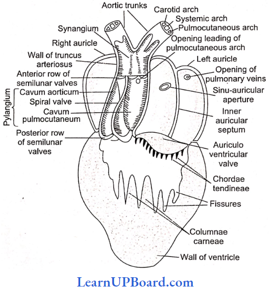
Nervous System
- The nervous system is organized into a central nervous system (brain and spinal cord), a peripheral nervous system (cranial and spinal nerves), and an autonomic nervous system (sympathetic and parasympathetic chains of ganglia).
- There are 10 pairs of cranial nerves.
- The brain is enclosed in a bony structure or brain box (cranium) which has two occipital condyles for attachment with the first vertebra (atlas).
- The brain is divided into forebrain, midbrain, and hindbrain.
- The forebrain includes olfactory lobes, paired cerebral hemispheres, and unpaired diencephalon. The midbrain is characterized by a pair of optic lobes.
- The hindbrain consists of the cerebellum and medulla oblongata.
- Medulla oblongata passes out through the foramen magnum and continues into the spinal cord which is contained in the vertebral column.
- Jacobson’s organ, also called the vomeronasal organ, opens into the nasal chamber and acts as an additional olfactory organ.
Sensory Organs
- Eye
- The eye is guarded by an immovable upper eyelid, movable low er eyelid, and transparent nictitating membrane.
- The outer sclerotic ring is cartilaginous and the cornea is the part exposed. In the middle, the highly vascular, pigmented layer is choroid.
- Iris is yellow-pigmented and perforated by a central aperture, the pupil.
- The retina is the innermost coat of the eyeball and consists of an inner pigmented layer and an outer receptor layer.
- Rods and cones are light-sensitive structures found in the retina.
- Rods have rhodopsin or visual purple meant for night vision while cones have iodopsin responsible for color vision in daylight.
- The anterior chamber present in the front of the lens is filled with aqueous humor while the chamber behind the lens is a posterior chamber, filled with vitreous humour.
- The eyeball is moved in the eye orbit by a set of six muscles—two are oblique and four are recti.
- Besides, there are retractor bulbs and levator bulbs muscles for intrusion or protrusion of the eyeball into the eye orbit, respectively.
- Ears
- The ear of frog has only a middle and internal ear.
- The tympanic membrane is present at the body surface.
- The middle ear has a single bone called columella auris.
- Its outer end is attached to the eardrum while the inner end is attached to the stapedial plate.
- The pressure of air in the middle ear is controlled by Eustachian tubes.
- The membranous labyrinth or internal car consists of a utriculus, sacculus, and semicircular canals. Endolynrph fills the membranous labyrinth.
- Excretory System
- The main organ of excretion is a pair of kidneys.
- These compact, dark red, and bean-like structures are situated a little posteriorly in the body cavity on both sides of the vertebral column.
- The frog excretes urea and thus is a ureotelic animal.
- Urea is carried by blood into the kidney where it is separated and excreted.
- Each kidney is composed of several structural and functional units called uriniferous tubules or nephrons.
- The ureter emerges from the kidney as urinogenital ducts in males.
- A common ureter opens into the cloaca.
- A thin-walled urinary bladder is present ventral to rectum which also opens in the cloaca.
Reproductive System: The male reproductive organs consist of a pair of yellowish ovoid testes, which are found to adhere to the upper part of the kidneys by a double fold of peritoneum called mesorchium.
- Vasa efferentia are 10-12 in number and, after arising from the testes, run through the mesorchium and enter the kidneys of their side.
- In kidneys, these open into Bidder’s canal which finally communicates with the urinogenital duct. This duct emerges from the kidneys and finally opens into the cloaca.
- The cloaca is a small, median chamber that is used to pass fecal matter, urine, and sperm to the exterior.
- A pair of ovaries, situated near the kidneys, comprises the female reproductive organs. However, these have no functional connection with the kidneys.
- A pair of oviducts opens into the cloaca, separately
- The release of ovum in females is termed as spawning.
- A mature female can lay 2500-3000 ova at a time.
- Fertilization is external and takes place in water.
- Development involves a larval stage called tadpole.
- Tadpole undergoes metamorphosis to form an adult.
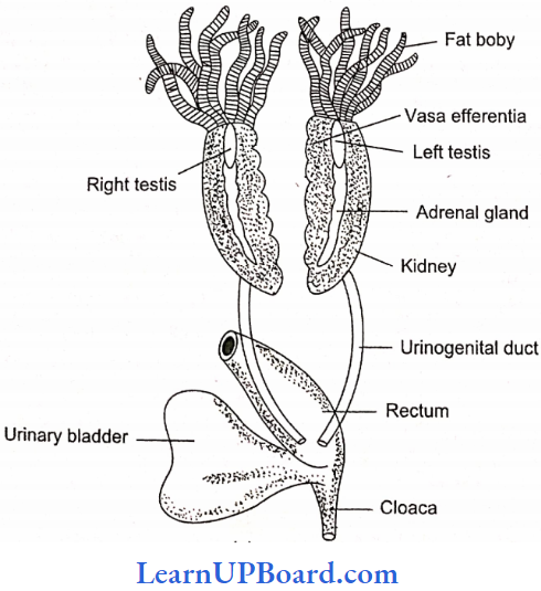
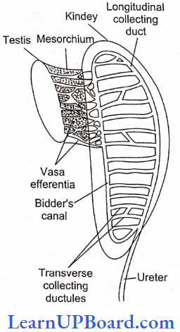
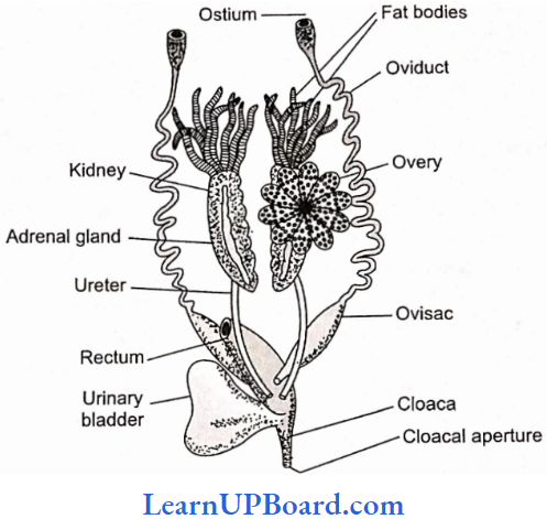
NEET Biology Notes Structural Organization In Animals Assertion Reasoning Questions And Answers
In the following questions, an Assertion (A) is followed by a corresponding Reason (R). Mark the correct answer.
- If both Assertion and Reason are true and the Reason is the correct explanation of the Assertion.
- If both Assertion and Reason are true, but the Reason is not the correct explanation of the Assertion.
- If Assertion is true, but Reason is false.
- If both Assertion and Reason are false.
Question 1. Assertion: Cardiac muscles have striations and fiber is nucleated and involuntary.
Reason: Intercalated discs form the three-dimensional network of cardiac muscle fiber.
Answer: 3. If Assertion is true, but Reason is false.
Question 2. Assertion: Multipolar neurons have several efferent processes.
Reason: Axons are the afferent processes of a neuron.
Answer: 4. If both Assertion and Reason are false.
Question 3. Assertion: Blood circulation is absent in epithelium tissue.
Reason: Blood vessels are unable to pierce the basement membrane.
Answer: 3. If Assertion is true, but Reason is false.
Question 4. Assertion: Reticular fibrous connective tissue is called as embryonic tissue.
Reason: Reticular fibrous connective tissue is mainly found in the embryonic stage.
Answer: 4. If both Assertion and Reason are false.
Question 5. Assertion: Epithelia are highly regenerative.
Reason: When epithelia get damaged, they regenerate more rapidly than other tissues.
Answer: 1. If both Assertion and Reason are true and the Reason is the correct explanation of the Assertion
Question 6. Assertion: Platelets play an important role in blood clotting.
Reason: In the blood oozing from an injury, the platelets disintegrate and release thromboplastin that initiates clotting.
Answer: 1. If both Assertion and Reason are true and the Reason is the correct explanation of the Assertion
Question 7. Assertion: Brown fat produces more energy.
Reason: Brown fat is composed of molecular adiposity
Answer: 3. If Assertion is true, but Reason is false.
Question 8. Assertion: Simple cuboidal epithelium is called as germinal epithelium.
Reason: The cuboidal cells of gonads form gametes.
Answer: 1. If both Assertion and Reason are true and the Reason is the correct explanation of the Assertion
Question 9. Assertion: Heparin is an anticoagulant found in mammals.
Reason: Heparin prevents the conversion of prothrombin to thrombin.
Answer: 1. If both Assertion and Reason are true and the Reason is the correct explanation of the Assertion
Question 10. Assertion: Epithelium cells get their nutrients from adjacent cells.
Reason: In epithelium tissues, large intercellular spaces are present.
Answer: 4. If both Assertion and Reason are false.
- If both Assertion and Reason are true and the Reason is the correct explanation of the Assertion.
- If both Assertion and Reason are true, but the Reason is not the correct explanation of the Assertion.
- If Assertion is tine, but Reason is false.
- If both Assertion and Reason are false.
Question 11. Assertion: Earthworm is brown or claycolored.
Reason: Because of the presence of pigment porphyrin.
Answer: 1. If both Assertion and Reason are true and the Reason is the correct explanation of the Assertion.
Question 12. Assertion: Chloragogen cells are considered analogous to the liver of vertebrates.
Reason: Because it is concerned with the storage of reserve food, deamination of proteins, formation of urea, etc.
Answer: 1. If both Assertion and Reason are true and the Reason is the correct explanation of the Assertion.
Question 13. Assertion: Earthworms is saprozoic.
Reason: Because it feeds on small insects.
Answer: 3. If Assertion is tine, but Reason is false.
Question 14. Assertion: Earthworm is hermaphrodite.
Reason: Because in earthworms both sexes are separate.
Answer: 3. If Assertion is tine, but Reason is false.
Question 15. Assertion: Earthworms are the enemy of fanner.
Reason: Because they destroy the crop in the field.
Answer: 4. If both Assertion and Reason are false.
Question 15. Assertion: In the body of earthworms, porphyrin pigment is found.
Reason: Because it protects earthworms from chemicals.
Answer: 3. If Assertion is tine, but Reason is false.
Question 16. Assertion: In earthworms, development is direct.
Reason: Because in development larval stage is not found.
Answer: 1. If both Assertion and Reason are true and the Reason is the correct explanation of the Assertion.
Question 17. Assertion: In the anus of earthworms, depressor muscles are found.
Reason: These muscles help in the elimination of excretion from the rectum.
Answer: 1. If both Assertion and Reason are true and the Reason is the correct explanation of the Assertion.
Question 18. Assertion: In cockroaches, inspiration is an active process.
Reason: It is due to the contraction of tergosternai muscle.
Answer: 4. If both Assertion and Reason are false.
Question 19. Assertion: In frogs, sinus venosus is present.
Reason: In mammals and birds, the remnant of sinus venosus has taken part in the formation of SA node.
Answer: 2. If both Assertion and Reason are true, but the Rea¬son is not the correct explanation of the Assertion.
Question 20. Assertion: Septal nephridia take part in osmoregulation.
Reason: They are entcronephric.
Answer: 1. If both Assertion and Reason are true and the Reason is the correct explanation of the Assertion.
Question 21. Assertion: In Periplcineta, only superposition or overlapping images are formed.
Reason: Retinal pigment sheath remains contracted throughout life.
Answer: 4. If both Assertion and Reason are false.
Question 22. Assertion: The pharyngeal gland of earthworms includes chromophil cells, which secrete sativa.
Reason: The salivary amylase of earthworms is essential to digest carbohydrates.
Answer: 3. If Assertion is tine, but Reason is false.
Question 23. Assertion: The head of the cockroach is hypognathus.
Reason: The proximal part of the lower lip of a cockroach is called the pastmentum.
Answer: 2. If both Assertion and Reason are true, but the Reason is not the correct explanation of the Assertion.
Question 24. Assertion: The heart of a cockroach is neurogenic.
Reason: The heartbeat rate in cockroaches is 49 per minute.
Answer: 2. If both Assertion and Reason are true, but the Rea¬son is not the correct explanation of the Assertion.