
NEET Biology Notes PDF Free Download
NEET Biology Notes For Biological Classification Introduction
Since the dawn of civilization, there have been many attempts to classify living organisms. Previously, it was done not using any scientific criteria, but on the basis of the need to use organisms for our own use, i.e., for food, shelter, and clothing, Aristotle was the earliest scientist who made an attempt scientifically to classify and use of characters of plants and animals for dividing them into various groups.
Read and Learn More NEET Biology Notes
NEET Biology Notes For Biological Classification Systems Of Classification Of Living Organisms
There are proposed five systems of classification.
Two Kingdom Classification: Linnaeus gave this system and divided all organisms into two kingdoms, viz., Plantae and Animalia.
- Shortcomings Of This System: Tunicatcs have cellulose and branching patterns like plants. Slime molds are amoeba-like structures but like fungi in reproduction.
Three Kingdom Classification: It was proposed by E. Haeckel. He separated out all unicellular organisms into a separate kingdom Protista (fungi, protozoans, algae, bacteria, and slime molds were included in Protista). Thus, he proposed three kingdoms, viz., Protista, Plantae, and Animalia.
- The protists are believed to have evolved from prokaryotic moncrans and the precursors from which higher eukaryotic kingdoms—Plantae, Fungi, and Animalia have evolved. The protists exhibit the following features:
- These have a typical eukaryotic cell organization- and possess a nucleus, mitochondria, endoplasmic reticulum, Golgi bodies, and in some organisms, plastids.
- Locomotion takes place with the help of pseudo-podia, cilia, or flagella. The flagella or cilia have 9 + 2 internal microtubular structures.
- These exhibit diverse lifestyles some are photosynthetic, some are predatory or parasitic, while some are saprobes, living on decaying organic matter.
Biological Classification
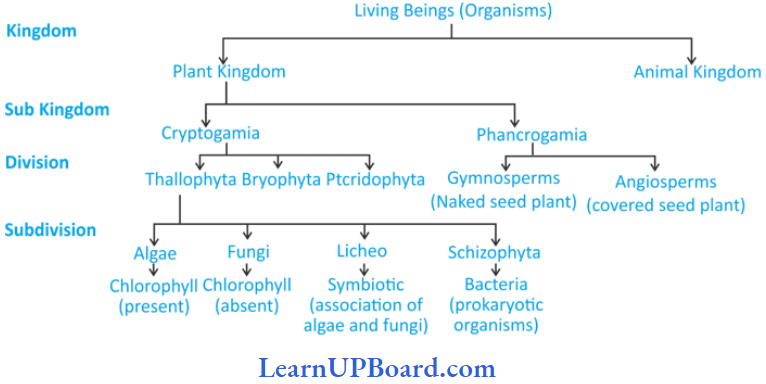
Four Kingdom Classification: Copeland (1956) gave four kingdom classifications and included Monera in them. Copeland originally called the kingdom Monera as mychota. It was called Monera by Daugherty and Allen. Kingdom Monera includes all the prokaryotic organisms, i.e., eubacteria (including cyanobacteria, formerly known as blue-green algae) and archaebacteria.
- Actinomycetes (filamentous bacteria) are also included in this kingdom. Hence, according to Copeland, there are four kingdoms, namely, Monera, Protista, Plantae, and Animalia. The monerans are characterized by the following features:
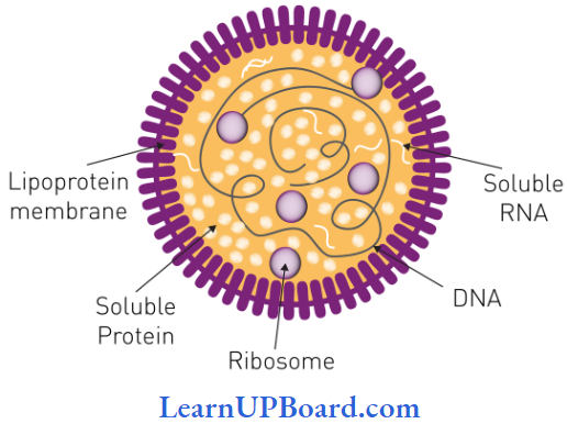
- All moneran cells are microscopic (1 mm to a few millimeters), do not contain an organized nucleus, and are membrane-bound organelles.
- Except for a few monerans (for example, Mycoplasma), the moneran cells are surrounded by a rigid cell wall.
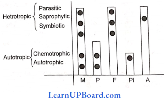
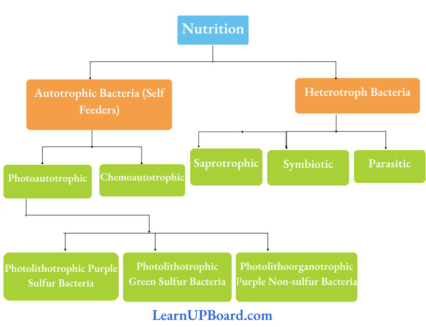
- Chemosynthesis, as a mode of nutrition, is found only in certain members of the kingdom Monera.
- Some monerans are autotrophs. These prepare their own food by reducing CO2, using either light energy (photoautotrophs), or energy derived from chemical reactions (chemoautotrophs). Many monerans are heterotrophs (parasites or saprotrophs). The saprotrophs obtain their food by decomposing dead organic matter and absorbing it in solution form as their wall prevents the ingestion of complex organic material. Some of these live symbiotically with other forms of life.
- These are cosmopolitan in distribution. Their representatives are found in all kinds of habitats, which can possibly support life. Some monerans (archaebacteria) can flourish under extreme environmental conditions such as the absence of oxygen, high salt concentration, high temperature, or acidic pH.
- Many monerans are important decomposers and mineralizers. Some of them are important nitrogen-fixers.
biological classification class 11 short notes
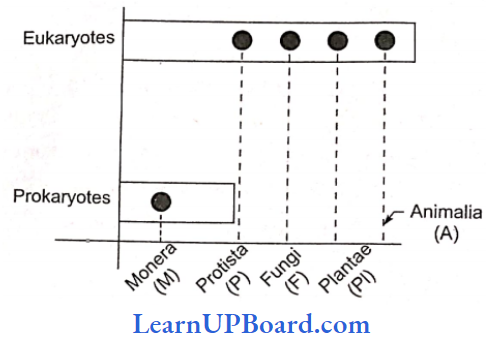
Five Kingdom Classification: It was developed by Whittaker. The five kingdoms proposed by Whittaker are Monera, Protista, Fungi, Plantae, and Animalia. It is a phylogenetic system that was based on the following criteria:
1. Complexity Of Cell Structure: Prokaryotic vs eukaryotic organization of cells.
2. Mode Of Nutrition: Autotrophic (holophytic) or heterotrophic [absorptive (saprozoic or parasitic) or ingestive (holozoic)]. The mode of nutrition is a major criterion of classification in this system.
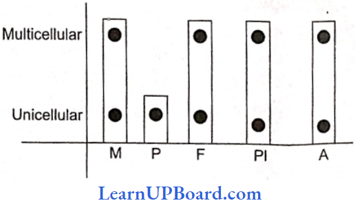
3. Complexity Of Body Organization: Unicellularity vs multicellularity. The five kingdoms include various organisms as mentioned below:
biological classification pdf
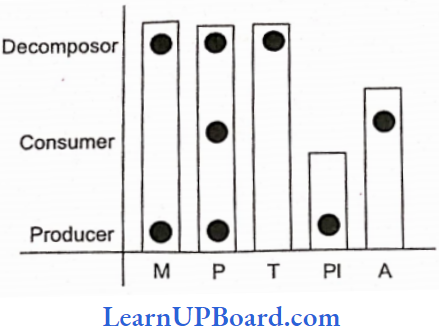
4. Major Ecological Role: Producers vs consumers vs decomposers
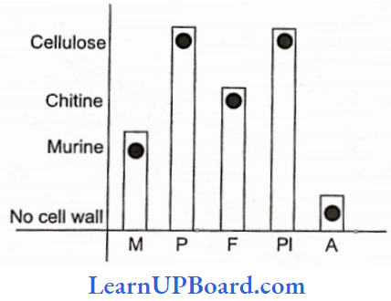
5. Cell Wall Decomposition: Cellulose vs chitin vs murine.
The Five Kingdoms Include Various Organisms As Mentioned Below:
- Kingdom Monera includes prokaryotic, autotrophic, and heterotrophic organisms.
- Kingdom Protista includes eukaryotic, unicellular, autotrophic, and heterotrophic organisms.
- Kingdom Fungi include eukaryotic, multicellular, heterotrophic organisms exhibiting absorptive (assimilative) types of nutrition; and sporeproducing.
- Kingdom Plantae or Metaphyta includes all chlorophyllous, multicellular, photosynthetic organisms (plants) that occur on land, on sea shores, in lakes as well as in streams, and on their non-green relatives. Their photoautotrophic nutrition is also termed holophytic nutrition. These include red, brown, and green algae, liverworts, mosses, ferns, and seed plants with or without flowers.
- Kingdom Animalia includes all multicellular holozoic or phagotrophic or ingestive eukaryotes. These are also known as Metazoa and include a wide variety of animal life such as sponges; cnidarians (Hydra and jellyfishes); worms, snails, and other mollusks; arthropods; insects; fishes; amphibians; reptiles; birds; and mammals.
Six Kingdom Classification: It was given by Carl Woese. The six kingdoms are Eubacteria and Archaebacteria. Protista, Fungi, Plantae, and Animalia. He separated archaebacteria from eubacteria on the basis of major differences such as the absence of peptidoglycan in the cell walls of the former and the occurrence of branched chain lipids (a monolayer) instead of a phospholipid bilayer in the membrane. Based on the sequence of 16S ribosomal RNA genes, Woese found that the six kingdoms naturally cluster into three main categories. He called these categories as domains. These domains are Bacteria, archaea, and Eukarya. All three domains have evolved from a common ancestor, the progenitor.
NEET Biology Notes For Biological Classification Eubacteria
Eubacteria History: Antoni van Leeuwenhoek discovered bacteria from stored rain water and teeth scum and called them “wild animalcules.” He is known as the discoverer of the microbial world or the wonder world of microbes. He used the term “darkens.”
- Ehrenberg gave the term “bacteria.”
- Nageli placed bacteria in schizomycetes, called “fission fungi.”
- Louis Pasteur proposed the germ theory of disease. He discovered bacteria causing chicken cholera and invented the antirabies vaccine. He gave the term “microorganism.” He is known as the father of modern microbiology and sterilization techniques.
- Robert Koch developed “Koch’s postulates.”
- Joseph Lister developed the technique of aseptic cul¬ture.
- D.A. Bergey gave the classification of bacteria in the “Manual of Determinative Bacteriology.”
- Sedillot used the term microbe for animalcules.
Eubacteria Habitat: These are cosmopolitan in distribution. They are present in water, soil, air, and in plant and animal bodies.
Eubacteria Size: It ranges from 0.1-1.5 m in diameter and 2-10 m in length.
- The smallest rod-shaped bacterium Dialisterpneumosintes (0.15-0.3 m long) present in the nasopharynx of man during the early stage of influenza.
- Spirillum Laidlaw, Epulopscium fishelsoni (600 m X 80 m), and Thiomargarita rainibiensis (750 m) are among the largest unicellular bacteria.
- The filamentous bacterium Beggiatoa mirabilis is the lamest bacterium (16-45 g diameter and up to several centimeters long).
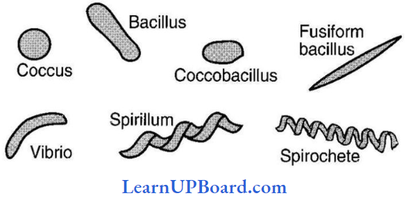
Eubacteria Shape: Cohn classified eubacteria into four types based on their shapes:
- Coccus (pl. cocci): Spherical or nearly spherical, small, and always non-flagellated.
- Micrococci: Occur singly, for example, Micrococcus hi tens, M. roseus.
- Diplococci: Found in pairs, for example, Diplococcus pneumoniae.
- Streptococci: Cells remain attached to form a chain, for example, Streptococcus lactis, Leptotricha buccal is.
- Staphylococci: Irregular bundles of cells or grape-like clusters, for example, Staphylococcus aureus.
- Sarcinae: Three-dimensional geometrical figures such as cubes, for example, Sarcina.
- Bacillus (pi. bacilli): Rod-shaped/cigarette-like with rounded or blunt ends. Most common shape. Motile/non-motile. They may occur as:
- Monobacillus: Occurs singly
- Diplobacillus: Occurs in the group of two
- Streptobacilli: Found in a chain, for example, Streptobacillus
- When the cells of the chain have a much larger area of contact with each other, these are said to have formed trichomes for example, Beggiatoa.
- If the cells are lined side by side like matchsticks and at angles to one another, the arrangement is said to be palisade-like, for example, Corynebacterium diphtheriae.
- In many bacteria (for example, Streptomyces), cells are arranged to form unicellular long, branched filaments called hyphae.
- Vibrio (Singular Vibrion): Bacteria with less than one complete twist or turn, and these resemble a comma (,) in appearance, for example, Vibrio cholerae.
- Spirilla (Singular Spirillum): Coiled tonus or bacteria exhibiting twists with one or more turns giving a spiral appearance, for example. Spirillum minus.
- Other Uncommon Shapes:
- The stalked bacterium, for example, Caulnbacter.
- The budding bacterium, for example, Rhoclomicrobium.
- Pleomorphic Occurs in more than one form, for example, Rhizobium, Corynebacteriurn, Azolobacter, and Mycobacterium.
Flagella: Instead of the “9 + 2” arrangement of tubulin-containing microtubules, there is simply a single filament of a globular protein called flagellin.
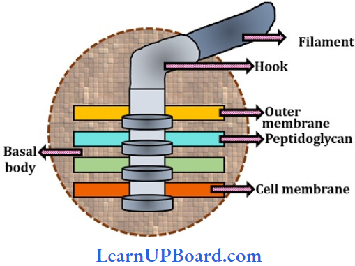
- Parts Of Flagellum
- Basal Body: It is the most complex portion of flagellum and has four rings (L, P, S, and M) in Gram-negative and two rings (S and M) in Gram-positive bacteria.
- Hook: It is made up of different protein subunits.
- Filament: The longest and most obvious portion of the flagellum. Protein molecules are arranged in a spiral manner. It is 20 nm wide and 1-70 nm long and consists of eight vertical rows of flagellin.
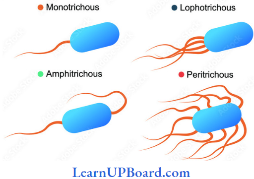
- Flagellar Arrangement:
Pllus And Fimbriae: They are small outgrowths of the plasma membrane. They help in sharing conjugation.
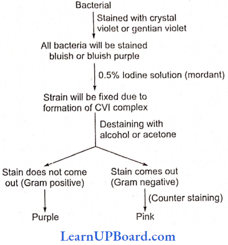
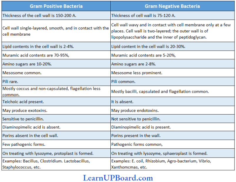
Gram Staining Technique: It was introduced by Christian Gram in 1884. Gram-negative bacteria become colorless on treatment with declining agents due to thin cell wails and more lipids in the cell wall.
NEET Biology Notes For Biological Classification Differences Between Gram Positive And Gram Negative Bacteria
Glycocalyx: The cell envelope consists of the outermost glycocalyx, the middle ono the cell wall, and the innermost cell membrane.
- Glycocalyx protects cells and also helps in adhesion. It is represented by either a slime layer or a capsule.
- The slime layer is composed of dextrin and levan, while the capsule is made of polysaccharides and D-glutamic acid.
- The slime layer protects the cells from loss of water and nutrients.
- Capsule provides gummy and sticky characters to the cell.
Cell Wall: It is made of peptidoglycan mucin or mucopcptide.
- The Glycan portion forms the backbone of peptidoglycan which is composed of alternating units of NAM (N- acctyl muramic acid) and NAG (A-acetyl glucosamine) joined together by 1,4-linkage.
- The tetrapeptide chain is attached to NAM.
- Teichoic acids are acidic polymers consisting of a carbohydrate (for example, glucose), phosphate, and an alcohol. It performs several functions such as binding metals, acting as receptor sites for some viruses, and maintaining cells at low pH to prevent degradation of cell walls by self-produced enzymes.
- Porins function as channels for the entry and exit of hydrophilic low molecular-weight substances.
- The outer layer of the cell wall in Gram-negative bacteria contains lipopolysaccharides that act as the main surface antigen in the cell wall.
Mesosonie (chondroid; Fitz James): Particularly in Gram-positive bacteria, the plasma membrane shows infoldings into the cell. Various types of mesosomes are:
- Central Mesosome: It holds the nucleoid, and helps in the separation of nucleoid and septa formation.
- Peripheral Mesosome: Help in storing respiratory enzymes such as succinic dehydrogenase, and cytochrome oxidase.
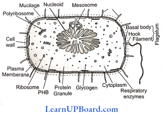
Plasmid (Ledcrbcrg And Hayes): In addition to the normal chromosomal DNA, some extrachromosomal genetic elements are often found in bacteria. These elements are called plasmids. In fact, these are circular pieces of DNA that have extra genes. These are capable of autonomous replication in the cytoplasm of the bacterial wall. Plasmid is circular, supercoiled double-stranded naked DNA. It is also called minichromosomes. Various types of plasmids arc as follows:
- Sex-plasmid: It carries the sex fertility factor responsible for the transfer of genetic material during conjugation.
- R-plasmid: Confers resistance to antibiotics, having resistance transfer factor (RTF).
- Col-plasmid: Produces special proteins colicins (bacteriocin) to kill other bacteria.
- Degradative Plasmid: Decompose hydrocarbons in petroleum.
- Ti-plasmid And Ri-plasmid: Tumor-inducing and rhizogene plasmids, respectively. These are large plasmids with about 200 kbp.
Plasmid (Ledcrbcrg And Hayes) Respiration: Depending upon the mode of respiration and their capability to perform alternate modes of respiration, bacteria are of the following types:
- Obligate Aerobes: Can perform only aerobic respiration, for example, Bacillus subtilis.
- Obligate Anaerobes: Can perform only anaerobic respiration, for example, Clostridium botulinum.
- Facultative Aerobes: Anaerobic forms but can live in the presence of O2, for example, Chlorobium.
- Facultative Anaerobes: Aerobic forms but can live an-aerobically also, for example, Pseudomonas.
- Aerotolerant Anaerobes: Bacteria continue to perform anaerobic respiration even in the presence of O2, for example, lactic acid bacteria.
- Anaerotolerant Aerobes: Aerobic bacteria continue to perform aerobic respiration even in the absence of free O2 by using O2 of oxidized salts, for example, denitrifying bacteria.
NEET Biology Notes For Biological Classification Plasmid (Ledcrbcrg And Hayes) Reproduction:
Plasmid (Ledcrbcrg And Hayes) Reproduction By Binary Fission
- It is the common method of reproduction under favorable conditions.
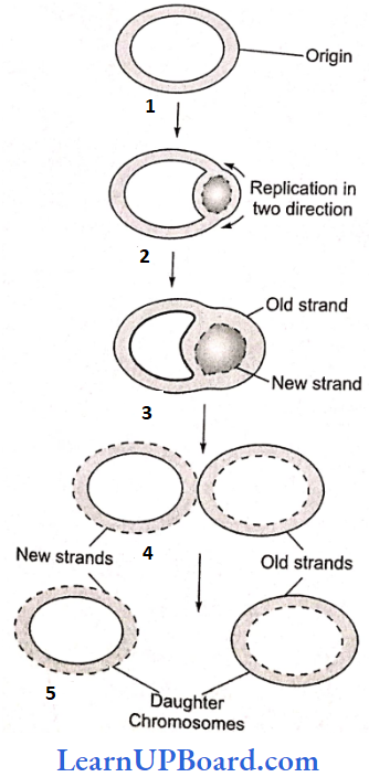
- The bacterial chromosome divides (replicates) during binary fission, resulting in the formation of two “circular” chromosomes. Since at one stage, the replicating chromosome appears like the Greek letter, this mode of replication is called the theta model. This mechanism of replication was suggested by Caim (1963) and is also known as Cairn’s model.
- Bacterial population multiplies by 2n (n = number of generations)
NEET Biology Notes For Biological Classification Plasmid (Ledcrbcrg And Hayes) Reproduction By Endosporeformation
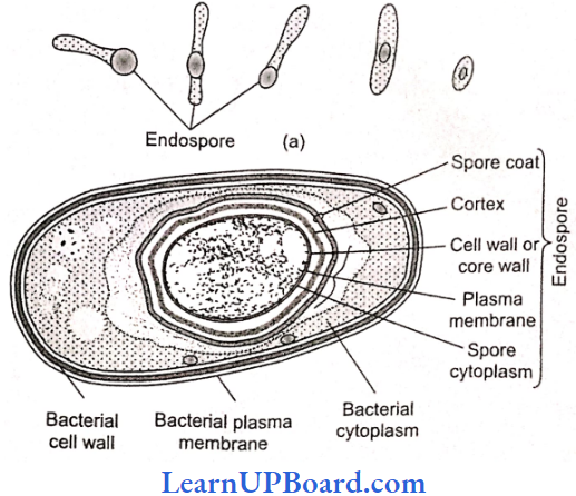
- Discovered by Cohn in hay bacteria (Bacillus subtilis).
- Endospores are thick-walled, highly dehydrated, and resistant spores formed under adverse conditions. Their wall is differentiated into 3-4 layers. The structure of the endospore is depicted.
- Endospores are, commonly formed in genera such as Bacillus and Clostridium. One endospore is formed per bacterial cell; so they are more a means of perennation than reproduction. Cortex and cytoplasm contain Ca2+ and an anticoagulant, dipicolinic acid, which prevents the protoplasm from coagulating at high temperatures.
Plasmid (Ledcrbcrg And Hayes) Reproduction Genetic Recombination Or Parasexuality: Three methods are known by which genetic recombination is achieved by bacteria. These are called parasexual because they do not involve syngamy and meiosis (true sexual reproduction). In the order of their discovery, these arc transformation, conjugation, and transduction.
Plasmid (Ledcrbcrg And Hayes) Reproduction Transformation: The donor and recipient do not come into contact. In this process, a bacterium picks up small DNA fragments of its dead relatives from the surrounding medium with the help of membrane receptors.
- The “competence” of a bacterium to pair up DNA and get transformed is present usually at the end of its active growth period. The process was discovered by Griffith. In 1928 while working with the bacteria Diplococcus (Pneumococcus) pneumoniae which causes pneumonia.
Plasmid (Ledcrbcrg And Hayes) Reproduction Conjugation: Involves DNA transfer between cells in direct contact and larger fractions of the donor DNA may be exchanged as compared to other means of genetic recombination. The process was first described by Laderberg and Tatum (1946) in Escherichia coli. It may involve the replication and transfer of the F plasmid (fertility factor) from donor (F+) to recipient (F–), thus also making the former a donor.
- Sometimes, the foreign plasmid integrates with the bacterial genome and such bacteria are termed as super male. When F is crossed with super male, the frequency of recombination is increased by 1000 times and that is why the super male is called Hfr or high frequency of recombination.
Plasmid (Ledcrbcrg And Hayes) Reproduction Transduction: A small double-stranded piece of DNA is transferred from donor to recipient by a bacteriophage. This mode of genetic recombination in bacteria was first demonstrated by Zinder and Lederberg in 1952 with Salmonella typhimurium. Following Are The Types Of Transduction:
- Generalized Transduction: Transducing bacteriophage can transfer any gene of the donor bacterium, for example, T4 bacteriophages.
- Restricted (Specialized) Transduction: Transducing bacteriophages can carry only a specific region of the bacterial DNA to a recipient, for example, bacteriophages.
- Abortive Transduction: DNA fragments from the donor bacterium is not integrated in the genome of the recipient bacterium and is lost after one or few generations.
NEET Biology Notes For Biological Classification Plasmid (Ledcrbcrg And Hayes) Reproduction Economic Importance
- Beneficial Activities:
- Sewage Disposal: E. coil, Clostridium, Pseudomonas, Streptococcus.
- Free Living Nitrogen Fixers: Beijerinckia, Clostridium
- Symbiotic Nitrogen Fixers: Rhizobium, Frankia, Xanthomonas.
- Ammonifying Bacteria: Bacillus mycoides, B. ramosus.
- Lactic Acid Production: Lactobacillus bulgaricus or L. delbrueckii convert ammoniated sugar solution into lactic acid.
- Vinegar Production: Acetobacter aceti, A. schizenbachi.
- Retting Of Fibers: Clostridium perfringens, Pseudomonas fluorescence.
- Curing Of Leaves: Curing of tea leaves by Micrococcus candisans and tobacco leaves by Bacillus megatherium.
- Vitamins: Riboflavin is prepared from Clostridium butyricum. Cobalamin (B12) is produced from B. megatherium.
- Single Cell Protein: Rhodopseudomonas capsulatci, Methylophilus methylotropus (source of protein).
- Pollution Control: Pseudomonas putida degrades petroleum wastes. Flavobacterium can decompose 2,4-D. DDT can be decomposed by Acetobacter aerogens. Ganges water contains Bdellovibrio bacteriovorus which maintains the purity of its water. Poly- P-hydroxybutyrate is used to produce biodegradable plastic.
- Harmful Activities:
- Spoilage Of Domestic Articles: Attack on domestic articles and resulting in their Spirochaete cytophaga and Cellulomonas.
- Denitrification: Thiobacillus denit rificans, Pseudomonas aeruginosa, Micrococcus denitrificans.
- Desulfunification: Desulphovibrio desulphur icons.
- Spoilage Of Alcoholic Beverages: Acetobacter Aceti.
- Diseases: About 90% of human diseases are bacterial.
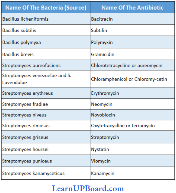
Antibiotics Produced By Various Bacteria
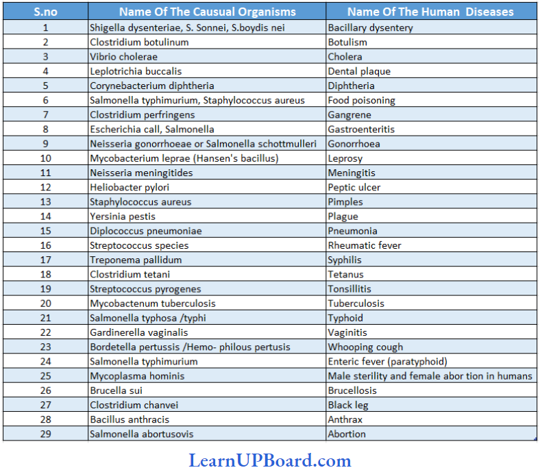
Human diseases caused by various bacteria
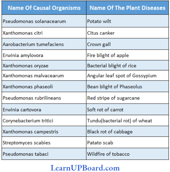
NEET Biology Notes For Biological Classification Plant Diseases Caused By Bacteria
NEET Biology Notes For Biological Classification Cyanobacteria (BGA)
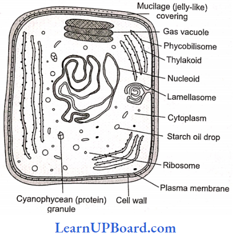
- Cyanobacteria are included into separate classes: Cyanophyceae or Myxophyceae (myxo = slime), according to phycologists.
- They are Gram-negative, oxygenic, photosynthetic monerans.
- They contain chlorophyll-a, carotene, a bit of myxo-xanthin, and phycobilin or phycobiliproteins. There are three types of phycobiliproteins: c-phycocyanin, c-phycoerythrin, and allophycocyanin.
- The cell wall is four-layered and the cell membrane lacks sterol with a 2:1 protein and phospholipid ratio.
- The lamellasome connects the nucleoid to the cell membrane.
- The reserve food material is cyanophycin protein, cynophycean starch or oc-granules, and p-granules (fat droplets).
- These are nonflagellate and movement (if it occurs) is by special gliding motion.
Sometimes, the same species, when grown under different wavelengths of light, exhibit variations in pigment composition. It is believed that by doing so, the alga is able to absorb the maximum available light for photosynthesis.
- This capacity to change color with a complementary effect toward light is known as the Gaidukov phenomenon (first given by Gaidukov) or complementary chromatic adaptation, for example, Trichodesmium erythreum causes red sea.
- Blue-green algae are the only monerans that are capable of performing oxygenic photosynthesis and in this respect are similar to the higher plants. Many of them are efficient nitrogen fixers. The existence of the processes of oxygenic photosynthesis and N2 fixation in some organisms is surprising since N2 fixation is an anaerobic process and the enzyme nitrogenase is inactivated by the presence of O2 even in low concentrations.
- Usually, but not always, N2-fixing cyanobacteria are filamentous and produce a specialized type of cell, called heterocyst, within which N2 fixation occurs.
- The heterocysts are distinct and thick-walled cells with pale-yellow homogenous contents. These may be terminal or intercalary. Under the light microscope, a heterocyst appears to be surrounded by a thick layer two-layered wall.
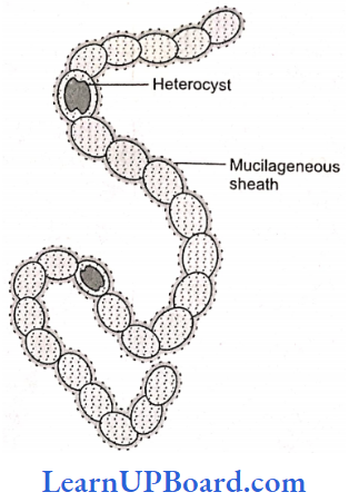
The outer thick layer is persistent, made up of pectin or cellulose only: one or two pores also perforate the heterocyst wall. The wall is thickened near the pores. A prominent granule called a polar granule is present at the pore of the heterocyst.
- Through the pores of heterocysts, protoplasmic connections are established with the neighboring cells. The heterocysts lack photosystem 2 and thus do not evolve oxygen.
- Some blue-green algae such as Anabaena, and Nostoc form a thick layer on the soil surface during the rainy season. These can be used for reclaiming usar soils.
- Many species of blue-green algae belonging to genera Anabciena, Anlosira, Nostoc, Scytonema, etc., are known to be able to fix atmospheric nitrogen. Anabaena, Tolypothrix, and Aulosira play an important role in enriching (up to 20%) rice fields with nitrogen.
- Nostoc commune is used as a food called “Yuyucho” in China and Japan.
- The excessive growth of BGA in water produces water blooms.
Mycoplasma: E. Nocard and E.R. Roux (1898), French scientists, discovered a new type of organism from the pleural fluids of cattle suffering from bovine pleuro-pneumonia. This new type was pleomorphic and was called PPLO (pleuro-pneumonia-like organisms). This organism was later given the name Astero-coccus mycoides by Bon-el et al. (1910) and later the name Mycoplasma by Nowak (1929).
basic biology notes
Mycoplasma Characters:
- Unicellular,
- Prokaryotic,
- Non-motile,
- Highly pleomorphic,
- Filterable through bacterial filters,
- Lack of cell wall,
- Are resistant to antibiotics such as penicillin which acts on cell walls,
- Are inhibited by tetracycline and similar antibiotics which act on metabolic pathways,
- Reproduce by the form on of elementary bodies,
- Tonn “fried egg” colonies in culture, and
- DNA is linear, double-stranded extending almost throughout the cell.
Mycoplasmas are also called jokers of the plant kingdom owing to their ability to have varying shapes; PPLO or MLO (molicute-like organisms) or MLBs (mycoplasma-like bodies). These are bacteria without cell walls, mesosomes, and flagella, and are pleomorphic.
Mycoplasma gallisepticum (0.3 to 0.5 m) is the smallest prokaryote. They are either saprophytic or cause diseases such as pleuropneumonia in domestic animals and potato witch’s broom, aster yellows, little leaf of brinjal, etc., in plants.
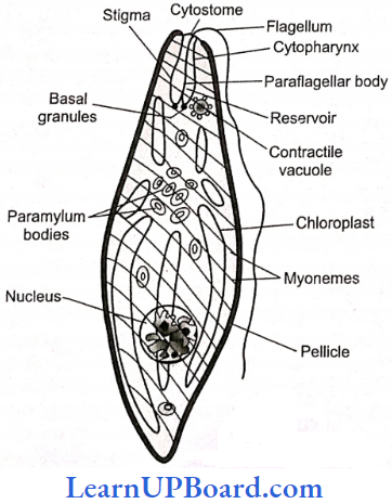
Euglenoids
- Unicellular and flagellate protists (rarely non-motile). May be in fresh waters and damp soils.
- The body of Euglena is spindle-shaped with a blunt anterior end and a pointed posterior end
- An elastic and flexible pellicle (periplast) composed primarily of elastic protein is present. The pellicle is composed of obliquely running parallel strips called myonemes (in epiplasm) underlying a delicate plasma membrane. The cell wall is absent.
- Two different flagella (heterokont) arise from blepharoplasty and arc united at their roots where the paraflagellar body occurs. The latter is photosensitive. The long flagellum is stichoncmatic (the tinsel type with a row of mastigoncmes). The other flagellum is very short. Euglenoids are also capable of wriggling movements by expansion and contraction of their body (metabolic).
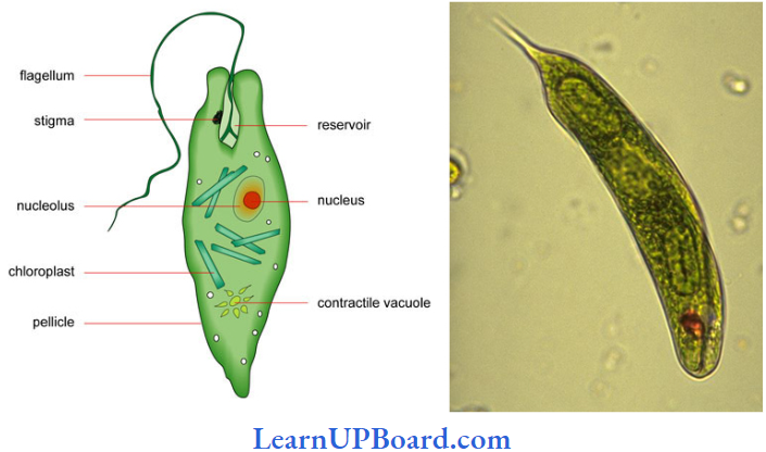
- An orange-red eye spot occurs attached to the membrane of the reservoir. It comprises the pigment astaxanthin and is a photoreceptive structure.
- It can be holophytic, holozoic (Peranema), saprobic (Rhabdomonas), or mixotrophic (Euglena).
- In holophytic forms, usually elongated or discoid plastids are present. They may possess pyrenoids (proteinaceous bodies). The photosynthetic pigments constitute chlorophylls a and b, carotene, and xanthophylls.
- The food reserve is paramylum bodies which store a 1,3-glucan called paramylon, which does not stain with iodine.
- A contractile vacuole occurs just below the reservoir and evacuates its contents into the latter. It is osmoregulatory in function.
Euglenoids Asexual Reproduction: It occurs by longitudinal binary fission. The nuclear membrane and nucleolus persist during division.
Euglenoids Palmella Stage: It is reported in some euglenoids at the advent of unfavorable conditions.
Euglenoids Sexual Reproduction: Not yet reported
Consumer-Decomposer Protists
- They were formerly included amongst metazoa or fungi.
- Slime molds are included in the division of gymnomycota by mycologists. Because of their protistan nature, they are also called protistan fungi.
- They do not have chlorophyll.
- They are surrounded by the plasma membrane only (somatic parts are without cell walls). However, the spores do not always have cell walls.
- At one stage of the life cycle, they have an amoeboid structure.
- They have phagotrophic or saprotrophic nutrition.
- There are of two types:
- Acellular slime molds (plasmodia, slime molds) and
- Cellular slime molds (communal).
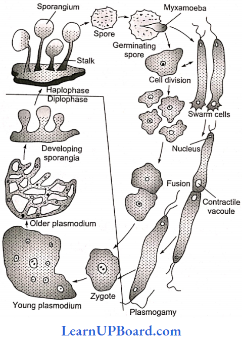
Acellular Slime Molds Salient Features
- Some Of The Common Acellular Slime Molds Are Physeuglena: structure arella and Physarum.
- Somatic bodies are free-living multinucleate, naked diploid protoplasmic masses called plasmodia.
- Acellular slime molds are holocarpic and polycentric.
- The sporangium usually contains a network of fine threads called capillitium.
- Acellular slime molds are present as slime masses on decaying leaves and lumber (disused article of timber wood for building).
- They move with the help of pseudopodia like amoebae.
- Sexual reproduction is an isogamous type.
Cellular Slime Mold Salient features
- Complete absence of flagellated cells in their life cycle.
- Presence of wall-less uninucleate myxamoebae.
- Formation of pseudoplasmodium by the aggregation of myxamoebae. The myxamoebae secrete acrasin (cAMP) and show chemotactic movements. They aggregate to form a pseudoplasmodium. These are holocarpic and monocentric. Capillitia are lacking in the sporangium.
- Presence of naked sporangia (without sporangial cover)
- Presence of cellulosic wall around spores
Cellular Slime Mold Anisogamous Sexual Reproduction: During sexual reproduction. one of the myxamoebae in the duster becomes larger by engulfing other myxamoebae. Karvogamy occurs inside the large myxamoeba. now called the macrocyst. Maerocvst later undergoes meiosis to release haploid myxamoebae.
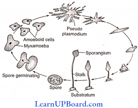
Cellular Slime Mold Sexual Reproduction Examples: Dictyostelium. Polysphondylium
Cellular Slime Mold Protozoan Protists: These protists have been discussed in the phylum Protozoa in the kingdom Animalia.
NEET Biology Notes For Biological Classification Yeast
- Unicellular with a size 3-15 m x 2-10m. Under conditions of rapid growth, they form temporary chains or pseudomycelia.
- The cell wall contains mannan, glucan, lipid, protein, and chitin.
- The cells are spherical to cylindrical in outline.
- These are facultative aerobes.
- A birth scar and a bud scar are present on opposite sides in budding yeast.
- True yeasts do not develop ascocarps.
- Yeast in which ascus formation is not reported are called false yeast, for example, Candida, Cryptococcus, Mycodernta, Geotrichum.
Types Of Yeast
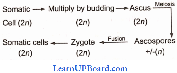
- Saccharomyces: They are budding yeast and show a diplohaplontic life cycle. There are four ascospores produced per ascus.
- Schizosaccharomyces: They are fission yeast, and the harmonic life cycle produces eight ascospores per ascus.
- Saccharomvcoides: They are global yeast and show a diplontic life cycle. There are four ascospores produced per ascus.
Importance Of Yeast
- Saccharomyces cerevisiae – Brewer’s/beer yeast or Baker’s yeast
- S. ellipsoids — Wine yeast
- Toru/opsis utilis and Endomyces vermalis are rich in proteins. Rhodotorula is rich in vitamin A and Ashbya gossypii is rich in vitamin.
- Yeasts are used in curing cocoa beans.
Diseases Caused By Yeast
- Candidiasis/Moniliasis: Candida albicans
- Blastomycosis: Blastomyces dermatitis
- Histoplasmosis: Histopiasma capsules
- Cryptococcosis: Cryptococcus neoformans
- Some yeasts reduce the yield of the silk industry by attacking silkworms.
- Species of Nematospora attack cotton, tomato, and beans.
NEET Biology Notes For Biological Classification Puccinia (Puccinia Graminis Tritici)
- It causes black rust of wheat.
- Heteroecious Fungus: Completing life cycle on two host plants, i.e., primary host (wheat) and secondary host of alternate host (barberry).
- The life cycle is macrocyclic and polymorphic.
- Five types of spores are formed.
- Uredospore: Unicellular, dikaryon (n + n), on wheat
- Teleutospore: Bicelled, dikaryon (n + n), on wheat
- Basidiospore: Unicellular, monokaryotic, formed in soil.
- Pycniospore: Unicellular, monokaryotic, formed on the upper surface of barberry
- Aeciospore: Unicellular, dikaryon, formed on the lower surface at the same barberry leaf.
Some More Basidiomycetes And Their Importance
- Rust Fungi: These fungi cause rust disease.
- Puccinia Striformis: It causes yellow/stripe rust of wheat.
- Puccinia Recondita: It causes leaf/brown rust of wheat.
- Puccinia Hordei: It causes brown/leaf rust of barley.
- Puccinia Graminis-Tritici: It causes the black rust of wheat.
- Smut Fungi: These fungi cause smut diseases. Few examples are given below
- Ustilago Tritici: It causes loose smut of wheat.
- Ustilago Avenae: It causes loose smut of oat.
- Ustilago Jensenii: It causes covered smut of barley.
- Ustilago Rnaydis: It causes smut of com.
- Ustilago Scitaminae: It causes whip smut of sugarcane.
- Toad Stools (Poisonous Mushrooms)
- Amanita caesarea (Caesarea’s mushroom)
- A. phalloides (death cup): Emperor Claudius Caesar was murdered by his wife by giving extract of this fungus which stops mRNA synthesis.
- Bracket Or Shelf Fungi: Example, Fames applant us, Polyporus, Ganoderma.
- Puffballs: Basidiocarp sends puffs of spores on ripening, for example, Lycoperdon.
- Stinkhorn: Example, Phallus impudicus (dead man’s finger). It gives a stinking odor and attracts flies.
Deuteromycetes (Fungi Imperfect)
- It includes all those fungi in which the sexual or perfect stage is either absent or not reported.
- Asexual reproduction commonly occurs by means of conidia.
- In any case, it is not possible for us, at present, to place these in any subdivision. Therefore, an artificial division is made.
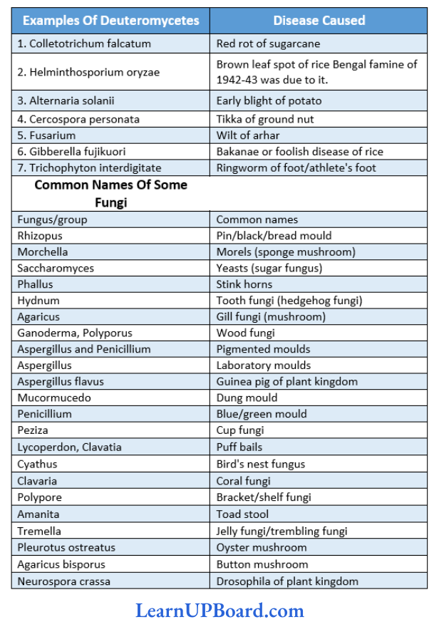
Common Deuteromycetes And Diseases Caused By Them
NEET Biology Notes For Biological Classification Common Fungicides And Their Composition
- Bordeaux Mixture (CuSO4 · Ca(OH)2 – H2O): First fungicide discovered by RMA Millardet. Commonly known as the holy water of plant pathology
- Burgandy Mixture: A mixture of CuSO4 + Na2CO3 + H2O; was discovered by mass or soda Bordeaux.
- Chestnut Mixture: Ammonium carbonate + Copper sulfate
Mycorrhiza
- It is a mutualistic symbiotic association of a fungus with the roots of a higher plant.
- Mycorrhizae can be divided into two groups: ectomycorrhiza and endomycorrhiza.
- In ectomycorrhiza, hyphae penetrate between the outermost cell layers of the host. The hyphae in intercellular space form a network called the Hartig net. Fungus partner is commonly basidiomycetes.
- In endomycorrhiza, most of the fungus is within the root and may be intercellular as well as intracellular. Fungus partners is commonly zygomycetes. In VAM (vesicular arbuscular mycorrhizae), the hyphae develop vesicles and arbuscules within the cortex of the root. VAM helps in phosphate absorption from soil.
- Fungus obtains shelter and food from roots. It helps the root in the dissolution and absorption of inorganic nutrients locked in the organic matter.
NEET Biology Notes For Biological Classification Kingdom Plantae
It includes all eukaryotic, chlorophyll-containing organisms commonly called as plants.
- The plant cells have eukaryotic structures with prominent chloroplasts, and cells are mainly made up of cellulose.
- The mode of nutrition is autotrophic but few members are partially heterotrophic such as insectivorous plants or parasites. The common examples of insectivorous plants are bladderwort and Venus fly trap, whereas a common example of a parasite is Cuscuta.
- They show distinct alternation of generation.
- Kingdom Plantae includes algae, bryophytes, pteridophytes, gymnosperms, and angiosperms.
NEET Biology Notes For Biological Classification Kingdom Animalia
It includes heterotrophic eukaryotic organisms that are multicellular.
- The cells lack cell walls.
- They directly or indirectly depend on plants for food.
- The food is atoned as a reserve in the form of glycogen or fat.
- The mode of nutrition is holozoic: by ingestion of food.
- Sexual reproduction is by copulation between males and females followed by embryological development.
- In that chapter, we shall also explain the unicellular protozoans which are placed normally in Kingdom Protista.
NEET Biology Notes For Biological Classification Lichens
They are composite organisms that are formed by a fungus partner (mycobiont) and an algal partner (phycobiont). Mycobiont is dominant forming 95-99% of total thallus and is responsible for reproduction. It belongs mostly to the ascomycete group (ascolichens, for example, Graphis, Cladonia, Parmelia, Usnea). Their fruiting body is perithecium or apothecium and is sometimes similar to basidiomycetes (basidiolichens, for example, Cora, and Corella).
- In 75% of lichens, phycobiont is mostly chlorophyceae (for example, Chlorella, Palmella, Protococcus, Trebouxia) or can be cyano-phyceae (for example, Chlorococcus, Nostoc, Scytonema, etc.).
- Lichens are called terricolous (growing in soil saxicolous (on stones/rocks), corticolous (on bark), and lignicolous (on wood).
- Soredia are the most efficient means of asexual reproduction. Soredia are composed of algal cells clasped and surrounded by fungal hyphae.
- There are three types of lichens on the basis of external morphology:
- Crustose Lichens: These form a thin crust closely adhered to the substratum, partly or wholly embedded into it, for example, Strigula, Graphis, Rhizocarpon.
- Foliose Lichens: These have flat, leaf-like, well-branched, or lobed thallus attached to the substratum usually with the help of rhizoid-like structures called rhizines, for example, Peltigcra, Parmelia.
- Fruticose Lichens: These are more or less bushy, usually much branched, and pendant to upright, for example, Bryonia, Usnea, Cladonia, etc.
Special Structures In The Thallus Of Lichen Are:
- Cyphellae: Help in the exchange of gases, present in lower context.
- Cephalodia: Helps to retain moisture and its algal partner fixes nitrogen also.
Breathing Pores: For aeration, present in the upper cortex of the thallus.
Economic Importance Of Lichen
- Pioneers Of Vegetation: They initiate weathering of rocks into soil particles.
- As Food And Fodder: Reindeer moss {Cladonia rangiferina) of the Arctic region is eaten by reindeer and cattle. Iceland moss (Cetraria islandica) is used as food by man in Iceland. Species of Parmelia are used as curry powder in India.
- In Cosmetics And Perfume: Some species of Evernia and Ramalina yield essential oils which are used in the manufacture of soap. Dye orchil or cudbear are obtained from the species of Roccella and Lecanora. Litmus is derived from Roccella.
- In Medicine: Lobaria pulmonaria is used for lung troubles, Usnea barbata for strengthening hair, and for uterine ailments. Xanhoria parietina is used in jaundice and Parmelia saxatilis for epilepsy. Peltigera canina is used against hydrophobia. Usnic acid is obtained from Usnea.
Indicators Of Air Pollution: Lichens are very sensitive to SO2 and die at higher levels of SO2.
NEET Biology Notes For Biological Classification Question And Answers
In the following questions, an Assertion (A) is followed by a corresponding Reason (R). Mark the correct answer.
- If both Assertion and Reason are true and the Rea¬son is the correct explanation of the Assertion.
- If both Assertion and Reason are true, but the Rea¬son is not the correct explanation of the Assertion.
- If Assertion is true, but Reason is false.
- If both Assertion and Reason are false.
Question 1.
Assertion: Cellular slime molds have the character of both plants and animals.
Reason: The reproductive phase is animal-like and the vegetative phase is plant-like.
Answer: 3. If both Assertion and Reason are false.
Question 2.
Assertion: An outer membrane is present in Rhizohlum.
Reason: The outer membrane contains lipopolysaccharides.
Answer: 2. If both Assertion and Reason are true, but the Rea¬son is not the correct explanation of the Assertion.
Question 3.
Assertion: Lichens do not grow in polluted areas having SO2.
Reason: Lichens secrete carbonic acid and oxalic acid on barren rocks.
Answer: 2. If both Assertion and Reason are true, but the Reason is not the correct explanation of the Assertion.
Question 4.
Assertion: The secondary mycelium of Agaric us is hi nucleated.
Reason: Secondary mycelium is formed by the somatogamy of primary mycelium.
Answer: 1. If both Assertion and Reason are true and the Rea¬son is the correct explanation of the Assertion.
Question 5.
Assertion: Cyphcllae help to retain moisture in lichens.
Reason: They contain a large number of hyaline cells.
Answer: 4. If both Assertion and Reason are false.
Question 6.
Assertion: Unicellular eukaryotes are included in monera.
Reason: Unicellular eukaryotes have 70S cytoribosomes.
Answer: 4. If both Assertion and Reason are false.
Question 7.
Assertion: Lamellasomc connects nucleoid to cell membrane.
Reason: Lamellasome is present in oxyphotobacteria.
Answer: 2. If both Assertion and Reason are true, but the Reason is not the correct explanation of the Assertion.
Question 8.
Assertion: Pseudomonas fluorescense is a cephslotrichous bacteria.
Reason: It is helpful in the retting of fibers.
Answer: 2. If both Assertion and Reason are true, but the Rea¬son is not the correct explanation of the Assertion.
Question 9.
Assertion: MLO is Gram-negative, pleomorphic monerans.
Reason: They are wall-less having acetylglucosamine as-sociated with cell membrane which is rich in cholesterol.
Answer: 2. If both Assertion and Reason are true, but the Rea¬son is not the correct explanation of the Assertion.
Question 10.
Assertion: Gram-positive bacteria detect and respond to chemicals by lipopolysaccharides.
Reason: They have high amounts of porins and lipids which act as antigens.
Answer: 4. If both Assertion and Reason are false.
Question 11.
Assertion: Holophytic protistans are important phytoplankton and contribute 80% of the total photosynthesis.
Reason: They lack chemosynthetic nutrition and utilize non-sulfur organic compounds as the source of carbon assimilation.
Answer: 3. If both Assertion and Reason are false.
Question 12.
Assertion: Sexual spores in pink mold are meiospores produced endogenously.
Reason: They develop a flask-shaped fruiting body in the sexual life cycle.
Answer: 2. If both Assertion and Reason are true, but the Rea¬son is not the correct explanation of the Assertion.
Question 13.
Assertion: Azotodesmic lichens are biofertilizers enriching nitrogen contents in soil.
Reason: This ability is due to the presence of heterocystous blue-green algae as a phycobiont component.
Answer: 1. If both Assertion and Reason are true and the Rea¬son is the correct explanation of the Assertion.