
NEET Biology Notes For Human Reproduction Introduction
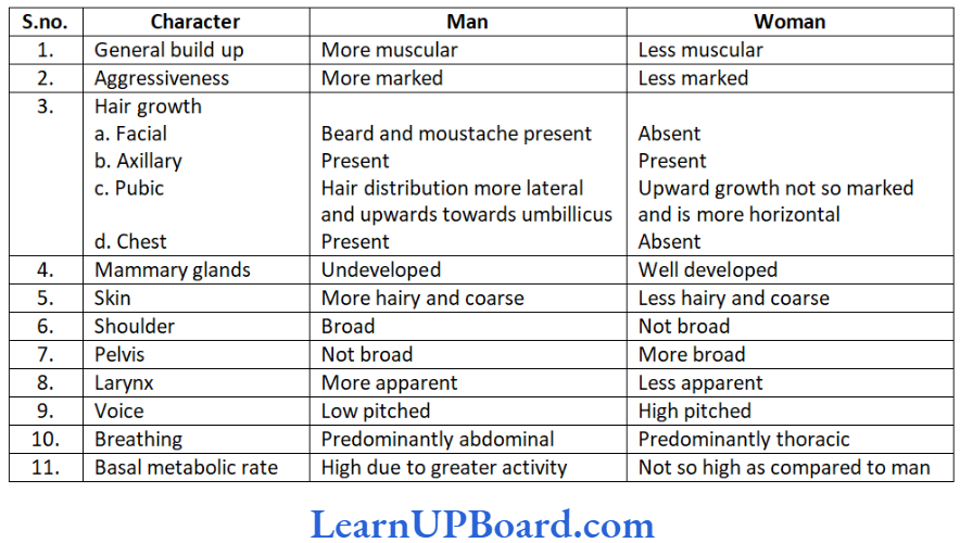
- Human beings are unisexual.
- The growth, maintenance, and functions of gonads are regulated by gonadotropins secreted from the anterior lobe of pituitary gland.
- The organs that neither produce gametes nor secrete sex hormones but perform important functions in reproduction are called secondary sex organs.
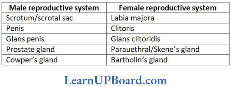
- These include prostate, seminal vesicles, vas deferens, and penis in males; and fallopian tubes, uterus, vagina, and mammary glands in females.
- The characters that distinguish a male from a female externally are called accessory or external sex characters or secondary sex characters.
human reproduction
NEET Biology Notes For Human Reproduction Male Reproductive System
- The male reproductive system of mammals consists of a pair of testes, several accessory glands, a duct system, and a mating organ called the penis. Testis is the primary male sex organ. It produces spermatozoa and secretes the male sex hormone testosterone.
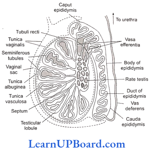
- A human testis measures about 5 cm, 3 cm, and 2.5 cm, respectively, in length, thickness, and width. It is covered by thick, fibrous, connective tissue called tunica albuginea.
- In a man, both testes normally remain suspended in a pouch called scrotum outside the abdominal cavity. This keeps the testes at a low temperature than the body temperature (about 2°C less). This is essential for the maintenance and normal functioning of the spermatogenic tissue of testes.
- Testes descend in the scrotal sac when fetus is about 7 months old. This occurs under the influence of follicle-stimulating hormone (FSH) and testosterone.
- If testes fail to descend, then the condition is called cryptorchidism. It leads to sterility. Scrotum remains connected with the abdomen or pelvic cavity by the inguinal canal. Blood vessels, nerves, and conducting tubes pass through the inguinal canal.
- Cremaster muscles and connective tissue form spermatic cord and surround all structures passing through the inguinal canal. Cremaster muscles and dartos muscles of the scrotal sac help in the positioning of testes.
- When the outside temperature is low, these contract to move the testes close to the abdominal cavity/pelvic cavity. When the outside temperature is high, these relax moving the testes away from the body.
- In some seasonally breeding mammals, testes descend into the scrotum during the breeding season but ascend back into the abdomen in the non-breeding season. For example, rats and bats.
Read and Learn More NEET Biology Notes
NEET Biology Notes For Human Reproduction Anatomy Of Testis
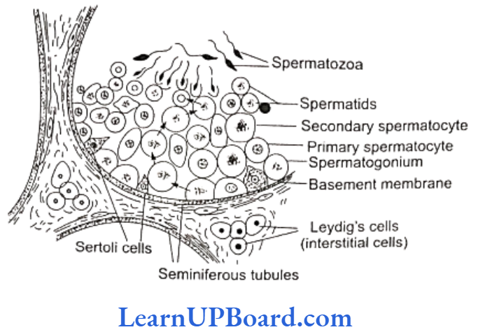
- Each testis contains numerous tiny, highly convoluted tubules, called seminiferous tubules. These constitute the spermatogenic tissue of the testis.
- Cells lining these tubules give rise to spermatozoa which are released into the lumen of the tubule.
- In between spermatogenic cells, Sertoli or sustentacular or nurse cells are present which provide nourishment to the developing spermatozoa and regulate spermatogenesis by releasing inhibin to check FSH overactivity.
- The other functions of Sertoli cells include the following:
- Providing nourishment to the developing spermatozoa.
“human reproductive system notes pdf “
- Absorbing the parts being shed by the developing spermatozoa.
- Releasing anti-Müllerian factor (AMF) to prevent the development of Müllerian duct/oviduct.
- Releasing androgen binding protein (ABP).
- Groups of polyhedral cells called interstitial cells, or the Leydig cells, are located in the connective tissue around the seminiferous tubules.
- They constitute the endocrine tissue of the testis. The Leydig cells secrete testosterone into the blood.
- Seminiferous tubules unite to form several straight tubules called tubuli recti which open into irregular cavities in the posterior part of the testis.
- Rete testis is a highly anastomosing labyrinth of cuboidal epithelium lined channels.
- Several tubes called vasa efferentia arise from it and conduct spermatozoa out from the testis. (Seminiferous tubule to vasa efferentia form intratesticular genital duct system).
- The extratesticular duct system consists of tubes which conduct sperms from the testes to the outside.
- It starts with vasa efferentia, which arise from each testis, and becomes confluent to form a folded and coiled tube called epididymis behind each testis.
- The epididymis consists of three parts: (a) Caput, (b) corpus, and (c) cauda. It stores the sperms temporarily.
- From cauda epididymis, a partially coiled tube called vas deferens ascends into the abdomen through the inguinal canal, passes over the urinary bladder, and receives the duct from the seminal vesicle behind the urinary bladder to from an ejaculatory duct.
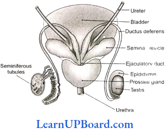
- Before entering prostate, the ductus deferens dilates to form ampulla. The final portion of ampulla passes through the prostate to open into the urethra shortly after its origin from the urinary bladder.
- Urethra receives the ducts of the prostate and Cowper’s glands, passes through the penis, and opens to the outside.
NEET Biology Notes For Human Reproduction Male External Genital Organ
Penis
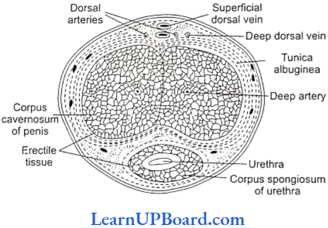
- Penis is the copulatory organ of man. It is a cylindrical and erectile, pendulous organ suspended from the pubic region in front of scrotum.
- It remains small and limp (or flaccid) but on sexual arousal, it becomes long, hard, and erect- ready for copulation (or coitus or intercourse). Erect human penis, on an average, is about 15 cm long.
“human reproduction project pdf class 12 “
- The penial mass is itself encased in a fibrous sheath, called tunica albuginea. The interior of penis is mostly formed of three cylindrical cords of spongy, erectile (cavernous) tissue.
- Two of these cords are thicker and situated parallely on the right and left sides, forming the thick part of penis that remains in the front when the penis is limp, but becomes superio-posterior when it is erect.
- These two cords are called corpora cavernosa.
- The fibers of tunica albuginea surround both cords jointly and form a separate sheath around each cord. Some fibers form a partition, called septum penis, between these cords.
- The third, smaller cord forms that part of penis which remains inferio-anterior in erect penis. Urethra runs through this cord. Hence, this cord is called corpus urethrae or spongiosum.
- The extended part of corpus spongiosum is enlarged, forming a bulged, conical structure called glans penis. The surface of glans is formed of a thin, smooth, shiny, and hairless skin.
- The base line of glans is referred to as the neck of the penis. The loose skin of penis folds over and is retractile on glans. It is called foreskin or prepuce.
- At the tip of glans penis is a slit-like external urethral orifice or meatus by which urethra opens out and discharges urine or semen.
- Preputial glands, present in the skin of the penis neck, secrete a white sebaceous substance, called smegma. Microbial infection in smegma causes irritation.
Glands
- Seminal vesicles: These are paired, tubular, coiled glands situated behind the urinary bladder. They secrete viscous fluid that constitutes the main part of the ejaculate. Seminal fluid contains fructose, citric acid, inositol, and prostaglandins.
- Prostate gland: It is a chestnut-shaped gland and is a collection of 30-40 tubuloalveolar glands. It lies at the base of the bladder and surrounds the base of the ure- thra. It contributes an alkaline substance to the seminal fluid.
- The substance of prostate helps the sperms to become active and counteract any adverse effects of urine on sperms. The prostatic fluid provides a characteristic odor to the seminal fluid. Prostate gland secretes citrate ion, calcium, phosphate ion, and profibrinolysin.
- Bulbourethral glands or Cowper’s glands: The two bulbourethral glands are pea-sized structures lying adjacent to the urethra at the base of penis. These secrete a viscous lubricant.
- Duct system, accessory glands, and penis are secondary male sex organs. Their growth, maintenance, and functions are promoted by testosterone secreted by the Leydig cells.
- On the other hand, the growth, maintenance, and functions of seminiferous tubules and the Leydig cells are regulated, respectively, by FSH and interstitial cell stimulating hormone (ICSH) of anterior pituitary.
Semen
- Semen is a mixture of sperms and the secretions of seminiferous tubules, seminal vesicles, prostate gland, and bulbourethral glands.
- The average volume of semen in an ejaculation is 2.5-5 ml, with a sperm count (concentration) of 200-300 million. For normal fertility, at least 60% sperms must have normal shape and size, and at least 40% of them must show vigorous motility. When the sperm count falls below 20 million/ml, the male is likely to be infertile.
- Semen has a slightly alkaline pH of 7.2-7.7. The prostatic secretion gives semen a milky appearance, whereas fluids from the seminal vesicles and bulbourethral glands give it a sticky consistency.
- Semen provides transportation medium and nutrients to sperms. It neutralizes the hostile acidic environment of the male urethra and the female vagina.
NEET Biology Notes For Human Reproduction Female Reproductive System
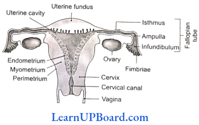
- The female reproductive system consists of a pair of ovaries, a duct system consisting of a pair of fallopian tubes (oviduct)-uterus, cervix, and vagina. The pair of mammary glands is accessory genital glands.
Ovaries
- Ovary is the primary female sex organ. It produces ova and secretes female sex hormones, estrogen, and progesterone that are responsible for the development of secondary female sex characters and regulate cyclic changes in uterine endometrium.
- Human ovaries are small, almond-like flat bodies about 3 cm in diameter.
Location
Ovaries are located near kidneys and remain attached to the lower abdominal cavity through mesovarium.
Structure
The free surface of the ovary is covered by a germinal epithelium made of a single layer of cubical cells. This epithelium is continuous with the mesothelium, called peritoneum. The epithelium encloses the ovarian stroma. The stroma is divided into two zones a peripheral cortex and an inner medulla. The cortex is covered by a connective tissue, called tunica albuginea, and it contains numerous spherical or oval, sac-like masses of cells, known as ovarian follicles. The medulla consists of loose connective tissue, elastic fibers, blood vessels, and smooth muscle fibers.
Ovarian Follicle
- Ovarian follicle carries a large, centrally placed ovum, surrounded by several layers of granular cells (follicular granulosa or discus proligerus or cumulus oophorus). It is suspended in a small cavity-the antrum.
- Antrum is filled with liquid folliculi. The secondary oocyte in the tertiary follicle forms a new membrane called zona pellucida. The follicle bulges on the surface of the ovary. Such a follicle is called mature Graafian follicle (after De Graaf, who reported them in 1672).
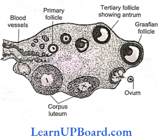
Corpus Luteum
- The ovum is shed from the ovary by rupturing the follicle. The release of ovum is called ovulation and occurs nearly 14 days before the onset of the next menstrual cycle.
- After the extrusion of the ovum, the Graafian follicle transforms into corpus luteum. Corpus luteum is filled with a yellow pigment, called lutein.
- Corpus luteum grows for a few days and if the ovum is fertilized and implantation occurs, then it continues to grow. But if the ovum is not fertilized, then corpus luteum persists only for about 14 days.
- It secretes progesterone. At the end of its functional life, it degenerates and gets converted to a mass of fibrous tissue, called corpusalbicans (white body). It remains as a scar in the ovary throughout the life of a female.
Fallopian Tubes (Oviducts)
- Oviducts are a pair of long (about 10 cm), ciliated, muscular, tubular structures that extend from ovaries to uterus. Each is suspended by mesosalpinx. Each fallopian tube is differentiated into four parts:
- Infundibulum: The part of oviduct closer to the ovary is the funnel shaped infundibulum. Its edges possess finger-like projections called fimbriae. Fimbriae help in the collection of the ovum after ovulation. Infundibulum opens in the abdominal cavity by an aperture called osteum.
- Ampulla: The infundibulum leads to a wider part of the oviduct called ampulla.
- Isthmus: It is the middle, narrow, ciliated part of the oviduct.
- Uterine part: It is the inner, narrow part which opens in the upper part of uterus. It is involved in the conduction of ovum or zygote towards the uterus by peristalsis and ciliary action. (Fertilization occurs at the junction of ampulla and isthmus.)
- Uterus: It is a large hollow, muscular, highly vascular, and pear-shaped structure present in the pelvis between the bladder and rectum. It is suspended by a mesentery-mesometrium. It is formed of three parts.
- Fundus: It is the upper dome-shaped part above the opening of fallopian tubes.
- Body: It is the middle and main part of the uterus.
- Cervix: It is the lower, narrow part which opens in the body of the uterus by internal os and in vagina below by external os. It is formed of the most powerful sphincter muscle in the body.
Its wall is formed of outer peritoneal layer (called perimetrium); middle muscular myometrium made of smooth muscle fibers, and inner, highly vascular and glandular endometrium.
It is the site of fetal growth during pregnancy. It also takes part in placenta formation and helps in pushing the baby out during parturition.
Vagina
- Vagina is a long (7.5 cm), fibro-muscular tube. It extends backward in the front of rectum and cervix to the vestibule. It is a vascular tube internally lined by mucus membrane and is raised into transverse folds called vaginal rugae.
- In a virgin female, vaginal orifice is closed by a membranous diaphragm called hymen which becomes centrally perforated at puberty for the discharge of menstrual flow (or menses).
- Vagina acts both as copulation canal (as it receives sperms from penis during copulation) and as birth canal during parturition.
NEET Biology Notes For Human Reproduction Female External Genital Organ
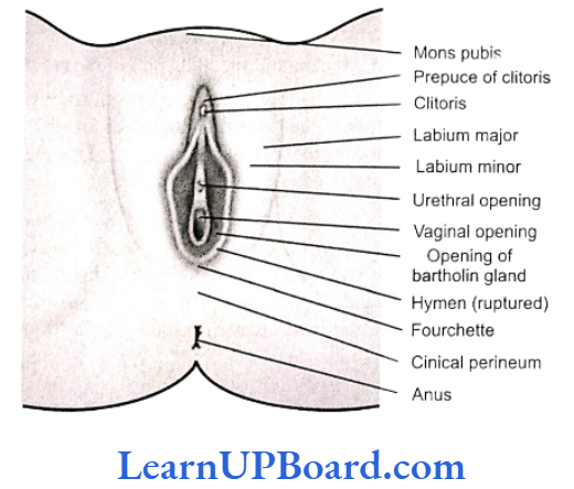
- Vulva: It is the external genitalia of females. It has a depression-the vestibule-in front of anus. A vestibule has two apertures-upper external urethral orifice of urethra and lower vaginal orifice of vagina.
- Mons pubis: It is a fleshy and fatty tissue covered by skin and pubic hair.
- Labia majora: It is a pair of fleshy folds which extend from mons pubis and surround the vaginal opening.
- Labia minora: It is another pair of tissue folds below the labia majora.
- Both labia majora and labia minora are provided with sebaceous glands.
- Hymen: It is a membrane that partially covers the vaginal opening. It gets torn during the first coitus.
- Clitoris: It is a tiny, erectile, finger-like structure present at the upper junction of the two labia minora above the urethral opening. The fold of skin that covers the clitoris is called prepuce. Clitoris is homologous to penis (as both are supported by corpora cavernosa).
NEET Biology Notes For Human Reproduction Glands
Vestibular Glands
Vestibular glands are of two types-greater and lesser.
- Greater vestibular or Bartholin’s glands are a pair of small reddish-yellow glands on each side of vaginal orifice. These secrete alkaline secretion for lubrication and neutralizing urinary acidity.
- Lesser vestibular or paraurethral or Skene’s glands are small mucus glands present between urethral and vaginal orifices.
Mammary Gland
- Each mammary gland consists of 15-25 lobules of the compound tubuloalveolar type. These lobules secrete milk to nourish the newborn babies.
- Each lobe is separated from the others by dense connective and adipose tissue and represents a gland. From each lobe, excretory lactiferous ducts emerge independently in the nipple, which has 15-25 openings, each about 0.5 mm in diameter.
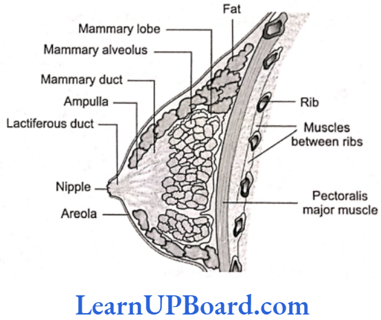
- However, the histological structure of mammary glands varies, depending on sex, age, and physiological state.
Path of Milk Ejection
Mammary alveolus → Mammary duct→ Ampulla → Lactiferous duct → Nipple
NEET Biology Notes For Human Reproduction Events In Human Reproduction
Gametogenesis→ Insemination → Fertilization → Implantation → Gestation → Parturition
NEET Biology Notes For Human Reproduction Formation Of Gametes
- Sexual reproduction requires the fusion of two haploid gametes to form a diploid individual. These haploid cells are produced through gametogenesis.
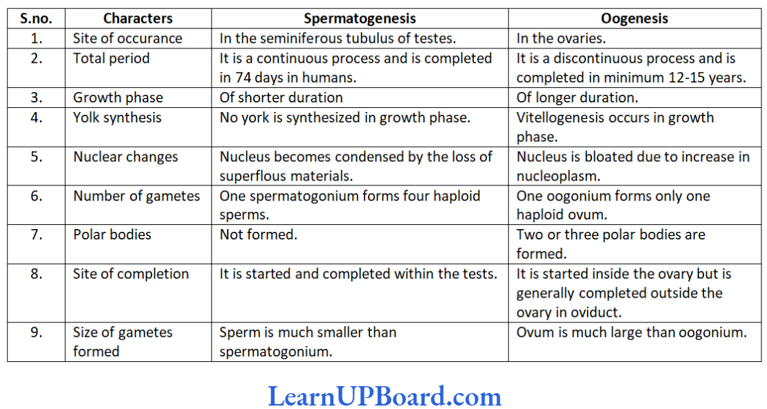
- As there are two types of gametes-spermatozoa and ova gametogenesis can be studied under two broad headings, namely spermatogenesis and oogenesis.
- Spermatogenesis is the formation of spermatozoa, whereas oogenesis is the formation of ova. Both spermatozoa and ova originate from primordial germ cells (PGCs), which are extragonadal in origin.
- In humans, the PGCs originate during early embryonic development from the extra-embryonic mesoderm. Eventually, they migrate to the yolk sac endoderm and, ultimately, to the gonads of the developing embryo, where they undergo further development.
NEET Biology Notes For Human Reproduction Spermatogenesis
- Spermatozoa are produced in the seminiferous tubules of testes. Spermatogenesis is the process of maturation of reproductive cells in the testes.
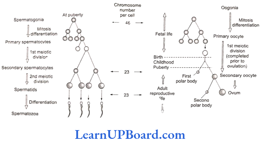
- Spermatogenesis includes two stages: (a) formation of spermatids and (b) metamorphosis of sperma- tids. Spermatids are formed in three phases, namely (a) phase of multiplication (mitosis), (b) phase of growth, and (c) phase of maturation (meiosis).
- During the phase of multiplication, the primordial germ cells divide repeatedly by mitosis to form diploid spermatogonia.
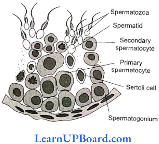
- During the phase of growth, spermatogonium enlarges in size to form primary spermatocyte and prepares to undergo maturation division.
- During the phase of maturation, the primary spermatocyte undergoes meiosis 1 giving rise to two haploid (n) secondary spermatocytes. The secondary spermatocytes undergo meiosis 2 resulting in the formation of four spermatids.
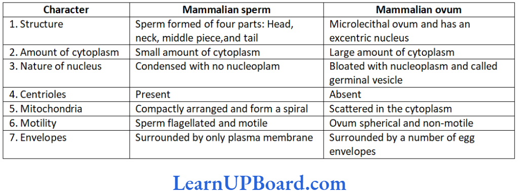
- The transformation of spermatid to sperms is termed as spermiogenesis. A spermatid is non-motile. It has organelles such as mitochondria, Golgi bodies, centrioles, and nucleus.
- During spermiogenesis, the weight of gamete is reduced along with the development of locomotory structures. The nucleus becomes compact forming the major part of head of spermatozoa.
- The Golgi complex of spermatid gives rise to acrosome. Acrosome forms a cap in front of the nucleus containing lytic agent. It dissolves egg membranes during fertilization.
- The acrosome of mammalian sperm produces sperm lysins. The two centrioles of spermatids become arranged one after the other behind the nucleus.
- The anterior one is known as proximal centriole. It is usually located on the neck of spermatozoan. During fertilization, it is introduced into the egg and is required for the first cleavage.
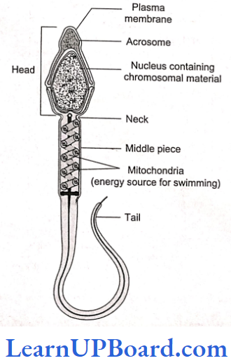
- The posterior centriole is known as distal centriole. It gives rise to the axial filament of the sperm. Mitochondria from different parts of spermatid get arranged in the middle piece around the axial filament. Mitochondria in the middle piece provide energy to the sperm for locomotion.
- A typical mammalian sperm is flagellated, consisting of four parts, namely head, neck, middle piece, and tail.
- The human sperm was first seen by Hamm and Leeu- wenhoek. Tail-less (non-flagellate), “amoeboid” sperm is found in the roundworm Ascaris.
Hormonal Control of Spermatogenesis
- Spermatogenesis is under the control of endocrine hormones. Hypothalamus produces gonadotropin releasing hormone (GnRH).
- It acts on anterior pituitary to produce gonadotropins, ICSH, and FSH. ICSH acts on the interstitial or Leydig’s cells which produce testosterone.
- Testosterone is essential for the formation of sperms, at least the spermiogenesis part by the Sertoli cells. Under the influence of FSH, the Sertoli cells develop ABP.
- The latter helps in concentrating testosterone in the seminiferous tubules.
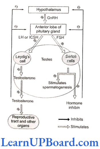
- Excess of testosterone inhibits LH/ICSH (luteinizing hormone/interstitial cell stimulating hormone) by ante- rior pituitary and GnRH production by hypothalamus.
- The Sertoli cells also produce a glycoprotein called inhibin. Inhibin suppresses FSH synthesis by anterior pituitary and GnRH synthesis by hypothalamus.
- Thus, the normal release of testosterone is under negative feedback control.
NEET Biology Notes For Human Reproduction Oogenesis
- Oogenesis is the process of maturation of reproductive cells in ovary. Oogenesis starts before birth. In 25-weeks-old female fetus, all oogonia are produced.
- Oogenesis is basically similar to spermatogenesis. It includes the phase of multiplication, the phase of growth, and the phase of maturation.
- During the phase of multiplication, the primordial cells in the ovary divide mitotically to form oogonia (egg mother cell). Each oogonium divides mitotically to form two primary oocytes.
- Primary oocytes undergo growth. The growth phase during oogenesis is comparatively longer.
- Primary oocytes begin the first step of meiosis 1 and proceed up to diakinesis.
- These oocytes resume their development at puberty. The primary oocyte (2n) completes meiosis 1 producing two haploid cells (n)-the larger one is secondary oocyte and the smaller one is first polar body.
- The secondary oocyte starts meiosis 2 and proceeds up to metaphase 2 only. Further development will start only after the arrival of spermatozoa.
- The entry of sperm restarts the cell cycle by breaking down MPF (M-phase promoting factor) and turning on APC (anaphase promoting complex). The completion of meiosis 2 results in the formation of functional egg or ovum and a second polar body.
“reproduction in humans “
Hormonal Control of Oogenesis
- In response to the production of GnRH, anterior pituitary secretes two hormones-FSH and LH .
- FSH stimulates follicular growth and maturation of oocyte. Granulosa cells of developing ovarian follicle produce estrogen.
- In the presence of high titer of both estrogen and LH, ovulation occurs. High concentration of estrogen inhibits secretion of both FSH and GnRH.
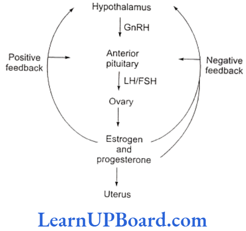
- This is negative feedback control. LH helps in converting ruptured Graafian follicle into corpus luteum.
- The latter secretes progesterone which prepares the uterus to receive fertilized ovum. High concentration of progesterone inhibits further release of LH from anterior pituitary and that of GnRH from hypothalamus.
NEET Biology Notes For Human Reproduction Menstrual Cycle
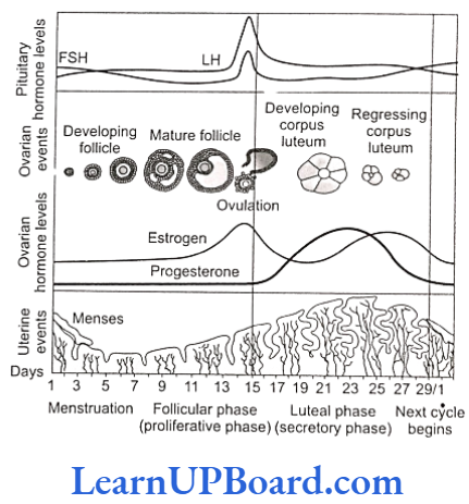
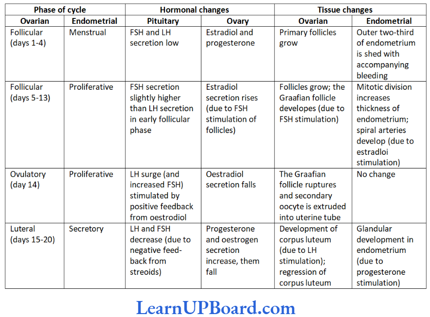
- Menstrual cycle is the cyclic changes in the reproductive tract of primate (man, monkey, and apes) females. Menstruation is the periodic shedding of the endometrium of uterus with bleeding. In healthy women, menstruation occurs at intervals of about 28-29 days.
- Menarche is the starting of menstruation in girls. It occurs at about 13 years of age. Menstrual cycle consists of menstrual phase, proliferative phase (follicular phase), and secretory phase (luteal phase).
- The proliferative phase (5th to 14th day of menstrual cycle) consists of the growth of the endometrium of uterus, the fallopian tube, and the vagina. In ovary, a Graafian follicle secretes estrogen during this phase.
- Estrogen is the hormone active during the prolifera- tive phase. Ovum is released from the follicle near the end of the proliferative phase, i.e., 14th day or midway during menstrual cycle.
- Ovulation occurs under the influence of LH from pituitary. The subsequent 14 days in which corpus luteum is active make up the secretory phase.
- Progesterone secreted by corpus luteum is active during the secretory phase. Uterine endometrium and glands grow further during this phase.
- At the end of the secretory phase, corpus luteum degenerates into corpus albicans in the ovary, progesterone secretion falls, the overgrown uterine endometrium breaks down, and mensturation takes place.
- Menstrual cycle is controlled by FSH, LH, estrogen, and progesterone. Menstrual cycle and menstruation remain suspended during pregnancy and lactation. Menopause (climacteric) is the period of life when menstruation naturally stops.
- Menopause occurs in females at the age of around 45-50 years. The ability to reproduce is lost in females after menopause.
NEET Biology Notes For Human Reproduction Estrous Cycle
- The estrous cycle consists of cyclic changes in the female reproductive system of non-primate mammals. There is no menstruation at the end of estrous cycle. The estrogen level in blood increases resulting in strong sex urge in the female. This is called the “period of heat.”
- The estrous cycles run only during breeding season these remain suspended in females during non-breed- ing season. The suspension of estrous cycles is called the state of anestrum.
- Animals that have only a single estrous during the breeding season are called monoestrous. For example, dog, fox, deer, bat, etc.
- Animals that have recurrence of estrous cycle during the breeding season are called polyestrous. For example, mouse, squirrel, cow, sheep, pig, horse, etc.
NEET Biology Notes For Human Reproduction Events In Mammalian Reproduction
Fertilization
- Ovum is released in the secondary oocyte stage (arrested in metaphase 2). Due to ciliary current produced by fimbriae of oviduct, ovum is drawn in through ostium.
- It reaches ampulla the site of fertilization by the ciliary action of ciliated columnar epithelium lining of oviduct.
- A human sperm can live for many weeks in the male genital duct. Once ejaculated, the sperm can live alive only for 24-48 h outside the body. Sperms move in the liquid medium secreted by the female genital tract (1.5-3 mm/min).
- Prostaglandins of semen help in the movement of sper- matozoa and finally reach the ampulla portion of the oviduct.
- Capacitation of sperm occurs in the female genital system due to the following factors:
- Removal of membrane cholesterol present over acrosome, weakening the membrane cover.
- Dilution of decapacitation factors.
- Entry of Ca2+ into sperms causing rapid whiplash motion of the tail.
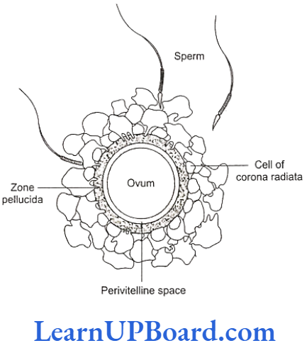
- Fusion of gametes/syngamy: The various steps are as follows:
- Acrosomal reaction: The sperms adhere to the surface of egg covers (agglutination). The acro- some starts releasing its hydrolytic enzymes (sperm lysins).
It includes the following:
- Acrosomal reaction: The sperms adhere to the surface of egg covers (agglutination). The acro- some starts releasing its hydrolytic enzymes (sperm lysins).
- Hyaluronidase: It dissolves the hyaluronic acid responsible for the cementing of follicle cells or granulosa cells.
- Corona digesting enzyme (CDE): It dissolves corona radiata.
- Zona lysin/acrosin: It digests zona pellucida. It involves zona pellucida compatibility reaction determined by the protein fertilizin over zona pellucida and antifertilizin in case of sperm.
- The contact of acrosome stimulates the development of an outgrowth by the oocyte called the fertilization cone or the cone of reception.
- As the sperm head gets in contact with the fertilization cone, it causes the opening up of Na* channel to cause the depolarization of membrane (fast block to check polyspermy) and Ca2+ wave inside the egg.
- Sperm and egg membranes dissolve. Male pronucleus and proximal centriole of sperm enter the cytoplasm of egg and the rest part is left out.
- Ca2+ wave causes the extrusion of cortical granules (cortical reaction) and the zona reaction makes zona pellucida impervious to the second sperm by destroying sperm receptors.
- Cortical reaction and zona reaction constitute slow block to check polyspermy.
- The entry of sperm causes the breakdown of metaphase promoting factor (MPF) and turns on anaphase promoting complex (APC). This results in oocyte completing its meiosis 2.
- Male and female pronuclei approach each other and, finally, mixing up of paternal and maternal chromosomes (amphimixis) occurs resulting in the formation of zygote/synkaryon.
Embryonic Development
Embryonic development includes cleavage, blastulation, implantation, gastrulation, and organogenesis.
Cleavage
- The first cleavage is completed after 30 h of fertiliza- tion. Cleavage furrow passes from the animal vegetal axis as well as the center of zygote (meridional cleavage).
- It divides the zygote into two blastomeres (holoblastic cleavage). The second cleavage is completed after 60 h of fertilization.
- It is also meridional but at right angle to the first one. It is completed earlier in one of the two blastomeres, resulting in transient three-celled stage.
- The third cleavage is horizontal forming 8 blastomeres. It is slightly unequal. Thereafter the rate and pattern of cleavage are not specific.
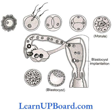
Morula
- Cleavage results in solid ball of celled morula with 16 cells (occasionally 32 cells). Zona pellucida is still present as the outer cover. Morula undergoes compaction.
- The outer/peripheral cells are small/flat with tight junction while the inner cell mass is slightly large round and with gap junction.
- Morula descends slowly towards the uterus in 4-6 days and corona radiata detaches during this period.
Blastulation or Blastocyst Formation
- Endometrium secretes a nutrient fluid and its mucosal cells become enlarged with stored nutrients. As the morula enters uterus, it obtains enriched supply of nu- trients.
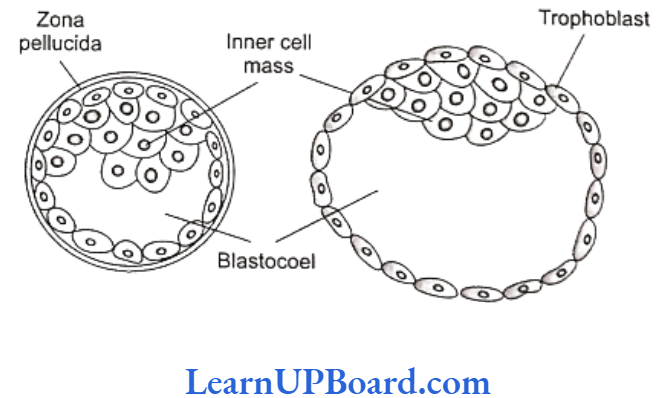
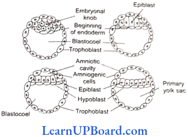
- The outer peripheral cells enlarge and flatten further. They form trophoblast or trophoectoderm. Trophoblast secretes a fluid into the interior. It creates a cavity called blastocoel.
- The inner cell mass now comes to lie on one side as embryonal knob.
- With the formation of blastocoel, morula is converted into blastula which is called blastocyst in mammals because of different nature of surface layer and eccentric inner cell mass.
- Due to the pressure of growing blastocyst, a slit is produced in zona pellucida. The growing blastocyst comes out. At times, it gets broken into two parts which then give rise to identical twins.
- Trophoblast cells in contact with the embryonal knob are called the cells of Rauber. The area of embryonal knob represents animal pole.
- The opposite side is embryonal pole. Soon embryonal knob shows rearrangement to form embryonal disc. The cells of trophoblast layer divide periclinally.
- This makes trophoblast two layered-outer syncyto trophoblast and inner cytotrophoblast. The two layers later form the chorion, amnion, and fetal part of placenta.
Implantation
- Implantation is the embedding of blastocyst into the endometrium of uterus.
- Blastocyst comes in contact with endometrium in the region of embryonal knob or embryonic disc. It adheres to the same.
- The surface cells of trophoblast secrete lytic enzymes which cause the corrosion of endometrial lining.
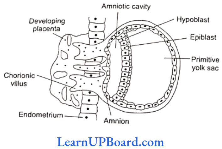
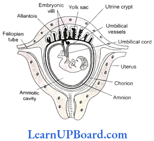
- They also give rise to finger-like outgrowths called villi. Villi not only help in fixation but also in the absorption of nourishment.
- Implantation causes nutrient enrichment, enlargement of cells, and formation of uterine part of placenta called decidua (L. deciduus-falling off).
- Decidua has three regions:
- Decidua basalis (basal decidua, tunica serotina): It is the part of decidua underlying the chorionic villi and overlying the myometrium.
- Decidua capsularis (decidua reflexa): It lies between the embryo and the lumen of uterus.
- Decidua parietalis (decidua vera): It is the part of decidua that lines the uterus at a place other than the site of attachment of embryo.
- Trophoblast secretes a hormone called human cho- rionic gonadotropin (hCG). The detection of hCG in urine is the basis of pregnancy/Gravindex test.
- hCG maintains the corpus luteum beyond its normal life. It continues to secrete progesterone which pre- vents menstruation and maintains the uterine lining in nutrient-rich state.
- Progesterone induces cervical glands to secrete viscous mucus for filling the cervical canal to form a protective plug.
- Progesterone is also called pregnancy hormone as it is essential for the maintenance of pregnancy. The hormone is secreted by placenta as well.
Gastrulation
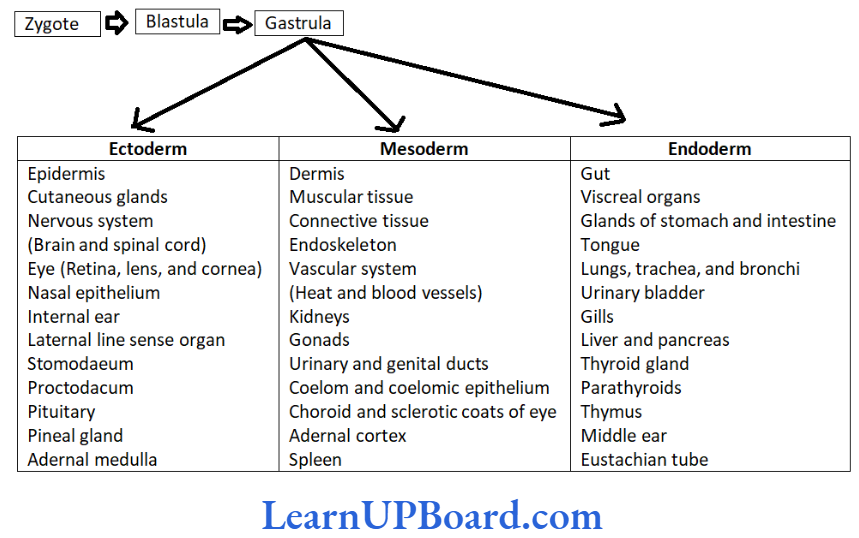
- Gastrulation is characterized by the movement of cells in small masses or sheets so as to form primary germinal layers. There are three primary germinal layers: (a) endoderm , (b) ectoderm, and (c) mesoderm.
- The cell movements that occur during gastrulation are called morphogenetic movements since they lead to the initiation of morphogenesis. The product of gastrulation is called gastrula.
Formation of Primary Germinal Layers
- The cells of inner cell mass or embryonal knob get rearranged to form a flat embryonic or germinal disc. The latter differentiates into two layers-outer epiblast of larger columnar cells and inner hypoblast of smaller cuboidal cells.
- Gastrulation begins with the formation of primitive streak on the surface of the epiblast.
- Cross-section through the cranial region of the streak after 15 days showing the invagination of epiblast cells. The first cells to move inward displace the hypoblast to create a definitive endoderm. Primitive node
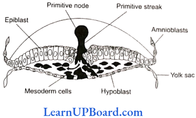
- Once the definitive endoderm is established, inwardly moving epiblast forms mesoderm.
- Cells remaining in the epiblast form ectoderm. Thus, epiblast is the source of all germ layers in the embryo.
NEET Biology Notes For Human Reproduction Placenta
- Placenta is an organ that connects the fetus and uterine wall.
- It is contributed by both maternal as well as fetal part although there is no blending of the maternal and fetal blood supplies. Placenta acts as an ultrafilter. Soluble inorganic and organic materials; nutrients; hormones; and antibodies against diphtheria, small pox, scarlet fever, measles, etc., can pass from the mother to the fetus.
- Placenta acts as an endocrine gland and synthesizes large quantities of proteins and some hormones, such as hCG, chorionic thyrotropin, chorionic corticotropin, chorionic somatomammotropin, estrogens, and pro- gesterone.
- hCG stimulates corpus luteum to secrete progesterone until the end of pregnancy. In addition, it secretes re- laxin that facilitates parturition by softening the con- nective tissue of symphysis pubica.
- The metabolic activity of the placenta is almost as great as that of the fetus itself. The umbilical cord con- nects the fetus to the placenta.
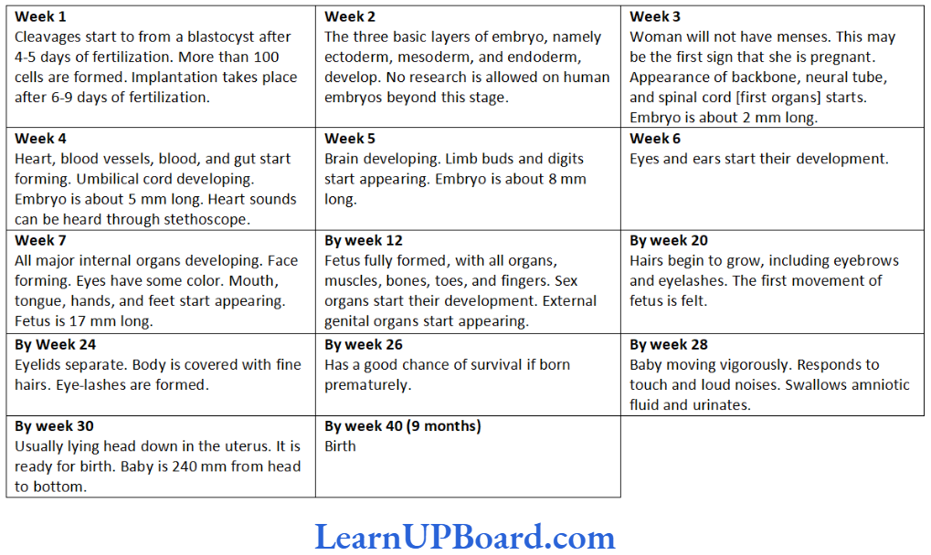
- During the first trimester (first 3 months) of pregnancy, the basic structure of the baby is formed.
- This involves cell division, cell migration, and the differentiation of cells into many types found in the baby. During this period, the developing baby-called fetus is very sensitive to anything that interferes with the steps involved.
- Virus infection of the mother, e.g., by rubella (German measles) virus or exposure to certain chemicals, may cause malformations in the developing embryo. Such agents are called teratogens (monster forming).
- After 3 months, all the systems of the baby have been formed, at least in a rudimentary form.
- From then, the development of fetus is primarily a matter of growth and minor structural modifications.
- The fetus is less susceptible to teratogens.
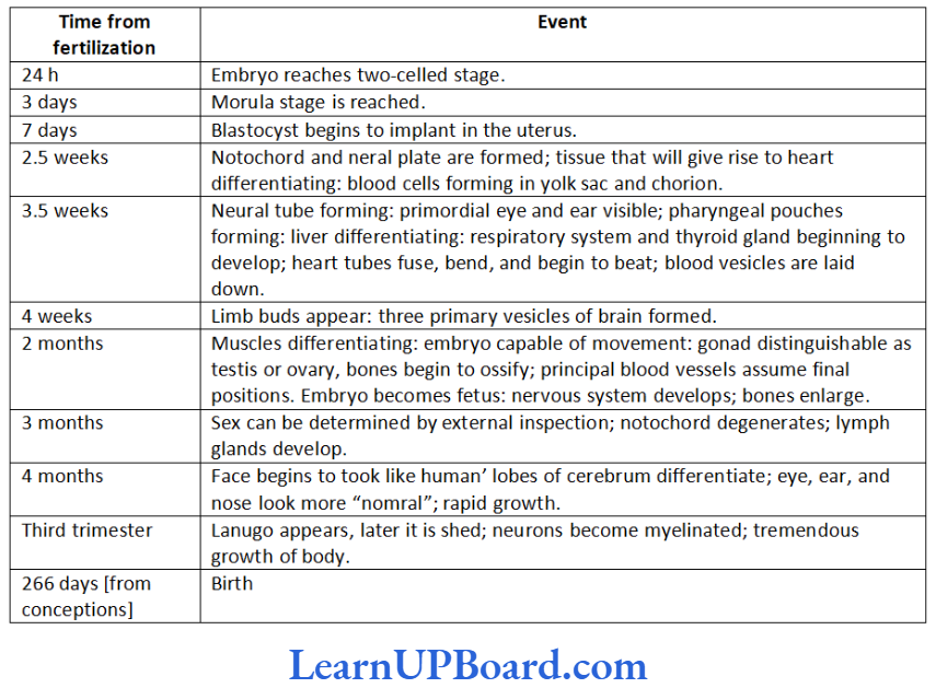
NEET Biology Notes For Human Reproduction Parturition
- The gestation period of a human is about 38 weeks/ 266 days followed by birth. The process of giving birth to a baby is called parturition.
- It starts with the rise in estrogen/progesterone ratio and increase in the level of oxytocin secretion by both— mother and fetus.
- It includes three stages.
- Dilation stage:
- Uterine contraction starts from the top and occurs at long intervals (once every 30 min). This forces the baby to push its head against the cervix.
- As a result, the cervix gets dilated with va- gina also showing similar dilation. The di- lation of cervix increases the stimulus for oxytocin secretion, further increasing the strength and frequency of contractions (1- 3 every minute).
- With continued powerful contractions, the amnion ruptures and the amniotic fluid flows out through vagina.
- Expulsion stage
- With further increase in the intensity of uterine and abdominal contraction, the baby comes out through the cervix and vagina with head coming out first.
- It may take 20-60 min. The umbilical cord is cut. The infant’s lungs expand and breathing begins. This requires a major switch-over in the circulatory system.
- Blood flow through the umbilical cord ductus arteriosus and foramen ovale ceases; the adult pattern of blood flow-through the heart, aorta, and pulmonary arteries-begins.
- In some infants, the switch-over is incomplete and blood flow through the pulmonary arteries is inadequate. Failure to synthesize enough nitric oxide is one cause.
- After birth: Within 10-15 min after delivery, the placenta and the remains of the umbilical cord, called “after birth,” are expelled out.
- Dilation stage:
Lactation
- Although estrogen and progesterone are essential for the physical development of breasts during pregnancy, a specific effect of both these hormones is to inhibit the actual secretion of milk. Conversely, the hormone prolactin has exactly the opposite effect on secretion- promotion of milk secretion.
- This hormone is secreted by the mother’s anterior pi- tuitary gland and its concentration in her blood rises steadily from the fifth week of pregnancy until the birth of the baby at that time it has risen 10-20 times the normal non-pregnant level.
- In addition, placenta secretes large quantities of human chorionic somatomammotropin, which probably also has lactogenic properties, thus, supporting the prolactin from the mother’s pituitary during pregnancy.
- The fluid that is secreted in the last few days before and in the first few days after parturition is called colostrums. It contains essentially the same concentrations of proteins and lactose as milk but almost no fat.
Ejection (or “Let-Down”) Process in Milk Secretion
- Milk is secreted continuously into the alveoli of the breasts, but it does not flow easily from the alveoli into the duct system and, therefore, does not continually leak from the breast nipples.
- Instead, milk must be ejected from the alveoli into the ducts before the baby can obtain it. This is caused by a combined neurogenic and hormonal reflex that in- volves the posterior pituitary hormone oxytocin.
- When the baby suckles, sensory impulses are trans- mitted through somatic nerves from the nipples to the mother’s spinal cord and then to her hypothalamus, there causing nerve signals that promote oxytocin secretion; at the same time they cause prolactin secretion.
- Oxytocin is carried in the blood to the breasts, where it causes myoepithelial cells (that surround the outer walls of the alveoli) to contract, thereby, expelling milk from the alveoli into the ducts.
NEET Biology Notes For Human Reproduction Assertion-Reasoning Questions
In the following questions, a statement of Assertion (A) is followed by a statement of Reason (R).
(1) If both Assertion and Reason are true and the reason is the correct explanation of the assertion, then mark (1).
(2) If both Assertion and Reason are true but the reason is not the correct explanation of the assertion, then mark (2).
(3) If Assertion is true but Reason is false, then mark (3).
(4) If both Assertion and Reason are false, then mark (4).
Question 1. Assertion: Scrotum provides optimum temperature conditions for spermatogenesis.
Reason: Dartos and cremaster muscles in scrotum contract and relax involuntarily in response to temperature.
Answer. 2. If both Assertion and Reason are true but the reason is not the correct explanation of the assertion, then mark (2).
Question 2. Assertion: The process of reproduction does not suffer if one ovary is removed.
Reason: The other ovary enlarges to take over the function of the missing ovary too.
Answer. 3. If Assertion is true but Reason is false, then mark (3).
Question 3. Assertion: “Nothing lives forever, but life continues.”
Reason: Death keeps the population growth under check.
Answer. 2. If both Assertion and Reason are true but the reason is not the correct explanation of the assertion, then mark (2).
Question 4. Assertion: Placenta is connected to the fetus by an umbilical cord.
Reason: Fetal components of placenta are derived from endometrium.
Answer. 3. If Assertion is true but Reason is false, then mark (3).
Question 5. Assertion: Placenta is contra-deciduate and even the fetal placenta is absorbed in mole.
Reason: Mole’s egg contains abundant yolk in ooplasm.
Answer. 3. If Assertion is true but Reason is false, then mark (3).
Question 6. Assertion: Polar bodies have small amount of cytoplasm.
Reason: It is formed by unequal mitotic division.
Answer. 3. If Assertion is true but Reason is false, then mark (3).
Question 7. Assertion: Ovulation takes place when the blood level of luteinizing hormone is high.
Reason: Leutinizing hormone is responsible for ovulation.
Answer. 1. If both Assertion and Reason are true and the reason is the correct explanation of the assertion, then mark (1).
Question 8. Assertion: Umbilical cord contains 100% fetal blood.
Reason: It has single umbilical artery and single umbilical vein.
Answer. 3. If Assertion is true but Reason is false, then mark (3).
Question 9. Assertion: The activation of sperm is called capacitation.
Reason: Capacitation takes about 5-6 h.
Answer. 2. If both Assertion and Reason are true but the reason is not the correct explanation of the assertion, then mark (2).
Question 10. Assertion: Before fusion, spermatozoa have to penetrate egg membrane,
Reason: The activated spermatozoa undergo acrosomal reactions and release sperm lysin.
Answer. 2. If both Assertion and Reason are true but the reason is not the correct explanation of the assertion, then mark (2).
Question 11. Assertion: In post natal life, oocyte development occurs in mature follicle,
Reason: After ovulation, the Graafian follicle transforms in corpus luteum.
Answer. 2. If both Assertion and Reason are true but the reason is not the correct explanation of the assertion, then mark (2).
Question 12. Assertion: Placenta is combined structure of fetal tissue and maternal tissue.
Reason: Placenta formation is completed before 6 weeks.
Answer. 3. If Assertion is true but Reason is false, then mark (3).
Question 13. Assertion: Seminal vesicle is known as the accessory sex organ of males.
Assertion: Seminal vesicle conserves sperm energy and provides fuel to sperm.
Answer. 1. If both Assertion and Reason are true and the reason is the correct explanation of the assertion, then mark (1).
Question 14. Assertion: Testes are retroperitoneal organ in man.
Reason: Peritoneal layer covers the testes on the dorsal side.
Answer. 4. If both Assertion and Reason are false, then mark (4).
Question 15. Assertion: Cervix contains the most weak sphincter muscle in the body.
Reason: Cervix opens into fallopian by os external.
Answer. 4. If both Assertion and Reason are false, then mark (4).
Question 16. Assertion: In ovarian cycle, corpus luteum is exocrine gland.
Reason: It secretes pheromones.
Answer. 4. If both Assertion and Reason are false, then mark (4).