
NEET Biology Notes Anatomy Of Flowering Plants Introduction
The branch of botany dealing with the internal organization of plants is called anatomy. N. Grew laid the foundation of plant anatomy. He coined the term tissue. He is known as the father of anatomy.
NEET Biology Notes Anatomy Of Flowering Plants Tissues
A group of similar or dissimilar cells that perform a common function and have a common origin is called tissue. Tissues are classified into two main groups: meristematic and permanent.
NEET Biology Notes Anatomy Of Flowering Plants Meristematic Tissues
- Mesistematic tissues consist of cells that retain the power of division.
- The protoplasm within the cell is dense, the vacuole is smaller or absent.
- These cells are isodiametric without intercellular spaces.
- The nucleus is bigger in size.
- These cells have thin cellulosic cell walls.
- These are metabolically active cells with high surface area per unit volume and nucleocytoplasmic ratio.
- Ergastic substances are absent.
- Colorless proplastids are present in the cells.
Classification Of Meristems
On The Basis Of Origin And Development
- Promeristems (Primordial Meristem): Promeristerms are a group of cells that represent the primary stages of meristematic cells. They are found at the apices of embryonic shoots and roots. They give rise to primary meristems.
- Primary Meristems: They originate from pro meristems. They are found at shoot and root apices, at the apex of leaves, and in intercalary parts. They give rise to primary permanent tissues.
- Secondary Meristems: They are not present from the beginning of the formation of an organ but develop at a later stage. They give rise to secondary permanent tissues. They develop from primary permanent tissues, for example, interfascicular cambium, cork cambium, and cambium in dicot roots.
On The Basis Of Position
- Apical Meristem: These cells or tissues are found at the apices of the stem and root. Due to continuous division, the root and stem increase in length. The apical meristem helps the plants to grow in length.
- Intercalary Meristem: The tissues are intercalated between permanent tissues. These are actually the parts of the apical meristem that get separated from it during the growth of the stem and root in length. The most characteristic example is the stem of grasses and Equisetum. Intercalary meristem is especially responsible for the increase in the length of the stems of grasses.
- Lateral Meristem: These meristems are present along the lateral side of the stem and root. They divide in a tangential plane, giving rise to secondary permanent tissues on the inner and outer sides and leading to the increase in thickness or girth of the plant body, for example, intrafascicular cambium, inter¬fascicular cambium, and cork cambium.
Read and Learn More NEET Biology Notes
On The Basis Of Plane Of Cell Division
- Mass Meristem: The cells divide anticlinally in all planes so that a mass of cells is formed. For example, the formation of spores, cortex, pith, endosperm, etc.
- Plate Meristem: The cells divide anticlinally in two planes, so the plate-like area is increased. For example, the formation of epidemics and lamina of leaves.
- Rib Or File Meristem: The cells divide anticlinally in one plane, so a row or column of cells is formed. For example, the formation of lateral roots.
On The Basis Of Function
- Protoderm: They are the outermost meristematic cells. They form the skin or epidermis of the plant and epidermal tissue system.
- Procambium: They are the innermost meristematic cells. They form the primary xylem, primary phloem, and cambium.
- Ground Meristem: They form ground or funda¬mental tissues such as hypodermis, cortex, pith, pericycle, etc.
Shoot Apex Organization: Shoot apex is present immediately above the youngest leaf primordia. It consists of meristematic cells. The lateral branches of the stem and leaves are formed by the activity of shoot apex. Many theories have been put forward to explain shoot apex such as
Apical Cell Theory: It was proposed by Hofmeister and Nageli. According to this theory, a single apical cell leads to the development of the entire plant body. This theory is applicable to algae, as well as to most of the bryophytes and pteridophytes.
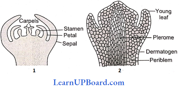
Histogen Theory: It was proposed by Hanstein. According to this theory, a shoot apex consists of the following halogens:
- Dermatogen: The outermost layer, it forms the epidermis (skin) and epidermal tissue system.
- Periblem: It gives rise to the tissues between the epidermis and the stele.
- Plerome: It is the innermost layer and the central mass of cells that give rise to the central stele.
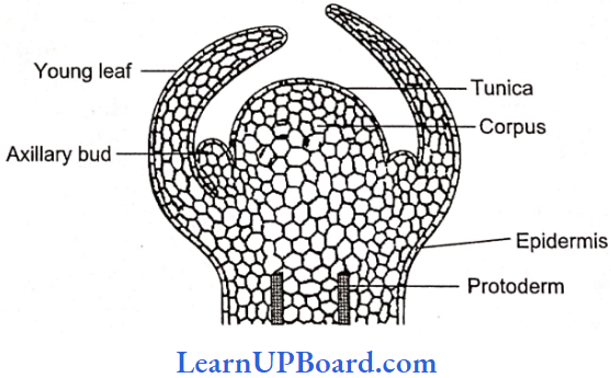
Tunica Corpus Theory: It was proposed by Schmidt (1924). It is based on the plane of division of the cells. According to this theory, the shoot apex consists of two distinct layers:
- Tunica: It is mostly single-layered and forms epider¬mis. The cells of tunica are smaller than the corpus and divide mostly by anticlinal divisions.
- Corpus: It represents the central core with larger cells. The cells divide periclinally. Sometimes tunica is multi-layered, only the outer layer forms epidermis, and the remaining layers with corpus form the cortex of the shoot.
” anatomy of flowering plants”
Root Apex Organization: The Root apex consists of a mass of meristematic cells. It is not responsible for the formation of lateral roots. Root cap or calyptrogen is present due to which the root meristem becomes subterminal in position. If the root cap is independent in origin, it arises from dermatogen. Regarding the organization of root apex, the following theories have been put forward
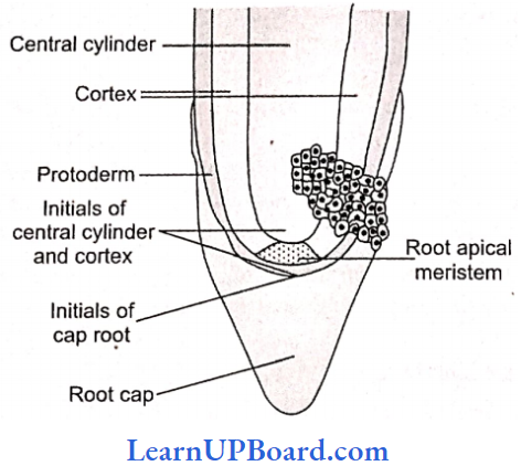
Korper-Kappe Theory: It was proposed b.y Schuepp (1917). This theory is comparable with the tunica corpus theory of shoot apex. Korper means body and kappe means cap.
Quiescent Center Theory: It was proposed by Clowes (1956-5 8). According to this theory, the root apex consists of an inverted cup-like structure, the quiescent center. The cells of this region are with very low mitotic activity (quiescent). They have low amounts of RNA, DNA, and protein. They are surrounded by a layer of actively dividing cells which are responsible for the formation of the different structures of roots.
NEET Biology Notes Anatomy Of Flowering Plants Permanent Tissue
Permanent tissues are composed of living or dead cells that are derived from the meristematic tissues but have lost their ability to divide. There are of three types: simple tissues, complex tissues, and secretory tissues.
Simple Tissues: They are made up of a single kind of cells performing similar functions. Simple tissues are mainly of three types: parenchyma, collenchyma, and sclerenchyma.
Parenchyma (Grew): These cells are found almost in all parts of plants such as roots, stems, leaves, fruits, and seeds. These cells are isodiametric, spherical, oval, or polygonal with intercellular spaces. These cells are living and with thin cellulosic cell wall.
Types Of Parenchyma
- Prosenchyma: Elongated parenchyma with tapering ends is called prosenchyma.
- Aerenchyma: The parenchyma which encloses the air cavity is called aerenchyma (hydrophytes)
- Chlorenchyma: The parenchyma containing chloroplasts is called chlorenchyma.
- Idioblast: Sometimes parenchyma stores secretory substances (orgastic substances) such as tannins, resins, and gum. They are called idioblasts.
Storage Parenchyma: Examples are fruits, and endosperm.
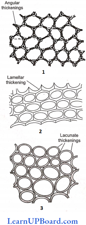
Collenchyma (Schleiden): Collenchyma has thickenings on the cell wall and in corners of intercellular spaces. They are not found in the roots and monocots. These cells form hypodermis in the stem and petiole. These are living mechanical tissues with high refractive index. The thickening material in the cell wall contains pectocellulose.
Types Of Collenchyma
- Angular Collenchyma: Angular walls thickened, for example, the stem of a marigold and tomato.
- Lamellate Collenchyma: Tangential walls thickened, for example, the stem of a sunflower.
- Lacunate Collenchyma: Lacunate thickening, intercellular spaces are present, for example, stem of cucurbita.
- Functions Of Collenchyma: They provide mechanical support, flexibility, and elasticity to the organs. Due to the peripheral position in stems they resist the bending and pulling action of wind. Collenchyma is especially useful for young plants and herbaceous organs where these act as important supporting tissues.
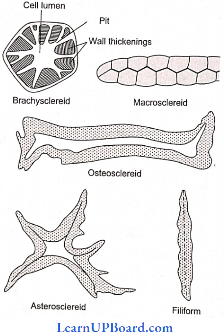
Sclerenchyma (Mettenius): Sclerenchyma has thickened secondary walls due to the deposition of lignin. At maturity they become dead. These cells have simple pits. They are of two types: Sclereids and sclerenchymatous fibers.
Sclereids: They may be spherical, oval, or cylindrical. They are lignified, and extremely thick-walled. So the lumen of the cells is almost obliterated. They are found in hard parts of the plant.
Types Of Sclereids
- Brachysclereids (Stone Cells): Grittiness in fruits is due to stone cells, for example, pear, and sapota.
- Macrosclereids (Rod Cells): Found in the seed coat of leguminous plants.
- Osteosclereids (Prop Cells): Found in the leaves and seed coat of many monocots and the sub-epidermis of legume seed coats.
- Astrosclereids (Star Cells): They are common in the stem and leaves of dicots, for example, tea leaves, petiole of lotus, etc.
- Trichosclereids (Internal Hair): Long, hair-like branched sclereids. They are common in hydrophytes. These are also present in the aerial roots of Monstera.
Sclei Enchymatous Fibers: They are long and tapering at ends. These are thick-walled cells (lignified). These fully developed fiber cells are always dead. The length of fiber varies from 72 to 550 mm in angiosperms and 1 to 12 mm in gymnosperms.
The fibers are present in the hypodermis of monocot stems, in the pericycle of many dicots, in the secondary wood, and in the vascular bundle sheath in monocot stems, for example, jute, flax, hemp, etc. Living fibers are found in tamarix.
Function Of Sclerenchyma: The main function of sclerenchyma is to provide mechanical strength.
Complex Tissues: A group of more than one type of cells having a common origin and working together as a unit is called complex tissue. These are made up of different types of cells: xylem and phloem.
Xylem (Nageli) Or Hadrome (Haberlandt): Xylem is the chief water-conducting element. It consists of the following types of cells
Tracheids: They are elongated cells with pointed chisel-like ends. Their wall is tough, thickened, lignified, and thickened or may be annular, spiral, reticulate, scalariform, or pitted. Cells are dead at maturity and have bordered pits.
In pteridophytes and gymnosperms, wood mainly consists of tracheids (no vessels). In angiosperms, tracheids are associated with vessels. The main function of tracheids is the conduction of water. Tracheids are the most primitive type of conducting elements in the xylem.
Vessels: Vessels are also elongated and tube-like. They are formed from a row of cells placed end to end. The partition walls are either perforated or disappear altogether resulting in an elongated tube. Walls are thickened, and lignified, and may have annular, spiral, reticulate, or scalariform thickening.
Vessels are dead at maturity and without nuclei. In pteridophytes and gymnosperms, vessels are absent (non-porous wood). Sometimes primitive vessels are present in gnetum and ephedra (Gnetales).
Vessels are characteristic of angiosperms (porous wood). Vesselless angiosperm families are Tetracentraceae, Trochodendraceae, and Winteraceae. The main function is the conduction of water. Vessels are an advanced type of conducting element.
On the basis of distribution and size of vessels, porous wood is of two types:
- Diffuse Porous Wood (Primitive): Vessels of the same size are uniformly distributed throughout the growth, for example, Pyrus, Betula.
- Ring Porbus Wood (Advanced): Large vessels are formed in early wood when the need of water is great and small vessels are formed in latewood, for example, Quercus, and morus.
Wood Or Xylem Fiber: These cells are elongated and pointed at both ends. Cell wall is highly lignified having simple pits. The air commonly found in the secondary xylem. They may lie
- Fiber Tracheids: Fiber such ns tracheids with bordered pits.
- Libriform Fiber: They have extremely thick walls and simple pits. They provide mechanical support.
Wood Or Xylem Parenchyma: They are living parenchymatous cells associated with xylem. They may occur as axial parenchyma or ray parenchyma. When parenchyma is diffused or not associated with vessels, they are called as apotracheal parenchyma and when parenchyma surrounds or is associated with vessels, it is called para tracheal parenchyma.
On The Basis Of Origin Xylem Is Of Two Types:
- Primary Xylem: It is derived from procambium during the formation of the primary plant body. It is differentiated into protoxylem (formed first and consists of tracheary elements and xylem parenchyma) and metaxylem (formed later and consists of tracheary elements, xylem parenchyma, and fiber). The cells of metaxylem are bigger in size than the protoxylem.
- Secondary Xylem: It is formed from cambium during secondary growth. It is well differentiated into two systems:
- Axial Or Vertical System
- Tracked elements (tracheids and vessels): For conduction of water
- Xylem or wood fiber: For support
- Xylem parenchyma: For storage of food
- Ray Or Horizontal System: Ray parenchyma: For storage of food.
- Axial Or Vertical System
Phloem (Nageli) Or Bast Or Leptome (Haberlandt): The main function of phloem is the transport organic food materials from leaves to stems and roots in down word direction. Phloem consists of the following types of cells
Sieve Element: The sieve elements in angiosperms are sieve tubes which are cylindrical tube like cells with perforated cross walls called sieve plates. Sieve tubes are associated with companion cells, and they are without nuclei. In pteridophytes and gymnosperms, the sieve elements have sieve plates on their lateral walls and companion cells are absent. They are called as sieve cells.
The walls of sieve tube elements are made up of cellulose and pectic substances. The cytoplasm is confined to a thin peripheral layer. P-proteins are proteinaceous structures present in sieve tubes and are believed to be responsible for
- The movement of materials and
- The sealing of pores after wounding.
In old sieve tubes, at the end of the growing season, a callose plug (made of callose carbohydrate) is deposited on the sieve plate which inhibits the activity of the sieve tubes. In the spring season, the callose plug gets dissolved.
Companion Cells: They are elongated living parenchymatous thin-walled cells. They are associated laterally to sieve tubes and have dense cytoplasm and nuclei. Companion cells are absent in pteridophytes and gymnosperms. Both sieve tubes and companion cells are related ontogenetically because both develop from the same mother cell. So these are sister cells.
Phloem Or Bast Fiber: They are absent or fewer in phloem and abundantly found in secondary phloem. They are sclerenchymatous fibers associated with phloem. Phloem fibers of plants such as jute, flax, and hemp are rotted in water and extracted for making ropes and coarse textiles.
Phloem Parenchyma: They are parenchymatous living cells with cellulosic cell walls and nuclei. The main function is the storage of food. They are not found in monocotyledonous plants.
Types Of Phloem
On The Basis Of Position
- External Phloem: It is of normal type and is present outside the xylem, for example, mostly angiosperms and gymnosperms.
- Internal Or Intraxylary Phloem: It originates from procambium and is the primary phloem which occurs on the innerside of primary xylem in bilateral bundles, for example, members of Apocynaceae, Asclepiadaceae, Convolvtilaceae, Solanaceae etc.
- Included Or Interxylary Phloem: It originates from cambium and is a secondary phloem that occurs in groups within the secondary xylem, for example, Leptadaenia, Salvadora, Chenopodiwn, Boerhaavia, Amaranthus, etc.
On The Basis Of Origin:
- Primary Phloem: It develops from procambium and does not have radial differentiation or rays are absent. It is differentiated into protophloem (consists of sieve elements and parenchyma) and metaphloem (develops after protophloem and consists of sieve elements, parenchyma, and fiber). During primary growth, the protophloem elements are crushed by the surrounding tissues and disappear. This process is known as obliteration.
- Secondary Phloem: It develops from cambium during secondary growth. It shows radial differentiation. It consists of two distinct systems such as
- Axial or vertical system
- Sieve Elements: Sieve tube and companion cells. For conduction of food.
- Bast fiber: For support.
- Bast Parenchyma: For storage of blood.
- Ray Or Horizontal System
- Ray Parenchyma: It is for the storage of food
- Axial or vertical system
Secretory Tissues: Secretory tissues perform special functions in plants. For example, secretion of resins, gums, oil, and later. These tissues are of two types laticiferous and glandular tissues.
Laticiferous Tissues: They contain colorless, milky, or yellow-colored juice called latex. These tissues are of two types
- Latex Cells: They do not fuse and do not form a network. Plants having such tissues are called simple or non-articulated laticifers, for example, Calotropis (Asclep Fabaceae), Nerium, Vinca (Apocyanaceae), Euphorbia (Euphorbiaceae), Ficus (Moraceae).
- Latex Vessels: They are formed due to the fusion of cells and form a network-like structure. Plants having such tissues are called compound or articulated laticifers, for example, Argemone, Pabaver (Papaveraccae), Sonchus (Compositae), Hevea, and Manihot (Euphorbiaceae).
” class 11 biology anatomy of flowering plants notes “
Glandular Tissues: They include different types of glands which secrete oils, gums, mucilage, tannins, and resins. They may be
- External Glands: Present as epidermal outgrowths. They are of many types
- Glandular Hair: With a stalk and head, for example, tobacoo, Plumbago, Boerhaavia.
- Stinging Hair: Secrete poisonous substances, for example, Urtica.
- Nectaries: Secrete sugary substance; maybe extra-floral present on the stem, leaves, etc. Examples: Nepenthes, Catheranthus, or floral, for example, Corchorus, Thea, Polygonum, Jatropha.
- Digestive Glands: Present in insectivorous plants, for example, Drosera, Nepenthes, etc.
- Internal Glands
- Oil Glands: Present in the mesophyll of leaves and cortex of stem fruit, for example, orange, lemon, etc.
- Mucilage-Secreting Glands: Leaves of piper betel.
- Gum, tannin, and resin-secreting glands or ducts are present in gymnosperms and angiosperms, for example, Pious resin ducts are schizogenous in origin.
NEET Biology Notes Anatomy Of Flowering Plants Tissue System
The various types of tissues present in the body of a plant perform different functions. Several tissues may collectively perform the same function. A collection of tissues performing the same general function is known as a “tissue system.” According to Sachs (1975), there are three major tissue systems in plants, namely the epidermal tissue system, the ground or fundamental tissue system, and the vascular tissue system.
Epidermal Tissue System: It consists of the epidermis and its associated structure such as hairs, trichomes cuticle, stomata, and bulliform cells. Mostly epidermis is single-layered parenchymatous but multilayered in Ficus, Nerium. The epidermis is mainly protective in nature.
In grasses, motor or bulliform cells are present in the upper epidermis. In grasses and Equisetum, silica is present in the epidermal cells. The epidermal cells containing cystoliths are called lithocysts.
Ground Or Fundamental Tissue System: It extends from the epidermis up to the center excluding vascular tissue. Ground tissues constitute the following parts
- Cortex: It lies between the epidermis and the pericycle. It is further differentiated into the following parts:
- Hypodermis: It is collenchymatous in the dicot stem and sclerenchymatous in the monocot stem. It provides strength.
- General Cortex: It consists of parenchymatous cells. Its main function is the storage of food.
- Endodermis (Starch Sheath): It is mostly single-layered and is made up of parenchymatous barrel-shaped compactly arranged cells. The inner and radial walls of endodermal cells have Casparian strips. In roots, thick-walled endodermal cells are interrupted by thin-walled cells just outside the protoxylem patches.
- These thin-walled endodermal cells are called passage cells. The endodermis behaves as water water-tight dam to check the loss of water and air tight dam to check the entry of air in xylem elements.
- Pericycle: It lies between endodermis and vascular tissue. It is parenchymatous in roots and sclerenchymatous or mixed with parenchyma in the stem. The pericycle cells just opposite the protoxylem are considered as seats for the origin of lateral roots. In dicot roots, the pericycle forms part of the cambium or the whole of the cork cambium.
- Pith: It occupies the central part in the dicot stem and monocot root. It is mostly made up of parenchymatous cells. In the dicot root, the pith is completely obliterated by the metaxylem elements. In the dicot stem, the pith cells between the vascular bundles become radially elongated and are known as primary medullary rays or pith rays. They help in lateral translocation.
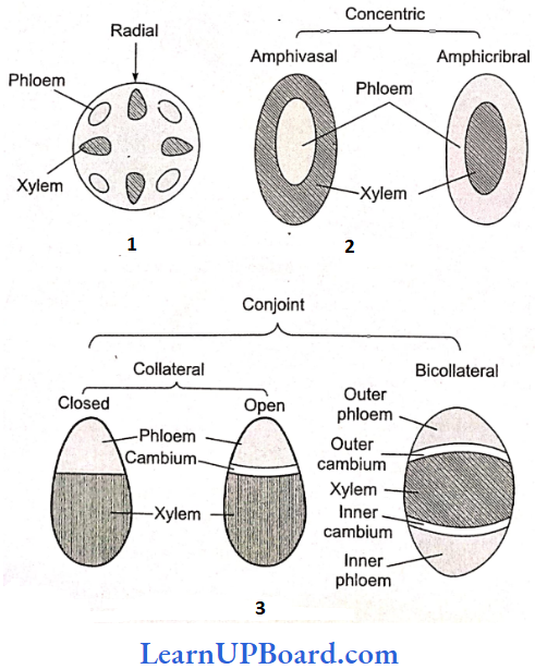
Vascular Tissue System: Vascular bundles found in the steer part constitute the vascular tissue system. Xylem. phloem and cambium are the major part of the vascular bundle. Vascular bundles may be of the following type:
- Radial: When the xylem and phloem are arranged on different radii alternating with each other, for example, roots.
- Conjoint: When xylem and phloem combine in the same bundles and are present on the same radius, for example, stem. Conjoint vascular bundles may be of the following types
- Collateral: The xylem is towards the inner side and the phloem, is towards the outer side.
- Open: Cambium is present between the xylem and phloem, for example, the dicot stem.
- Closed: Cambium is absent between the xylem and phloem, for example, monocot stem.
- Bicollateral: When the xylem has cambium and phloem on both sides, for example, members of Cucurbitaceae, Solanaceae, Apocyanaceae, etc.
- Collateral: The xylem is towards the inner side and the phloem, is towards the outer side.
- Concentric: When one vascular tissue surrounds the other. They are of two types
- Amphicribral Or Hadrocentric: The xylem is surrounded on all sides by phloem, for example, ferns.
- Amphivasal Or Leptocentric: The phloem is surrounded on all sides by the xylem, for example, Yucca, and Dracaena.
NEET Biology Notes Anatomy Of Flowering Plants Internal Structures Of Dicot And Monocot Plants
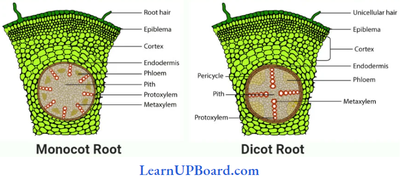
Anatomy Of Root: The three zones that can be distinguished in a root are
- Epidermis (Epiblema/Rhizodermis): It is single-layered (uniseriate) and consists of tightly placed, thin-walled unscrutinized cells. This epidermis layer is called as epiblema, piliferous layer, or rhizodermis. Epiblema in younger roots bears unicellular root hairs (water-absorbing organs).
- Cortex: It consists of thin-walled parenchymatous cells with intercellular spaces. In most monocots and some dicots, the cortex layer below the epidermis becomes suberized to form protective tissue called exodermis.
- The cells of the cortex store food material (for example, carrots). The innermost layer of the cortex develops into endodermis. It is made up of closely packed living cells characterized by the presence of band-like thickening; made of lignin and suberin on their radial and transverse walls.
- These bands or strips are called Casparian bands or strips. Some cells of endodermis lying opposite to the protoxylem remain thin-walled and are called passage cells which allow radial diffusion of water.
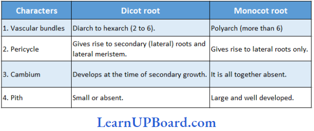
- Vascular Bundles: Vascular bundles are radial and exarch. The center of the monocot root is occupied by parenchymatous cells called piths.
Some basic differences between a monocot and a dicot root are given.
” plant anatomy ncert”
Differences Between Dicot And Monocot Root
Anatomy Of Stem: The Primary structure of the diet stem consists of the following layers
- Epidermis: It is the outermost layer consisting of a single layer of closely arranged cells with cuticles (cutinized). It bears multicellular hairs.
- Cortex: It is differentiated into hypodermis, general cortex, and endodermis. Hypodennis is collenchymatous. The general cortex bundles consist of the phloem and xylem.
- Vascular Bundles: Vascular bundles are conjoint, collateral, or collateral, open and endarch, and are arranged in a ring (eustele).
- Pith: It is the central portion of the stem consisting of parenchymatous cells with narrow, radially elongated parenchymatous cells extending from the pith toward the periphery called medullary rays. The main function of pith is food storage.
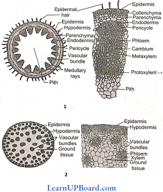
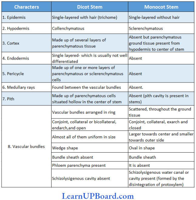
The primary structure of a monocot stem consists of the following layers:
- Epidermis: It is the outermost layer and consists of compactly arranged parenchymatous cells which are usually covered with cuticles.
- Hypodennis: The cells of hypodermis are sclerenchymatous providing mechanical strength to the stem.
- Ground tissue: All the tissues internal to hypodermis represent the ground tissue. It is made up of parenchymatous cells rich in food reserves such as starch.
- Vascular bundles: They lie scattered in the ground tissue. Each vascular bundle is surrounded by a two- or three-layered sclercnchymatous sheath called bundle sheath. The vascular bundles are conjoint, collateral, closed, and endarch (atactostelc). Vessels are arranged in a V-shaped manner. Schizolysigenous water cavity or canal arc present below protoxylem.
Shows the transverse section of the dicot stem and monocot stem. The differences between dicot stem and monocot stem are given.
Anatomy Of Leaf: In the cross-section of a dorsiventral leaf (dicot), the following parts can be made out
Epidermis: The upper and lower surfaces are covered by the epidermis. The cells of the epidermis are parenchymatous and are closely packed together without any intercellular spaces. Mostly the stomata are restricted to the lower surface of the leaf such leaf is called hypostomatic. The outer walls of the epidermal cells are thickened and cutinized (cuticle) which prevents the loss of water.
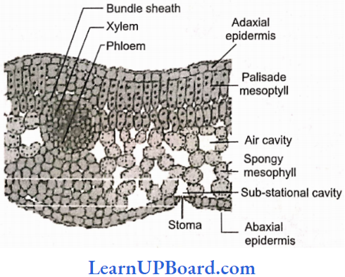
Mesophyll: Between the two epidermal layers, there are numerous chlorenchyma cells that constitute the mesophyll. In dicots, there are two distinct layers of mesophyll, the palisade (the upper layer consisting of closely arranged column-shaped cells containing abundant chloroplasts) and spongy tissue (the lower layer of irregularly shaped cells containing fewer chloroplasts).
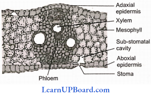
Vascular Bundles: Vascular bundles in the leaf are located in the midrib and the veins. Vascular bundles are conjoint, collateral, and closed. Bundles are surrounded by a compact layer of parenchymatous cells which is called bundle sheath. The xylem (protoxylem) is towards the upper epidermis (adaxial) and the phloem is on the lower side (abaxial). Like the dicot leaf, an isobilateral leaf (monocot) can also be differentiated into the following types of tissues
NEET Biology Notes Anatomy Of Flowering Plants Secondary Growth
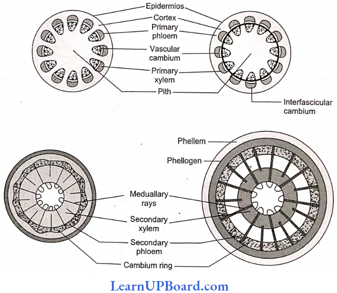
Secondary growth is the increase in girth thickness or diameter of the axis due to the formation of new tissues as a result of the joint activity of vascular cambium and cork cambium in stellar and extrasolar regions, respectively. It occurs in the roots and stems of gymnosperms and dicots. Secondary growth in the dicot stem is completed in the following steps:
Formation Of Vascular Cambium Ring
- Intrafascicular Cambium: It is primary in origin, present in between the primary phloem and primary xylem.
- Interfascicular Cambium: It is a true secondary meristem. It originates from the parenchyma cells of the medullary rays region. It lies in between the vascular bundles.
- Vascular Cambium Ring: Both intrafascicular and interfascicular cambium join together and form cambium ring.
Cambium Cells Are Of Two Types:
- Fusiform Initials: They form tracheids, vessels, fibers, and axial parenchyma in the secondary xylem and sieve tubes, companion cells, fibers, and axial parenchyma in the secondary phloem.
- Ray Initials: These are isodiametric and form ray parenchyma and vascular rays.
- The Periclinal division of the vascular cambium ring helps in the formation of secondary phloem tout side the vascular cambium) and secondary xylem firmer to vascular cambium). The amount of secondary xylem produced is H 10 times greater than secondary phloem.
Fate Of Primary Phloem And Primary Xylem: The primary phloem is crushed to death, known as obliteration. The primary xylem, being dead and lignified, is replaced in the pith region.
Formation Of Secondary Structures
- Annual Rings: These are formed by the seasonal activity of vascular cambium. Cambium does not stay active uniformly throughout the year. In spring or summer, cambium is more active and forms large-sized xylem elements (vessels) which constitute spring or early wood.
- In autumn or winter, cambium stays less active and cuts off small-sized xylem elements (vessels) and constitutes autumn wood or latewood. Both autumn and spring wood constitute a growth or annual ring. In one year, only one growth ring is formed. In successive years, numerous growth rings are formed. Thus, by counting the number of annual rings in the main stem at the base, we can determine the age of a tree. This branch of science is known as dendrochronology.
- Growth rings are distinct or sharply demarcated in the plants of temperate climates, for example, Shimla, Nainital, Mussoorie, etc., due to the presence of contrasting seasonal variations. Growth rings are not distinct or sharply demarcated in the trees of tropical climates (near the equator) for example, Calcutta, Bombay, and Madras, due to the absence of contrast¬ing seasonal variations.
- Heart Wood And Sapwood: The young elements of the secondary xylem in the peripheral region constitute sapwood or alburnum. It is light in color and physiologically active. The water conduction takes place through sapwood.
- Sapwood is converted into heartwood or duramen in the central region. It is darker in color—due to the deposition of tannins, gums, and resins, and physiologically inactive (almost dead). It provides mechanical support only
- During the conversion of sapwood into heartwood, the most important change is the development of tyloses in the heartwood. Tyloses are balloon-like structures, that develop from xylem parenchyma.
- These tyloses block the passage of xylem vessels, hence so also called tracheal plug. The heart wood is commercially used as wood. When the plant is made hollow, it does not die because the water conduction takes place through sapwood.
- The heartwood is well developed in Moms alba (mulberry). The heartwood is absent in Populus and salix plants. The wood of Tectona grandis is termite-resistant. As a tree grows older, the thickness of heartwood increases and sapwood remains the same.
- Heartwood is much more durable and resistant to microorganisms, insects, pests, etc. than sapwood. The wood of dicot trees is called porous or hardwood because it consists of vessels (pores). The wood of gymnosperms does not contain vessels (pores) and is known as soft or non-porous wood. Such wood consists of 90 to 95% tracheids and 5 to 10% of ray cells. Sapwood will decay faster if exposed freely to the air.
- Formation Of Cork Cambium: Cork cambium or phellogen develops from the outer layer of the cortex. It produces a secondary cortex or phelloderm on the inner side and cork or phellem on the outer side. The cells of phellem are dead, suberized, and impervious to water.
- Cork cells are airtight and used as bottle stoppers or corks. The bottle cork is prepared from the cork of Quercus suber (oak tree). The cells of the phelloderm are thin-walled, living, and store food. Phellem, phellogen, and phelloderm are collectively called periderm. The periderm is a secondary protective tissue.
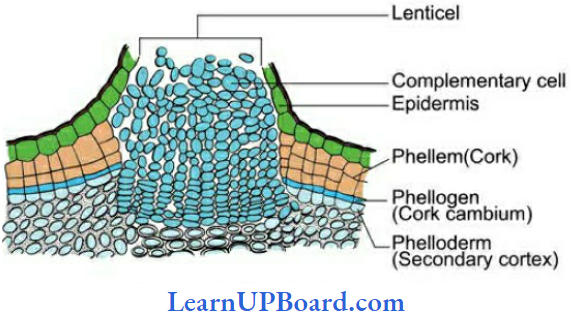
- Due to the pressure of the secondary xylem, the epidermis ruptures arid cortex is largely lost after two or three years of secondary growth. In the cork layer (bark), the lenticels are present which are meant for gaseous exchange.
- In cork, lenticels have loosely arranged cells called complementary cells with intercellular spaces. For bottle corks, the cork is processed in such a manner so that lenticels come in the vertical direction.
Bark includes all the dead and living tissues outside the vascular cambium. Bark may be of two types:
” flower anatomy”
- Scaly Bark: When develops in strips, for example, Eucalyptus, Psidium.
- Ring Bark: When develops in the form of a sheet or ring, for example, Betula (bhojpatra). The outermost layer of bark is dead and called as rhytidome. The bark of Betula was used as a substitute of paper in ancient times to write manuscripts.
Secondary Growth In Dicot Root: Vascular bundles in the dicot root are radial, exarch, and mostly triarch. Vascular cambium is formed secondary) from conjunctive parenchyma cells lying just below each phloem strand. Thus, the number of cambium strips formed equals the number of phloem strands. The cells of the pericycle lying outside the protoxylem also become meristematic to form part of strips of cambium.
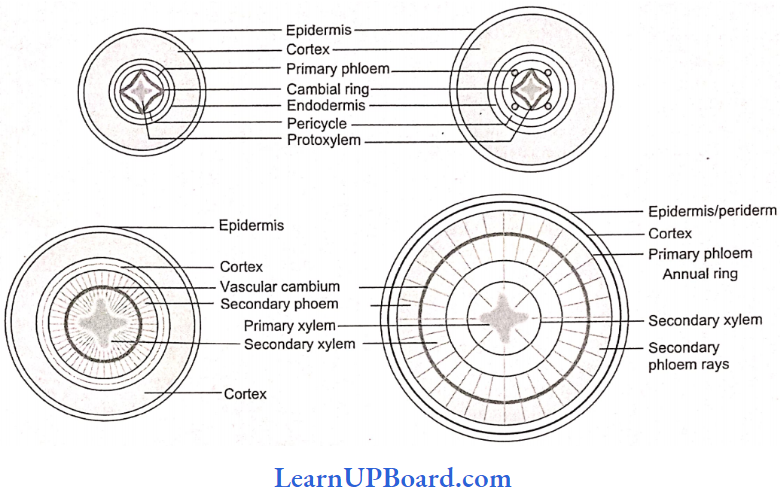
These cambial strips join the first formed cambium strips to form a complete but wavy ring of vascular cambium. This cambium ring produces a secondary xylem on the inner side and a secondary phloem on the outer side.
In roots, the growth rings are not distinct, because there is no seasonal variation under the soil. From the outer layers of the pericycle arises the phellogen which cuts phellem (cork) or the outer side and secondary cortex or phelloderm toward the inner side.
NEET Biology Notes Anatomy Of Flowering Plants Point To Remember
Examples of dicots with scattered vascular bundles are Podophyllum, Peperomia, Piper, and Popover. Examples of cortical vascular bundles are Nyctanthus, Kalanchoe, and Casuarina.
- Examples of medullary bundles are Mivabilis, Bougainvillea, Amaranthus, and Achyranthus. Examples of polystelic conditions are Primula and Dianthera.
- Anomalous or abnormal secondary growth occurs in Bougainvillea, Boerhaavia, Chenopodium, and Aristolchia.
- Some monocots show abnormal secondary growth by meristematic tissue which develops around vascular bundles, for example, Dracaena, Yucca, Agave, etc. Virgin cork is the first formed periderm.
Wound Cork: It is the secondary meristem; formed below the injured cell, it forms a cork on the outer side and a callus below which heals the wound.
- Abnormal secondary growth in dicot root occurs in beetroot (Beta vulgaris) and sweet potato (Ipomoea batatas) by the formation of numerous accessory rings of cambium which cut more storage—parenchyma in secondary phloem and less secondary xylem.
- Homoxylous wood is the wood of vessels dicots, for example, Ranales (Winteraceae, Tetracentraceae Trochodendraceae).
- Heteroxylous wood is the wood of vessel-bearing dicots.
The latex of some plants is of great commercial importance such as
- The source of commercial rubber is the latex of Hevea brasiliensis, Ficus elastica, Ciyptostegia, and Manihot glaziovii.
- The source of chewing or chuckle gum is the latex of Achras sapota.
- The source of the enzyme papain is the latex of Carica papaya.
- The source of alkaloid opium is the latex of Popover somnific (poppy).
Polyderm is a special type of protective tissue that occurs in roots and underground stems of the members of Rosaceae and Myrtaceae. Its outermost layer is dead and suberized.
NEET Biology Notes Anatomy Of Flowering Plants Assertion Reasoning Questions And Answers
In the following questions, an Assertion (A) is followed by a corresponding Reason (R). Mark the correct answer.
- If both Assertion and Reason are true and the Reason is the correct explanation of the Assertion.
- If both Assertion and Reason are true, but the Reason is not the correct explanation of the Assertion.
- If Assertion is true, but Reason is false.
- If both Assertion and Reason are false.
” anatomy of flower “
Question 1. Assertion: In maize stem, endodermis is present between the general cortex and pericycle.
Reasoning: Eustele is present in maize Stem.
Answer: 4. If both Assertion and Reason are false.
Question 2. Assertion: In the Cucurbita stem, vascular bundles are conjoint, bicollateral, and either open or closed.
Reasoning: The outer and inner cambium are present and only the inner cambium is functional in the Cucurbita stem.
Answer: 4. If both Assertion and Reason are false.
Question 3. Assertion: Fusiform cells are elongated and tapering cells.
Reasoning: These cells form an axial system consisting of vascular rays.
Answer: 3. If Assertion is true, but Reason is false.
Question 4. Assertion: Septa less tracheids are absent in Trochodendron.
Reasoning: Heteroxylous wood is present in Trochodendron.
Answer: 3. If Assertion is true, but Reason is false.
Question 5. Assertion: According to Hanstein, there are three halogens in a monocot root.
Reasoning: In monocot roots, the outermost groups of initials form both root cap and dermatogen.
Answer: 4. If both Assertion and Reason are false.
Question 6. Assertion: The apical meristem is always protected.
Reasoning: A root cap is present above the meristem in roots.
Answer: 2. If both Assertion and Reason are true, but the Reason is not the correct explanation of the Assertion.
Question 7. Assertion: The stem in herbaceous plants do not develop cracks during severe wind and is used to bond under these conditions.
Reasoning: Sclerenchyma is peripheral in position and provides flexibility to the herbaceous stems.
Answer: 3. If Assertion is true, but Reason is false.
Question 8. Assertion: The death of a companion cell leads to the death of a sieve cell also.
Reasoning: Both companion and sieve cells are phloem cells.
Answer: 2. If both Assertion and Reason are true, but the Reason is not the correct explanation of the Assertion.
Question 9. Assertion: Dicot roots are mostly tetrach.
Reasoning: There occur four phloem bundles forming rays.
Answer: 4. If both Assertion and Reason are false.
Question 10. Assertion: Heartwood is not involved in the conduction function.
Reasoning: Tyloses and depositions of tannins, resins, and gums is common in duramen cells.
Answer: 1. If both Assertion and Reason are true and the Reason is the correct explanation of the Assertion.
Question 11. Assertion: Vascular cambium appears wavy in dicot roots.
Reasoning: Vascular cambium is formed by conjunctive tissue in dicot roots which is found located inside the xylem and outside phloem strands.
Answer: 3. If Assertion is true, but Reason is false.
Question 12. Assertion: Velamen is hygroscopic in nature and absorbs environmental moisture.
Reasoning: Velamen is common in orchids which are epiphytes.
Answer: 2. If both Assertion and Reason are true, but the Reason is not the correct explanation of the Assertion.