
NEET Biology Notes Body Fluids And Circulation Introduction
A circulatory system consists of a heart (pumping organ), arteries and arterioles, venules (bringing blood back to the heart), and capillaries (connecting arterioles and venules). Circulation is of two types:
- Single circulation: When blood flows through the heart only once during its course of circulation, for example., invertebrates and fish.
- Double circulation: When blood flows through the heart twice during its course of circulation, for example, amphibians, reptiles, birds, and mammals.
In amphibians and reptiles, the left atrium receives oxygenated blood from the gills/lungs/skin, and the right atrium gets deoxygenated blood from other body parts. However, they get mixed up in a single ventricle which pumps out mixed blood (incomplete double circulation).
In birds and mammals, oxygenated and deoxygenated blood received by the left and right atria, respectively, passes onto the ventricles of the same sides. The ventricles pump it out without any mixing up, i.e., the two separate circulatory pathways are present in these organisms; hence, these have complete double circulation.
Depending upon the medium of transportation, the circulatory system is divided into a water vascular system and a blood vascular system.
Water vascular system: Water is the medium of transportation, for example, in sponges (water canal system), in Hydra (gastrovas- cular system), and in starfish (ambulacral system).
Blood vascular system: Blood is the medium of transportation. It is of two types:
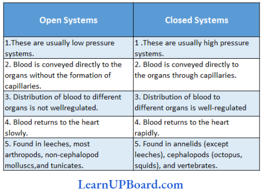
- Open circulatory system: Blood flows through vessels that open into tissue spaces or membrane-lined sinuses, for example, arthropods, non-cephalopod mollusks, and tunicates.
- Closed circulatory system: Blood flows through the heart, vessels, and finely branched capillaries, without coming in direct contact with body tissues or body cavities. It was discovered by William Harvey. A comparison between open and closed circulatory systems is mentioned.
Read and Learn More NEET Biology Notes
Comparison Between Open And Closed Circulatory Systems:
NEET Biology Notes Body Fluids And Circulation Types Of Heart
- On the basis of type of blood:
- Venous heart: Receives deoxygenated blood only. For example, fish (as the heart receives deoxygenated blood from all over the body except gills).
- Arteriovenous heart: Receives deoxygenated blood from the body and oxygenated blood from the lungs/gills. For example, amphibians, reptiles, birds, mammals, etc.
- On the basis of the origin of impulse:
- Myogenic heart: The impulse for a heartbeat originates in heart muscles (pacemakers), for example. chordates and mollusks (octopus).
- Neurogenic heart: The impulse for a heartbeat is brought about by nerves, for example, in most invertebrates.
- Based on structure, the hearts are of the following types:
- Tubular hearts: For example, insects (cockroach: 13-chambered heart)
- Pulsating vessels: Annelids, holothurians, and Amphi- oxus.
- Chambered hearts: Hearts of vertebrates and mollusks.
- Ampullar accessory hearts: Branchial hearts of cephalopods (for example, octopus), insects, heart bulbils of Amphioxus, and lymph hearts of frogs.
body fluids and circulation
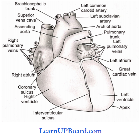
NEET Biology Notes Body Fluids And Circulation Structure Of The Human Heart
The human heart is situated in the pericardial cavity, in mediastinal space. Its covering is called pericardium which is double-walled—outer parietal pericardium and inner visceral pericardium.
- In between these two is the present pericardial cavity, filled with pericardial fluid.
- The wall of the heart is made up of three layers. The outermost is the epicardium, the middle myocardium (consisting of cardiac muscles), and the innermost lining is the endocardium.
- The cardiac muscles are syncytial in functional terms because of intercalated discs. These are function points between two muscle spindles with an electrical resistance of 1/400th of any membrane.
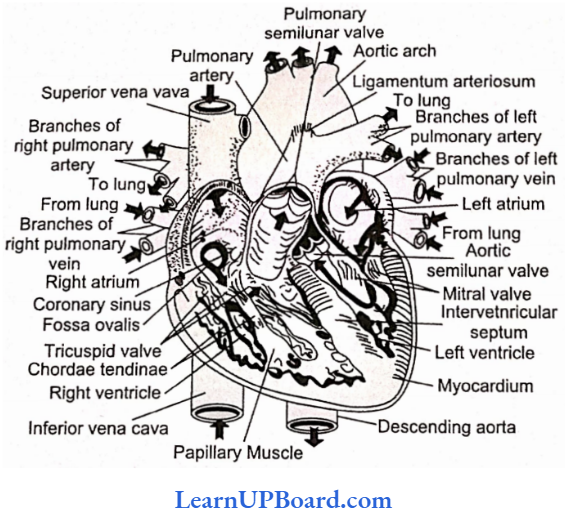
- The human heart is four-chambered.
- Sinus venous and conus arteriosus are not present in the human heart.
- The auricle or atrium is divided by an interatrial septum into right and left auricles.
- On this septum, a depression fossa ovalis is present which is a remnant of embryonic foramen ovale, an aperture present between right and left auricles.
- Three large veins pour blood into the right auricle by separate pores.
- Eustachian valve guards the opening of post caval.
- A coronary or Thebesian valve is present at the opening of the coronary sinus.
- Two pulmonary veins bring oxygenated blood into the left auricle.
- The internal structure of the human heart
- The opening of the pulmonary vein is without any valve as its opening is oblique, which prevents the backflow of blood.
- The ventricle is also divided by an interventricular septum.
- Auricles open into ventricles by separate atrioventricular apertures.
- On the right side is present a tricuspid valve and on the left side is present a bicuspid or mitral valve.
- The cuspid valves are connected below to the walls of ventricles by chordae tendinae which terminate on papillary muscles.
- The right ventricle receives the deoxygenated blood from the right atrium and pumps it into the pulmonary trunk arising from it.
- The pulmonary trunk bifurcates to form right and left pulmonary arteries which supply deoxygenated blood to the lungs of the respective side.
- The left ventricle receives oxygenated blood from the left atrium and sends it into the ascending aorta which puts blood into the coronary arteries and the systemic circulation of the body.
- The origin of the pulmonary trunk and ascending aorta is guarded by a set of three semilunar valves in each.
- This internal structure of the human heart is to prevent the backflow of blood.
- The inner surface of the ventricle has a number of irregular muscular ridges called trabeculae camae or colum- nae camae.
body fluids
- Large protrusions—papillary muscles—are also present. These are inserted at a ventricular wall at one end and continued at the other end with collagenous cords called chordae tendinae.
- These prevent the pushing of flaps into the atrium during ventricular contraction.
- In the right ventricle, a muscular band connects the interventricular septum with the parietal wall of the ventricle. It is called the moderator band which is a component of larger septomarginal trabecula.
- The left ventricle has the thickest muscles because it pumps blood to the whole body.
NEET Biology Notes Body Fluids And Circulation Working Of The Human Heart
The heart shows alternate contraction and dilation of its chambers. Contraction is known as systole, while dilation is called diastole.
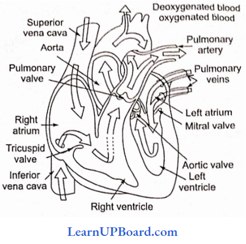
- The heart is said to be in the state of joint diastole when all its four chambers are in a relaxed state. In this state, the atria receive blood. Atrial systole forces the blood from atria into the ventricles; the left ventricle receives oxygenated blood whereas the right ventricle receives deoxygenated blood.
- As the ventricular systole starts, the cuspid valves close, and the blood from the ventricles is forced into the great arteries.
- Oxygenated blood from the left ventricle enters the aortic arch and is carried to all parts of the body except the lungs.
- Deoxygenated blood from the right ventricle reaches the pulmonary arch and is carried to the lungs.
- As the ventricles begin to relax, the semilunar valves close and prevent the return of blood from the two arches.
- The action potential of heart muscles differs from that of skeletal muscles.
- One heartbeat completes in 0.8 s (atrial systole = 0.1 s, atrial diastole = 0.7 s, ventricular systole = 0.3 s, and ventricular diastole = 0.5 s). Thus, there are 72 heartbeats per minute on average when a person is performing normal work.
NEET Biology Notes Body Fluids And Circulation Cardiac Cycle
During a heartbeat, there is contraction and relaxation of auricles and ventricles in a specific sequence. The contraction phase is known as systole, while the relaxation phase is known as diastole. A series of events that occur during a heartbeat is known as the cardiac cycle.
- During the joint diastole phase, the blood flows into the right auricle from the superior vena cava and inferior vena cava.
- The blood also flows from the auricles to their respective ventricles through the atrioventricular valves.
- There is no flow of blood from the ventricles to the aorta and its main arteries as the semilunar valves remain closed in this phase.
- At the end of joint diastole, the auricles contract or they enter into the systolic phase. In this phase, it now forces most of its blood into the ventricles which is still in the diastolic phase.
- During auricular systole, the blood cannot pass back into the superior and inferior vena cava because they are compressed by the atrial contraction. Thus, the atrium acts as a main vessel to collect and pump venous blood into the ventricles. Thus, at the end of atrial systole, the atria get empty.
- After the atrial systole is over, the atrial muscles relax and the blood enters into the atrial diastolic phase.
- During atrial diastole, it again gets filled up with venous blood coming from the superior and inferior vena cava.
- Along with the atrial diastole, the ventricular systole starts. This results in an increased pressure of blood in the ventricle and it rises more than the pressure of blood in the atria. Soon the atrioventricular valves are closed, and thus, the backflow of blood is prevented. This closure of AV valves at the beginning of ventricular systole produces a sound “lub” and is known as the first heart sound. Initially, when the ventricle starts contracting, the pressure of blood within it is lower than the pressure of blood within the aorta and so the semilunar valves do not open. Therefore, the ventricles now contract as closed chambers. As the ventricular systole progresses, the pressure of blood within the ventricles increases more than that of the aorta. As a result, the semilunar valves now open, and blood flows (with a speed) into the aorta and its branches.
- The period between the closure of the AV valve and the opening of the semilunar valve is called isovolumetric systole/contraction.
- The backflow of blood in the atria is prevented as the AV valves remain closed.
- Now at the end of ventricular systole, ventricular diastole starts.
- As the atria are still in diastole, all four chambers are now in diastole. This is known as joint diastole.
- In the ventricular diastolic phase, the pressure of blood in the ventricles falls below the pressure of blood in the aorta. So the semilunar valves get closed to prevent the backflow of blood from the aorta to the ventricles. This closure of semilunar valves at the beginning of ventricular diastole produces a sound “dupp” and is known as the second heart sound.
- After the closure of the semilunar valves, the ventricles become closed chambers again. Also, as the ventricular pressure is more than the atrial pressure, the AV valves remain closed. However, as the ventricular diastole continues, the pressure of blood in the ventricles falls below the pressure of blood in the atria. At this point, the AV valves open and blood starts flowing again from the relaxed atria to the relaxed ventricles.
- The duration between the closure of semilunar valves and the opening of AV valves is called isovolumetric diastole/relaxation.
- When the joint diastole is over, the atrial systole starts and the blood is pumped into the ventricles.
The ventricular filling of the blood can be divided into three phases:
- First rapid filling: With the beginning of ventricular diastole, intraventricular pressure declines, and AV valves open. Due to this, blood already stored in the atria rushes into the ventricles rapidly. This also creates a sound, the third sound of heart.
- Diastasis/slow filling: After the first rapid rushing of blood, the blood keeps on entering the ventricles, though at a slow rate (diastasis).
- Second rapid filling: This occurs with the atrial systole which again causes rapid squeezing of blood into the ventricles. This creates the foolish sound of the heart.
NEET Biology Notes Body Fluids And Circulation Points To Remember
Cardiac output rises during exercise. During severe exercise, it may rise to even 20 L/min, about four to fivefold the normal resting value of about 5 L/min. The rise in the cardiac output helps the body during exercise by enhancing manifold the supply of nutrients and oxygen to the contracting muscles.
- HDLs (high-density lipoproteins): High levels of HDL in the blood may help to reduce our risk of coronary heart disease. HDLs are good lipoproteins. They contain 40-45% proteins, 5-10% triglycerides, 30% phospholipids, and 20% cholesterol. They remove excess cholesterol from body cells and the blood and transport it to the liver for elimination. Because HDLs prevent the accumulation of cholesterol in the blood, a high HDL level is associated with a decreased risk of coronary artery disease.
- LDLs (low-density lipoproteins): LDLs contain 25% protein, 5% triglycerides, 20% phospholipids, and 50% cholesterol. When present in excessive numbers, LDLs deposit cholesterol in and around smooth muscle fibers in arteries, forming “fatty plaques” that increase the risk of coronary artery disease. For this reason, the cholesterol in LDL is known as “bad” cholesterol.
” body fluid definition”
NEET Biology Notes Body Fluids And Circulation Heartbeat
Physiological Properties of Cardiac (Heart) Muscle
- Excitability and contractility.
- It obeys the all-or-none principle.
- It has a longer refractory period, therefore it never develops fatigue.
- It does not show summation or tetanus.
- It shows conductivity and rhythmicity.
Origin Of Heartbeat
- The mammalian heart is a myogenic heart, i.e., the heartbeat originates from the muscles. However, it is regulated by nerves.
- In the right atrium, near the region where the superior vena cava opens, a specialized group of muscle fibers called sinus-auricular node (SA-node) is present from where the heartbeat originates.
- It is also called a pacemaker and is richly supplied with blood capillaries. A wave of contraction (systole) originates from it and spreads over to the whole heart.
Conduction Of Heartbeat
- At the junction of the right atrium and right ventricle, a tissue called auriculo-ventricular node (AV-node) is present that picks up the wave of contraction propagated by the SA-node.
This is also known as the bundle of His. - Its branches spread over the ventricles forming the Purkinje system.
- The wave of contraction spreads over the ventricle through the AV node and its Purkinje system.
Regulation Of Heartbeat
- The normal activities of the heart are regulated intrinsically, i.e., auto-regulated by specialized muscles (nodal tissue), hence the heart is called myogenic.
- A special neural center in the medulla oblongata can moderate cardiac function through the autonomic nervous system (ANS).
- Neural signals through the sympathetic nerves (part of ANS) can increase the rate of heartbeat, the strength of ventricular contraction, and thereby the cardiac output.
- On the other hand, parasympathetic neural signals (another component of ANS) decrease the rate of heartbeat, speed of conduction of action potential, and thereby the cardiac output. This happens because these nerves release chemicals (hormones) when stimulated. Adrenal medullary hormones can also increase the cardiac output.
- High levels of potassium and sodium ions decrease heart rate and strength of contraction.
- An excess of calcium ions increases heart rate.
- Increased body temperature during fever increases heart rate.
- Strong emotions such as fear, anger, and anxiety increase heart rate, resulting in increased blood pressure.
- Mental states such as depression and grief decrease heart rate.
- The heartbeat is somewhat faster in females.
- The heartbeat is fastest at birth, moderately fast in youth, average in adulthood, and above average in old age.
Heart Sounds: Heart sounds are caused due to the sudden closure of the heart valves. There are mainly two sounds:
First sound: Occurs at the onset of ventricular systole and is caused due to the sudden closure of AV valves and the ejection of blood from the ventricles. It is dull and pronounced as L-U-B.
Second sound: Occurs at the onset of ventricular diastole and is caused by the sudden closure of the semilunar va ves o e aorta and pulmonary artery- It is short and sharp like the word
D-U-B.
The sequence of both these sounds is like first sound → second sound → pause → first sound → second sound → pause and so on. If damage occurs, as in rheumatic fever, blood may leak out through the valves, and a characteristic sound murmur is produced.
Pulse Rate: The blood is pumped from the ventricles of the heart into the aorta to be distributed to all the body parts. This happens during the ventricular systole and is repeated every 0.8 seconds.
- The blood from the aorta then goes to other arteries of the body. This causes a rhythmic contraction of the aorta and its main arteries and is felt as regular jerks or pulse in them.
- It can be felt in the regions where arteries are present superficially such as the wrist, neck, and temples.
- The pulse rate is, therefore, the same as that of the heartbeat rate.
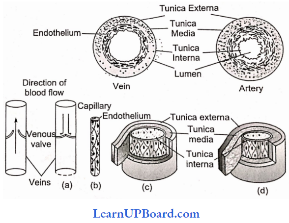
NEET Biology Notes Body Fluids And Circulation Blood Vessels And Course Of Blood CircuLation
Arterioles: Arterioles are small arteries that deliver blood to capillaries. Arterioles also have smooth muscles on their walls.
Contraction and relaxation of these muscles alter the diameter of arterioles and thereby, respectively, reduce and increase the blood flow through them.
Capillaries: Capillaries were discovered by Malpighi in 1661.
- Capillaries are the smallest blood vessels in the body.
- A capillary has no muscular wall.
- Its wall is made of a single layer of flat endothelial cells and is consequently permeable to water and small solutes, but not to proteins and other macromolecules.
- The diameter of the capillary lumen is from 7.5 pm to 75 pm. Only about 5-7% of the total blood volume is contained in the capillaries.
Venules: Venules are small vessels that continue from capillaries and merge to form veins. They drain blood from capillaries into veins.
Veins: Veins have less elastic tissues and smooth muscles than arteries.
- One major difference between an artery and a vein is that a vein has a thin muscular wall.
- Veins contain valves to prevent the backflow of blood.
- Valves are necessary in veins but not in arteries because the pressure in veins is too low to push the blood.
- Weak valves can lead to varicose veins or hemorrhoids.
- All veins carry deoxygenated blood except pulmonary veins.
- Pulmonary veins carry oxygenated blood from the lungs back to the heart.
- Blood vessels that carry blood from the lungs to the heart are called pulmonary veins.
- The wall of veins is collapsible (non-collapsible in arteries).
- The lumen of veins is wider and narrower than the arteries.
” circulatory pathway class 11″
- Vasa vasorum: A network of small blood vessels that supply blood to large blood vessels.
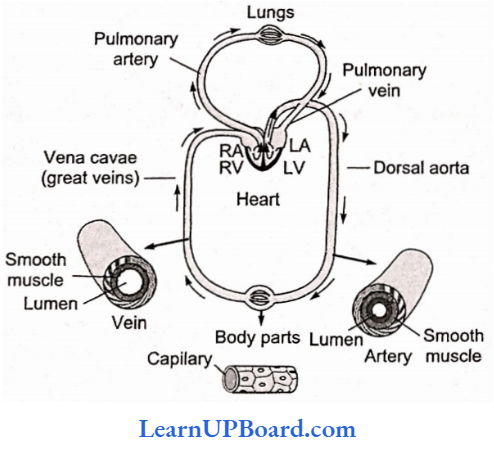
The course of Blood Circulation: There are two types of blood circulation pathways in the human body:
Pulmonary circulation: The circulation starts with the pumping of deoxygenated blood by the right ventricle which is carried to the lungs where it is oxygenated and returned to the left atrium.
Systemic circulation: In systemic circulation, the oxygenated blood is pumped by the left ventricle to the aorta. The oxygenated blood is carried to all the body tissues and the deoxygenated blood from there is collected by the veins and returned to the right atrium.
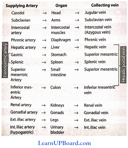
Main Arteries and Veins in the Human Body
Our human body has a network of different types of arteries and veins running across all organs, providing nourishment to them through oxygenated blood, and bringing back the deoxygenated blood to the heart. Table 18.2 describes all sorts of arteries and veins present in the human body.
Main Arteries And Veins Present In the Human Body:
Coronary Circulation: Two coronary arteries—right and left—branch from the ascending aorta. The left coronary artery passes inferiorly to the left auricle and divides into the anterior
interventricular and circumflex branches.
- The anterior interventricular branch or left anterior descending (LAD) artery is in the anterior interventricular sulcus and supplies oxygenated blood to the walls of both ventricles and the interventricular septum.
- The circumflex branch lies in the coronary sulcus and distributes oxygenated blood to the walls of the left ventricle and left atrium.
- The right coronary artery supplies small branches (atrial branches) to the right atrium.
- It continues inferiorly to the right auricle and divides into the posterior interventricular and marginal branches.
- The posterior interventricular branch follows the posterior interventricular sulcus and supplies the walls of the two ventricles and the interventricular septum with oxygenated blood.
- The marginal branch in the coronary sulcus transports oxygenated blood to the myocardium of the right ventricle.
- Most parts of the body receive branches from more than one artery, and where two or more arteries supply the same region, they usually connect.
The connections called anastomoses provide alternate routes for blood to reach a particular organ or tissue. - The myocardium contains many anastomoses, connecting branches of one coronary artery or extending between branches of different coronary arteries.
- In a resting person, heart muscles can remain alive if they receive as little as 10-15% of their normal blood supply, but the person may have little ability to engage in activities.
Coronary Veins: As blood passes through the coronary circulation, it delivers oxygen and nutrients and collects carbon dioxide and wastes.
- It then drains into a large vascular sinus on the posterior surface of the heart called the coronary sinus, which empties into the right atrium.
- A vascular sinus is a venous space with a thin wall that has no smooth muscle to alter its diameter.
- The principal tributaries carrying blood into the coronary sinus are the great cardiac vein, which drains the anterior aspect of the heart, and the middle cardiac vein, which drains the posterior aspect of the heart.
NEET Biology Notes Body Fluids And Circulation Blood Pressure
If a person is persistent above 140 mm Hg systolic and above 90 mm Hg diastolic blood pressure, he/she is said to have high blood pressure or hypertension.
- There are a number of factors responsible for hypertension.
- Over-eating and obesity are the most important factors for hypertension.
- Physical and emotional stresses such as fear, worry, anxiety, sorrow, etc., also cause hypertension.
- People living in big cities usually suffer from hypertension. Smoking is also a cause.
- Physical and mental rest are essential for a patient suffering from hypertension.
- Sometimes, cholesterol starts depositing on the walls of blood vessels when there are high levels of cholesterol in the blood. This causes the arteries to lose their elasticity and they become stiff. This is known as arteriosclerosis or hardening of arteries and as a result, the blood pressure rises. This is one of the main causes of heart attack.
- Frank-Starling law: Two great physiologists Frank and Starling said that the greater the heart muscle is stretched during the filling phase, the greater will be the quantity of blood pumped into the aorta.
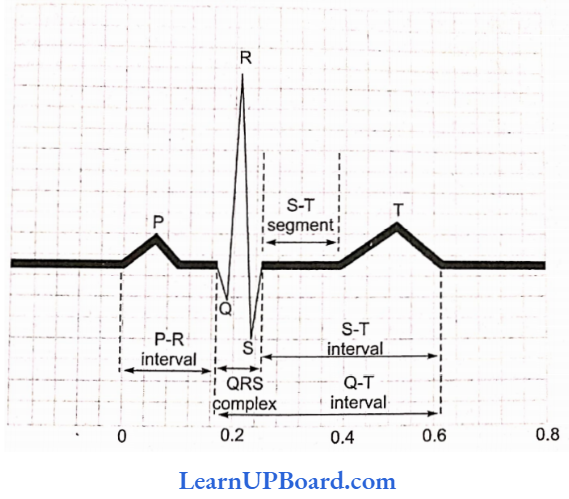
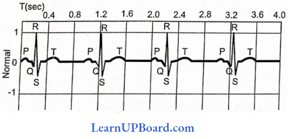
Electrocardiogram (ECG): Electric changes in the cardiac chambers follow a specific sequence. These changes can be recorded with the help of an apparatus—electrocardiograph.
- The record is called ECG.
- It is represented as PQRST, where P stands for depolarization of atria, QRS depolarization of ventricles, and T repolarization of ventricles.
- Defects in cardiac function or structure are recorded in the ECG.
- For the purpose of recording, metal electrodes or leads are attached to each arm and leg with the help of straps after cleaning and putting a special jelly, which improves electrical conduction.
- An additional electrode is placed on the chest with the help of a rubber suction cup.
- Then the electrocardiograph is switched on.
- The electrical current of the heart is detected and amplified by the machine and is transmitted to
the recording pen that draws a wavy line, called the deflection waves (electrocardiogram). - A normal electrocardiogram is composed of a P wave, a QRS complex, and a T wave.
- The QRS complex has three separate Q, R, and S waves.
- The P wave is a small upward wave that indicates the depolarization of the atria or the spread of impulse from the sinus node throughout the atria.
- The second wave, i.e., the QRS complex, begins after a fraction of a second of the P wave.
- It begins as a small downward deflection (Q) and continues as a large upright (R) and triangular wave, ending as a downward wave (S) at the base. This is the expression of the ventricular depolarization.
- The potential generated by the recovery of the ventricle from the depolarization state is called the repolarization wave.
- In electrocardiography, the PQ interval (also called PR interval) is the time taken by the impulse to travel through the atria, AV node, and the rest of the conducting tissues.
- During rheumatic fever and in arteriosclerotic heart disease (i.e., the formation of plaques and calcification), the PQ interval lengthens. This is due to the inflammation of the atria and atrioventricular node.
- The normal PR interval lasts for 0.16s.
- The enlarged Q and R waves are an indication of myocardial infarction.
- The ST interval is the representation of time between the end of the spread of impulse through ventricles and its repolarization.
- The ST segment is elevated in acute myocardial infarction and depressed in a condition when the heart muscles receive insufficient oxygen.
- The ventricular repolarization is represented as a T wave.
- When the heart muscles receive insufficient oxygen, then the T wave is flattened.
NEET Biology Notes Body Fluids And Circulation Pacemaker
During the pumping action of the heart, the atria and ventricles contract rhythmically. The impulse of this wave of contraction begins every time from the SA node (sinus node) present in the right atrium. Thus, it can be said that the SA node controls the heartbeat, and hence, it is the natural pacemaker of the heart.
- The pacemaker is the rhythmic center, which establishes a pace of activity.
- Sometimes, a component of the impulse conduction system is disrupted, causing irregularity in the heart rhythm, such as failure to receive the atrial impulse by the ventricle or completely independent contraction of the atria and ventricles. Such types of patients are provided with an artificial electronic device, which regularly sends a small amount of electrical charge to maintain the rhythmicity of the heart. This device is known as an artificial pacemaker, which is implanted subcutaneously in the upper thoracic region having a connection with the heart.
- In patients having the symptoms of ventricular escape (Stokes, Adams syndrome), in which the atrial impulse suddenly fails to be transmitted to the ventricle, which may last for a few seconds to a few hours even, the artificial pacemaker is connected to the right ventricle for controlling its rhythm.
- The artificial pacemaker consists of a pulse-generator containing a cell (solid state lithium cell) to produce electrical impulse, the lead in the form of a wire, which transmits the impulse, and an electrode, which is connected to the portion of the heart where the impulse is to be transmitted.
NEET Biology Notes Body Fluids And Circulation Portal Systems
Hepatic portal system: Inferior mesenteric, superior mesenteric, duodenal, and lienogastric veins join to form a hepatic portal vein. It pours blood from the digestive system into the liver. This blood is collected by hepatic veins and poured into the canal to be returned to the heart.
- Renal portal system: In fishes and amphibians, the renal portal system is also found which is reduced in reptiles and birds and is absent in mammals.
- Hypophyseal portal system: A hypophyseal portal vein collects blood from the hypothalamus and enters the anterior lobe of the pituitary.
NEET Biology Notes Body Fluids And Circulation Lymphatic System
The lymphatic system comprises lymph, lymphatic capillaries, lymphatic vessels, lymphatic ducts, and lymphatic nodes.
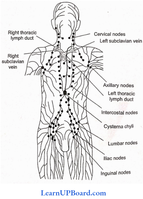
- Lymphatic capillaries: They lie close to the blood capillaries but end blindly. They have extremely thin walls. They are composed of a single layer of endothelial cells.
- Lymphatic vessels: The lymphatic capillaries unite to form larger lymphatic vessels. They are composed of an outer coat of fibrous tissue, a middle coat of muscular tissue, and an inner lining of endothelial cells. The lymphatic vessels have numerous valves. The lymph vessels of intestinal regions absorb the digested fats. They are milky in appearance and are called lacteals.
- Thoracic duct: The lymphatic vessel of the left side begins at the cisterna chyli, present at the level of (anterior to) the first and second lumbar vertebrae. It discharges its lymph into the left subclavian vein.
- Right lymphatic duct: The lymphatic vessels of the right side of the thorax, head, and neck unite to form the right lymphatic duct. It discharges its lymph into the right subclavian vein.
- Lymphatic nodes: The lymphatic vessels bear lymph nodes at intervals and are abundant in the neck, armpit, and groin. The lymph is filtered through lymph nodes which contain phagocytic white blood corpuscles and macrophages which eat harmful microorganisms and foreign particles from the lymph. Lymph nodes also add lymphocytes and antibodies.
- Lymph movement: The lymph flows slowly and moves from lymphatic vessels to lymphatic ducts to the venous system. Blocking of lymph flow causes edema.
- Lymphoid organs: The organs which secrete lymph are called lymphoid organs. Besides the lymph nodes, tonsils, thymus gland, Peyer’s patches, liver, and spleen are the other lymphoid organs that secrete lymph.
Functions of Lymph: Lymph acts as a “middle man” that transports various proteins, hormones, etc., to the body cells brings carbon dioxide and other metabolic wastes from the body cells, and finally pours the same into the venous system.
- Lymph nodes produce lymphocytes. Lymph takes lymphocytes and antibodies from the lymph nodes to the blood.
- It absorbs and transports fat and fat-soluble vitamins from the intestine. Lymph capillaries present in the intestinal villi are called lacteals which are associated with the absorption and transportation of fat and fat-soluble vitamins.
- It brings plasma protein macromolecules synthesized in the liver cells and hormones produced, in the endocrine glands to the blood. These molecules cannot pass into the narrow blood capillaries but can diffuse into the lymphatic capillaries.
- The lymph maintains the blood volume. As soon as the blood volume reduces in the blood vascular system, the lymph rushes from the lymphatic system to the blood vascular system.
NEET Biology Notes Body Fluids And Circulation Diseases Of Heart
High Blood Pressure (Hypertension): Hypertension is the term for blood pressure that is higher than normal (120/80).
- In this measurement, 120 mm Hg (millimeters of mercury pressure) is the systolic or pumping pressure and 80 mm Hg is the diastolic or resting pressure.
- If the repeated checks of blood pressure of an individual are about 140/90 (140 over 90) or higher, it shows hypertension.
- High blood pressure leads to heart diseases and also affects vital organs such as the brain and kidneys.
Coronary Artery Disease: Coronary artery disease (CAD), often referred to as atherosclerosis, affects the vessels that supply blood to the heart muscle,
- It is caused by the deposits of calcium, fat, cholesterol, and fibrous tissues, which make the lumen of arteries narrower.
Angina: Angina is also called angina pectoris.
- A symptom of acute chest pain appears when enough oxygen does not reach the heart muscles.
- Angina can occur in men and women of any age, but it is more common among the middle-aged and elderly.
- It occurs due to conditions that affect the blood flow.
Heart Failure: Heart failure means the state of the heart when it is not pumping blood effectively enough to meet the needs of the body.
- It is sometimes called congestive heart failure because congestion of the lungs is one of the main symptoms of this disease,
- Heart failure is not the same as cardiac arrest (when the heart stops beating) or a heart attack (when the heart muscle is suddenly damaged by an inadequate blood supply).
NEET Biology Notes Body Fluids And Circulation Points To Remember
- The lowest level of glucose is in the hepatic vein.
- The highest levels of amino acids are present in the hepatic vein.
- The highest level of urea is in the hepatic vein and the lowest in the renal vein.
- The largest vein in the human body is the inferior vena cava.
- The largest artery in the human body is the aorta.
- The smallest blood vessel in the body is the blood capillary.
- A giraffe’s blood pressure may be the highest of all animals because it has to pump blood to the head through a long neck.
- One species of Antarctic fish is the only fish known to have white blood. It has no red pigment in its blood.
- Frog has two pairs of lymph hearts to pump the lymph back into veins.
- Thrombopenia: Decrease in blood platelet count.
- Erythropoietin: Hormone secreted by the juxta-glo- merular cells of the kidneys.
- Circulatory shunts in the fetus: The fetus bypasses the pulmonary system through two shunts—foramen ovale (opening in the interatrial septum) and ductus arteriosus (the connection between dorsal aorta and pulmonary arch). Shunts are sealed after birth.
- Lungfish have three-chambered hearts—two auricles and one ventricle.
- Crocodiles, alligators, and gavialis have four-chambered hearts—two auricles and two ventricles.
- The heart of a fish is called a venous heart because it receives and pumps deoxygenated blood.
- In the human heart, auricles are called atria (singular atrium).
- Nereis and Amphioxus do not have hearts. The heart of the prawn contains oxygenated blood.
- There is single blood circulation in fish hearts. Hearts of amphibians, reptiles, birds, and mammals have double blood circulation. Foramen of Panizzae is
present in between two systemic arches (they arise from the heart) of the heart of lizards and crocodiles. - An average human heart is about 12 cm. The average weight of a male human heart is 300 g, and that of a . female heart is 250 g. The opening of the coronary sinus into the right atrium is guarded by the Thebesian valve. The opening of the inferior vena cava into the fight atrium has an Eustachian valve. Fossa
this is a depression on the interatrial septum. - Coronary angiography: When the contrast medium dye is injected into coronary arteries (arteries of the heart) and pictures are taken, it is known as coronary angiography.
ess calcium ions cause an increased heartbeat. - RBCs fail to mature if there is a deficiency of vitamin Bj2 and folic acid.
- Papillary muscles are found in the heart of mammals.
- Keber’s organs or pericardial glands discharge excretory products into the pericardial cavity in the freshwater mussel.
- “Blue baby” is the name given to an abnormal human baby who has a hole in the auricular or ventricular septum and hence the oxygenated and deoxygenated blood gets mixed in the heart.
- An insect larva has red blood. The larva of the genus Chi-ronomusis is called a “blood worm.”
- The red color of his larva is due to hemoglobin, which has the power to attract and store oxygen and give it off to the tissues as they require it. Such larvae are able to live in burrows constructed by them in the mud.
- Vasa vasorum are small blood vessels that supply blood to the larger blood vessels.
- A blue whale has the largest heart.
- Cardiomegaly is heart enlargement.
- Angiology: The study of blood vascular and lymphatic systems.
- Venoms of bees and cobra contain Jecithinase which when injected into the bloodstream by sting or bite breaks down lecithins and produces iysolechhins which in turn cause rupturing of the RBC cell membrane (cell lysis).
- Marey’s law: Heart rate is inversely related to systemic blood pressure.
NEET Biology Notes Body Fluids And Circulation Assertion-Reasoning Questions
In the following questions, an Assertion (A) is followed by a corresponding Reason (R). Mark the correct answer.
- If both Assertion and Reason are true and the Reason is the correct explanation of the Assertion.
- If both Assertion and Reason are true, but the Reason is not the correct explanation of the Assertion.
- If Assertion is true, but Reason is false.
- If both Assertion and Reason are false.
Question 1. Assertion: The cardiac impulse that originates from the SA node in the mammalian heart cannot spread directly from the atria to the ventricles.
Reasoning: In the mammalian heart, there is no continuity between cardiac muscle fibers of atria and those of ventricles except AV bundles.
Answer: 1. If both Assertion and Reason are true and the Reason is the correct explanation of the Assertion.
Question 2. Assertion: The first phase of ventricular filling is rapid and causes the third sound of the heart.
Reasoning: It is because of auricular systole.
Answer: 3. If Assertion is true, but Reason is false.
Question 3. Assertion: “Dub” is a long and sharp sound.
Reasoning: It is caused by the closing of AV valves.
Answer: 4. If both Assertion and Reason are false.
Question 4. Assertion: The portal system consists of veins that start from capillaries and end into capillaries.
Reasoning: All vertebrates have a hepatic portal system.
Answer: 2. If both Assertion and Reason are true, but the Reason is not the correct explanation of the Assertion.
Question 5. Assertion: Arterioles possess smooth muscles on their walls.
Reasoning: These smooth muscles help in regulating blood volume flowing through a tissue or organ.
Answer: 1. If both Assertion and Reason are true and the Reason is the correct explanation of the Assertion.
Question 6. Assertion: The open circulatory system is more efficient than the closed circulatory system.
Reasoning: The blood flows far more rapidly in the open circulatory system than in the closed one.
Answer: 4. If both Assertion and Reason are false.
Question 7. Assertion: The heart of a fish contains only deoxygenated blood.
Reasoning: Oxygenated blood does not return back to the heart in fishes.
Answer: 1. If both Assertion and Reason are true and the Reason is the correct explanation of the Assertion.
Question 8. Assertion: The cardiac impulse is said to be myogenic.
Reasoning: The rate of formation and conduction of cardiac impulses can be changed by the action of nerves.
Answer: 2. If both Assertion and Reason are true, but the Reason is not the correct explanation of the Assertion.
Question 9. Assertion: The left ventricle of the heart has a thinner wall than that of the right ventricle.
Reasoning: The left ventricle needs to pump blood to nearby lungs only,
Answer: 4. If both Assertion and Reason are false.
Question 10. Assertion: The AV bundle is essential for the conduction of cardiac impulses.
Reasoning: There is no continuity between the cardiac muscle fibers of the auricles and those of the ventricles.
Answer: 1. If both Assertion and Reason are true and the Reason is the correct explanation of the Assertion.
Question 11. Assertion: The AV node is also called the pacemaker of the heart.
Reasoning: It is because of the fact that the AV node determines the rate of heartbeat.
Answer: 4. If both Assertion and Reason are false.
Question 12. Assertion: There is no mixing of oxygenated and deoxygenated blood in the human heart.
Reasoning: Valves are present in the heart, which allow the movement of blood in one direction only.
Answer: 2. If both Assertion and Reason are true, but the Reason is not the correct explanation of the Assertion.
Question 13. Assertion: Hypotension is observed in arteriosclerotic patients.
Reasoning: In the condition of arteriosclerosis, arteries gain their elasticity and get stiffened.
Answer: 2. If both Assertion and Reason are true, but the Reason is not the correct explanation of the Assertion.
Question 14. Assertion: ECG is of immense diagnostic value in cardiac diseases.
Reasoning: Defects in cardiac functions can be reflected in changes in the pattern of electrical
potential recorded in the ECG.
Answer: 4. If both Assertion and Reason are false.
Question 15. Assertion: An artificial pacemaker can replace the SA node of the heart.
Reasoning: This is because an artificial pacemaker is capable of stimulating the heart electrically to maintain its beats.
Answer: 1. If both Assertion and Reason are true and the Reason is the correct explanation of the Assertion.