
NEET Biology Notes Cell The Unit Of Life Introduction
Cell Structure And Functions Introduction: The cell is the fundamental unit of structure and function of all life. It is a unit of biological activity delimited by a differentially permeable membrane and is capable of self-reproduction.
NEET Biology Notes Cell The Unit Of Life What Is Cell
The cell is the structural and functional unit of a living organism. The widely accepted definition of the cell was given by Lowey and Sikewitz.
- Cell is the unit of biological activity container and nucleus and is able to divide in a medium free from other living organisms.
- Cytology is the study of the state of different components of a cell.
- Cell biology is the study of the structure, function, and reproduction of cells.
- The cell was first observed by Marcello Malpighi and he called it saccade.
NEET Biology Notes Cell The Unit Of Life Historical Details
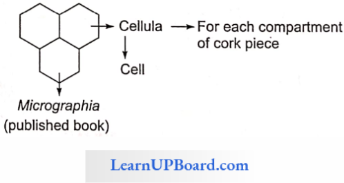
The father of cytology is Robert Hooke and the father of cell biology is Swanson. The term “cell” was first coined by Robert Hooke in his book Micrographia. Robert Hook observed cell wall in the dead cork cell. The first living cell was observed by Leeuwenhoek.
- Alfonso Corti first observed the living substance of a cell.
- This living substance was called protoplasm by Purkinje in animal cell and by Von Mohl in plant cell.
- Hammerling was the first to call the nucleus as the brain or master or controlling center of the cell by using a grafting experiment with Acetabiilarici (unicellular marine green algae) which is the largest unicellular organism among the plants.
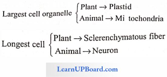
- The largest cell in animals is the egg of an ostrich.
- The largest cellular component is the nucleus.
- The smallest cell organelle is ribosomes.
- The term cytoplasm was coined by Strassburgcr.
- The term hyaloplasm was coined by Preffer.
” cell unit of life”
NEET Biology Notes Cell The Unit Of Life Cell Theory
Cell theory was proposed by Schleiden (botanist) and Schwan (zoologist) in 1839. According to the cell theory, All living organisms are composed of cells.
- The cell is the unit of structure and function of life form.
- All cells are basically similar in chemical composition and metabolism.
- A cell is bound by a membrane and contains a nucleus.
- The activity of a multicellular organism is the result of the interaction and activities of its component cells.
Read and Learn More NEET Biology Notes
Cell Lineage Theory or Common Ancestry Theory
- Proposed by Rudolf Virchow.
- Omnis cellula e cellulcr. All cells originate from pre-existing cells.
- Virchow and Nageli suggested the importance of the reproduction of cells and suggested that it is responsible for the continuity of life.
Exception of Cell Theory: Viruses/viroids do not follow cell theory but mycoplasma, bacteria, Vaucheria, Mucor, and Rhizopus also do not follow cell theory in certain points.
NEET Biology Notes Cell The Unit Of Life Overview Of Cell
You have earlier observed cells in an onion peel and/or human cheek cells under the microscope. Let us recollect their structure. The onion cell which is a typical plant cell has a distinct cell wall as its outer boundary and just within it is the cell membrane.
- The cells of the human cheek have an outer membrane as the delimiting structure of the cell. Inside each cell is a dense membrane-bound structure called the nucleus. This nucleus contains chromosomes which in turn contain the genetic material, DNA.
- Cells that have membrane-bound nuclei are called eukaryotic whereas cells that lack a membrane-bound nucleus are called prokaryotic. In both prokaryotic and eukaryotic cells, a semi-fluid matrix called cytoplasm occupies the volume of the cell.
- The cytoplasm is the main arena of cellular activities in both plant and animal cells. Various chemical reactions occur in it to keep the cell in the “living state.”
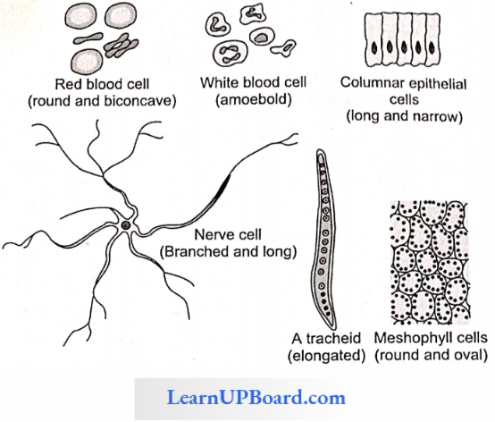
NEET Biology Notes Cell The Unit Of Life Cell Shape And Size
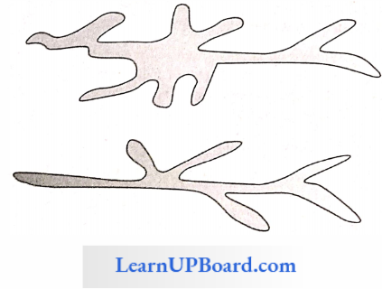
Depicts various forms of cell shapes and sizes.
Type Of Cells
- Based On Differentiation
- Undifferentiated Cell: Cells having dividing abilities, i.e., meristem in plants and stem cells in animals.
- Differentiated Cell: Post-mitotic cell formed by division of undifferentiated cells. For example, parenchyma, collenchyma, tracheids, vessels, etc.
- Dedifferentiated Cell: If a differentiated cell reverts back into an undifferentiated cell, it is known as a dedifferentiated cell. For example, the formation of cork cambium and cambium in dicot root.
- Re-differentiated Cell: A cell formed by the division of a differentiated cell, for example, the formation of a secondary cortex and cork, is called a re-differentiated cell.
- Based On Structure
- Prokaryotic Cell (Daugherty): Nucleus not well organized and membrane-bound cell organelles absent. For example, monerans.
- Eukaryotic Cell (Daugherty): Nucleus well organized and membrane-bound cell organelles present.
- Mesokaryotic Cell (Dodge): The Nucleus well organized, but DNA lacks histone protein.
NEET Biology Notes Cell The Unit Of Life Prokaryotic Cell (Eubacteria)
Historical Detail: Antony Von Leeuwenhoek discovered bacteria from stored rain water and teeth scum and called them wild animalcules. He is known as the discoverer of the microbial world or the wonder world of microbes. He used the term “Dicrkens.”
” cell biology notes”
- Ehrenberg gave the term “bacteria.”
- Nageli placed bacteria in Schizomycetes, called “fission fungi.”
- Louis Pasteur proposed the germ theory of disease. He discovered that bacteria is the causing agent of chicken cholera.
- Pasteur invented antirabies vaccine. He gave the term “microorganism.” He is known to be the father of modem microbiology and sterilization techniques.
- Robert Koch gave “Koch’s Postulates.”
- Joseph Lister developed the technique of aseptic culture.
- D. A. Bergey gave a classification of bacteria in the Manual of Determinative Bacteriology.
- Se’dillot used the term “microbe” for animalcules.
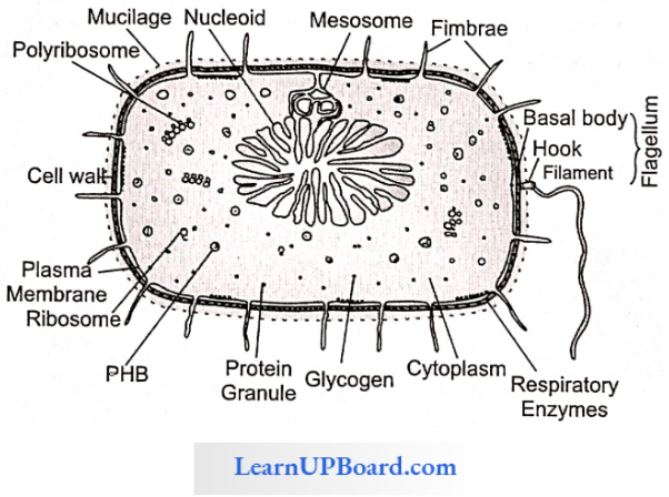
Habitat: Eubacteria are cosmopolitan in distribution. They are present in water, soil, air, and in plant and animal bodies.
Habitat Size: Bacteria range from 0.1-1.5 μm in diameter and 2-10 μm in length. The smallest bacteria is rod-shaped Dialister pneumosintes (0.15-0.3 μm long) present in the nasopharynx of man during the early stage of influenza.
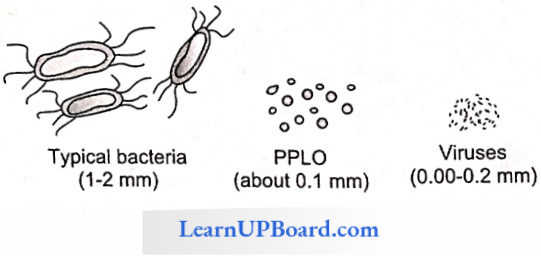
Spirillum Laidlaw, Epulopsciumfishelsoni (600 μm x 80 μm), and Thiomargarita ramibiensis (750 μm) are among the largest unicellular bacteria. The filamentous bacterium Beggiatoa mirabilis is the largest bacterium (16-15 μm diameter and up to several centimeters long).
Habitat Shape: Cohn classified bacteria into four types based on their shapes
- Coccus (pl. cocci): Spherical or nearly sphericaL small, and always non-flagellated.
- Micrococcus: Occurs singly, for example, Micrococcus lutens. M. roseus.
- Diplococci: Found in pairs, for example, Diplococcus pneumonia.
- Streptococci: Cells remain attached to form a chain. For example, Streptococcus lactis, Leptotricha buccalis.
- Staphylococci: Irregular bundles of cells or grape-like clusters, for example, Staphylococcus aureus.
- Sarcina: Three-dimensional geometrical figures like cubes, for example, Sarcina.
- Bacillus (pl. bacilli): Rod-shaped or cigarette-like with rounded or blunt ends. Most common shape. Motile or non-motile. They may occur as:
- Monobacillus: Single bacillus
- Diplobacillus: Occurs in a group of two bacilli
- Streptobacilli: Found in a chain, for example, Streptobacillus. When the cells form a chain and have a much larger area of contact with each other, these are said to have formed trichomes.
- For example, Beggiatoa. If the cells are lined side by side like matchsticks and at angles to one another, the arrangement is said to be palisade-like.
- For example, Corynebacterium diphtheria. In many bacteria (for example, Streptomyces), cells are arranged to form unicellular long, branched filaments called hyphae.
- Vibrio (Singular Vibrion): Bacteria with less than one complete twist or turn. These resemble a comma (,) in appearance, for example, Vibrio cholerae.
- Spirilla (Singular Spirillum): Coiled forms of bacteria exhibiting twists with one or more turns giving a spiral appearance, for example, Spirillum minus.
Other Uncommon Shapes
- Stalked Bacterium: For example, Caulobacter.
- Budding Bacterium: For example, Rhodomicrobium.
- Pleomorphic: Occurs in more than one form, for example, Rhizobium, Coiynebacteriurm, Azotobacter, and Mycobacterium.
Flagella Structure: Instead of the “9 + 2” arrangement of tubulin-containing microtubules, there is simply a single filament of a globular protein called flagellin.
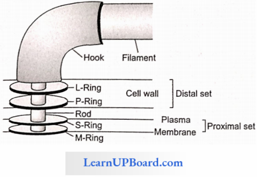
Parts Of A Flagellum
Flagellum Basal Body: It is the most complex portion of the flagellum and has four rings (L, P, S, and M) in gram-negative bacteria and two rings (S and M) in gram-positive bacteria.
Flagellum Hook: It is made up of different protein subunits.
Flagellum Filament: The longest and the most obvious portion of the flagellum. The protein molecules are arranged in a spiral manner. The filament is 20 nm wide and 1-70 nm long and consists of eight vertical rows of flagellin.
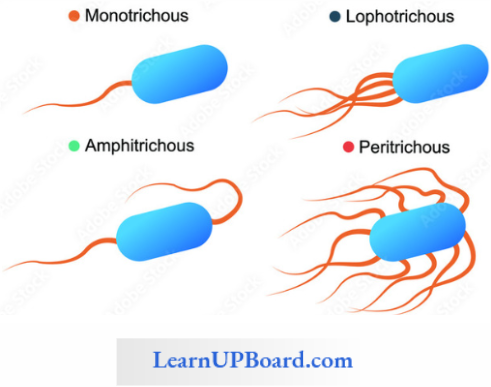
Flagellar Arrangement
- Atrichous: Flagella absent, for example, Lactobacillus, and Pasteurella.
- Monotrichous: Bacteria with single flagella, for example, Vibrio, Thiobacillus.
- Cephalotrichous: Bacteria with many flagella attached at one end, for example, Pseudomonasfluoresce.
- Lophotrichous: Bacteria with many flagella attached at both ends, for example, Spirillum volitions.
- Amphitrichous: Bacteria with a single flagellum at each end, for example, Nitrosomonas
- Peritrichous: Bacteria with flagella all over the body, for example. E, coil, Clostridium tetani.
“cell the unit of life structure and function “
Pilus And Fimbriae: Pilus is an elongated, tubular, sharing structure for conjugation between two bacteria. They are. made up of pilin protein and are smaller than flagella. Fimbriae are small brush-hair-like fibers that are supporting structures for attachment of the bacterial cell
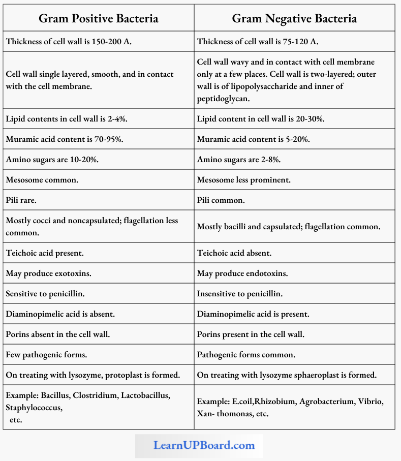
Gram Staining Technique: The gram staining technique was introduced by Christian Gram in 1884. The stain works on the cell wall of bacteria. depicts the procedure for Gram staining.
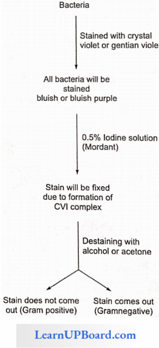
Gram-negative bacteria become colorless on treatment with a destaining agent due to a thin cell wall and more lipids in the cell wall. The table shows the basic difference between the structures of Gram-positive and Gram-negative bacteria.
Difference Between Gram Positive And Gram-negative Bacteria
Bacterial Cell Envelope
- The cell envelope consists of the outermost glycocalyx, middle cell wall, and innermost cell membrane.
- Glycocalyx protects cells and also helps in adhesion. It is represented by either a slime layer or a capsule.
- The slime layer is composed of dextrin, dextran, and levan, while the capsule is made of polysaccharides and D-glutamic acid.
- The slime layer protects the cells from loss of water and nutrients.
- Capsule provides gummy and sticky characters to the cell.
Bacterial Cell Wall
- Cell wall is made up of peptidoglycan, mucin, or mucopeptide.
- The Glycan portion forms the backbone of peptidoglycan which is composed of alternating units of NAM (N- acetyl muramic acid) and NAG (N-acetyl glucosa¬mine) joined together by β-1,4-linkage.
- The tetrapeptide chain is attached with NAM.
- leichoic acid is an acidic polymer consisting of a carbohydrate (glucose), phosphate, and an alcohol. It performs several functions such as binding metals and acting as a receptor site for some viruses.
- Teichoic acid maintains cells at low pH to prevent the degradation of cell walls by self-produced enzymes.
- Porins function as channels for the entry and exit of hydrophilic low molecular-weight substances.
- The outer layer of the cell wall in gram-negative bacteria contains lipopolysaccharides (LPS) that act as the main surface antigens in the cell wall.
“what is the basic unit of life “
Mesosome (Chondroid; Fitz James): The plasma membrane shows enfolding into the cell and this enfolding is called mesosome. It is particularly found in Gram-positive bacteria. The various types of mesosomes are as follows:
- Central Mesosome: It holds the nucleoid, and helps in the separation of nucleoid and septa formation.
- Peripheral Mesosome: It helps in storing respiratory enzymes such as succinic dehydrogenase, cytochrome oxidase, etc.
Plasmid (Lederberg And Hayes): In addition to the normal chromosomal DNA, some extrachromosomal genetic elements are often found in bacteria. These elements are called plasmids. In fact, these are circular pieces of DNA that have extra genes. These are capable of autonomous replication in the cytoplasm of the bacterial wall. Plasmid is circular, supercoiled, double-stranded naked DNA. It is also called mini chromosomes.
Types Of Plasmids
- Sex-plasmid: It carries sex fertility factor responsible for the transfer of genetic material during conjugation.
- R-plasmid: Confers resistance to antibiotics, having resistance transfer factor (RTF).
- Col-plasmid: Produces special proteins colicins (bacteriocin) to kill other bacteria.
- Degradative Plasmid: Decomposes hydrocarbons in petroleum.
- Ti- and Ri-plasmid: “Ti” refers to tumor-inducing plasmids and “Ri” refers to rhizogene plasmids. These are large plasmids with about 200 kbp.
Plasmids Respiration: Depending upon the mode of respiration and their capability to perform alternate mode of respiration, bacteria arc of the following types
- Obligate Aerobes: These can perform only aerobic respiration, for example, Bacillus subtilis.
- Obligate Anaerobes: These can perform only anaerobic respiration, for example, Clostridium botulinum.
- Facultative Aerobes: These are anaerobic forms but can live in the presence of O2, for example, Chlorobium.
- Facultative Anaerobes: These are aerobic forms but can live anaerobically also, for example, Pseudomonas.
- Aerotolcrant Anaerobes: These bacteria continue to perform anaerobic respiration even in the presence of O2, for example, lactic acid bacteria.
- Anaerotolerant Aerobes: Aerobic bacteria continue to perform aerobic respiration even in the absence of free O2 by using O2 of oxidized salts, for example, denitrifying bacteria.
Plasmids Reproduction
- By Binary Fission: It is the common method of reproduction under favorable conditions.
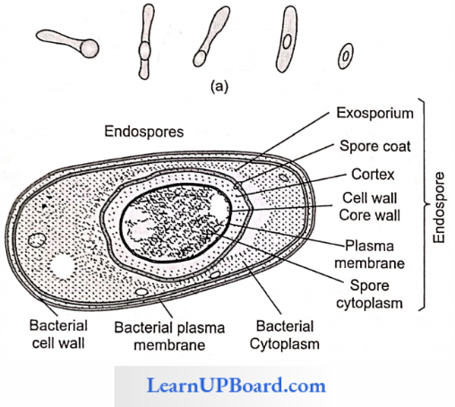
- By Endospore Formation: Discovered by Cohn in hay bacteria (Bacillus subtilis), endospores are thick-wailed, and highly dehydrated. and resistant spores formed under adverse conditions. Their wall is differentiated into three to four layers.
- Endospores are commonly formed in genera such as Bacillus and Clostridium. One endospore is formed per bacterial cell. So they are more a means of perennation than reproduction. Cortex and cytoplasm contain Ca2+ and an anticoagulant dipicolinic acid, which prevents the protoplasm from coagulating at high temperatures.
- Inclusion Bodies: Reserve material in prokaryotic cell are stored in cytoplasm in the form of inclusion bodies. These are not bounded by any membrane. For example, phosphate granules, glycogen granules, etc.
NEET Biology Notes Cell The Unit Of Life Eukaryotic Cell
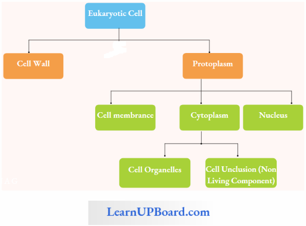
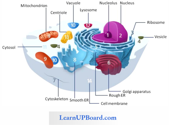
Cell Organelles
- Plastids
- Mitochondria
- ER
- Golgi body
- Lysosome
- Ribosomes
- Microbodies
- Cytoskeleton
Cell Inclusion
- Secretory Product: enzyme nectar
- Excretory Product: essential oils, alkaloids. gums, latex
- Storage Product: carbohydrates, fats, nitrogenous substances
Hyaloplasm = Cytoplasm – (Cell organelles + Cell inclusion)
Protoplasm: Life is impossible without protoplasm, which is the living substance of a cell. Protoplasm is present in all living cells and performs all vital functions of a cell. Hence, J. Huxley rightly defined it as the “physical basis of life.” Dujardin named it as sarcode. Purkinje renamed it as protoplasm.
Protoplasm Cell Wall
- Discovered by Robert hook.
- Present in the kingdom Plantae, Fungi, Monera, and most of the members of kingdom Protista.
- Absent in Animalia, Mycoplasma, gametes, and zoo-spores.
- The cell wall is the non-living component and protective layer of cell.
- Originated during cytokinesis from the vesicle of the Golgi body (phragmoplast).
Protoplasm Components Of Cell Wall
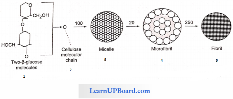
- Matrix: It is the non-cellulosic component mainly composed of H2O(30-60%), pectin (2-8%), hemicellulose (5-15%), glycoprotein (1-2%), and lipid (0.53%).
- Fiber: Mainly composed of cellulose.
- Deposition: Deposition only takes place on the secondary wall in the form of silica, pectin, suberin, cutin, lignin, etc.
Protoplasm Structure Of Cell Wall
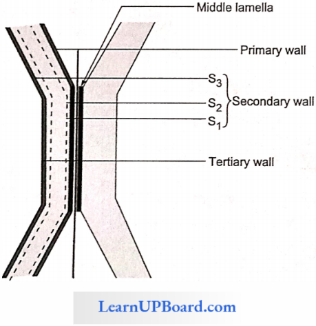
- Middle Lamella: It is the cementing layer between two cells which is formed first among all the layers during cytokinesis.
- Chemically composed of calcium pectate, but a small amount of magnesium pectate may be present or absent.
- Absent in the free surface of the cell.
- The softening of fruits and retting of fibers is due to the dissolution of middle lamella by the pectolytic enzyme.
- Primary Wall: First layer of cell, thickness 1-3 pm and elastic.
- Cellulosic fibers are loosely scattered in the matrix.
- It is only a permanent layer in the parenchyma and meristem.
- The process of formation of the primary wall is called into susceptions.
- Secondary Wall: The second wall is more thick and present inside the primary wall. The thickness is 3-10 pm.
- Mainly composed of cellulose and arranged in three layers (S1 – S3) and shows a threeply structure.
- Matrix is present between these layers.
- Deposition of suberin, and lignin takes place on the secondary wall.
- The process of the formation of a secondary wall is known as accretion (apposition).
- Tertiary Wall: The tertiary wall is only present in the tracheids of gymnosperms and composed of xylan and cellulose.
Structure Associated with Cell Wall
Plasmodesmata: It is the cytoplasmic connection between two neighboring plant cells. The term “plasmodesmata” was coined by Strassburger and discovered by Tangle.
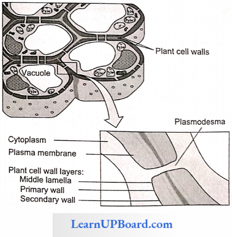
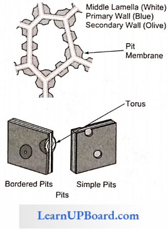
Pits: Depression present on cell wall lacking secondary wall.
Components Of Pits
- Pit Membrane: Made up of primary wall and middle lamella. It is permeable.
- Pit Chamber: It is the area where the secondary cell is absent.
- Pit Pore: It is the open passage between two cells.
“the basic unit of life “
Types Of Pit:
- Simple pit and
- Border pit.
Cell Membrane (Plasma Membrane, Plasmalemma): The Protoplasm of all types of cell is surrounded by a double-layered, living, selectively permeable membrane called cell membrane.
- Cell Membrane Origin: Cell membrane originates de-novo (independently synthesized) from its constituent chemical but does not originate from any other structure.
- Cell Membrane Chemical Composition: Cell membrane is chemically composed of phospholipids proteins and carbohydrates.
- Protein = 20-70%
- Lipid = 20-79%
- Carbohydrate = 1.5%
- Water = 20%
- Enzymes
- Amino acid
Cell Membrane Note:
- Lipids are found in the form of phospholipids, glycolipids, and sterols.
- Phospholipid forms the basic component of cell membrane.
- Upids show flip-flop movement (movement of lipids inside and outside of cell membrane).
- It provides stability, elatedly, and fluidity of the membrane.
- The fluidity of the membrane is due to the hydrophobic non-polar tail part of fatty acid.
- Phospholipids is amphipathic in nature due to the presence of both hydrophilic and hydrophobic nature.
Models Of Cell Membrance
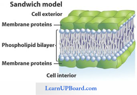
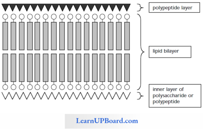
- Trilamellar Model Or Sandwich Model
- This model was given by Danielli and Davson.
- The arrangement of lipid (L) and protein (P) molecules is called PLLP.
- The bimolecular lipid layer is sandwiched between the single layer of protein on both sides.
- Protein is globular and α type.
- This is a hypothetical model which was proposed before the discovery of the electron microscope.
- Unit Membrance Concept
- This model was given by Robertson.
- All membranous systems of a cell are composed of lipoprotein.
- The molecular organization and chemical composition is similar to that of the lamellar model.
- This model is different from the trilamellar model, that is, the protein is of β-type.
- Robertson also proposed measurements of this membrane. The total thickness of the membrane is equal to
- Both inner and outer proteins are different in nature. The outer protein is muco protein and the inner protein is nonmuco protein.
- A core of 8Å diameter is also present in the protein layer which helps in transportation.
- Fluid Mosaic Model
- This model was given by Singer and Nicholson.
- Widely accepted model which clearly explains selectively permeable proportions of cell membranes.
- This model can be explained as a protein iceberg in the sea of lipids.
- Lipids are present in two layers in which the head of fatty acids faces towards the outside and tail towards the inner side.
- Protein is of α-type and is present in two forms.
- Extrinsic Protein: Small size protein present outside the lipid layer and can be easily separated. It forms 30% of the total protein.
- Intrisic Protein; large-sized protein embedded in lipid molecules and cannot be easily separated. It forms 70% of the total protein.
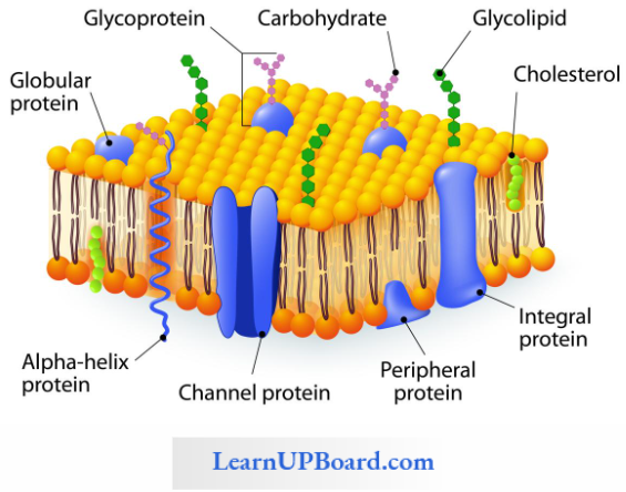
Asymmetry Of Plasma Membrane: Two surfaces of the plasma membrane are not similar because
- Extrinsic proteins present more towards the inner side.
- Lecithin (lipid) is present on the outer side.
- Cephalin (lipid) is present on the inner side.
- The carbohydrate of oligosaccharide nature is present towards the outer side of the membrane and attaches to both lipid and protein called glycolipid and glycoprotein. respectively, and collectivity is called glycocalyx. Glycocalyx is the eye and ear of the membrane, i.e., the recognition center.
Structures Associated With Plasma Membrane
- Microvilli: Finger-like structure developing from the cell membrane in the alimentary canal.
- Mesosome: Enfolding of the cell membrane in bacteria. Lomasome: Enfolding of the cell membrane in fungi.
- Lamellosome: Enfolding of the cell membrane in blue-green algae (BGA).
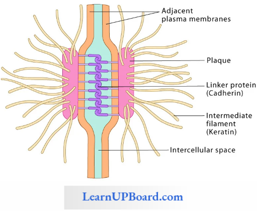
- Desmosome: Two animal membranes are separated by each other by intercellular spaces in which the cell membrane develops thickening due to the deposition of protein from which tonofibrils originate. This constitutes a desmosome.
- Hemidesmosome: Thickening present only on one membrane.
- Terminal Bars: Desmosomes without tonofibril.
- Tight Junction: Two animal membranes are closely attached together. Hence, no intercellular space.
- Gap Junction: Two animal cells are connected with each other by protein pipes.
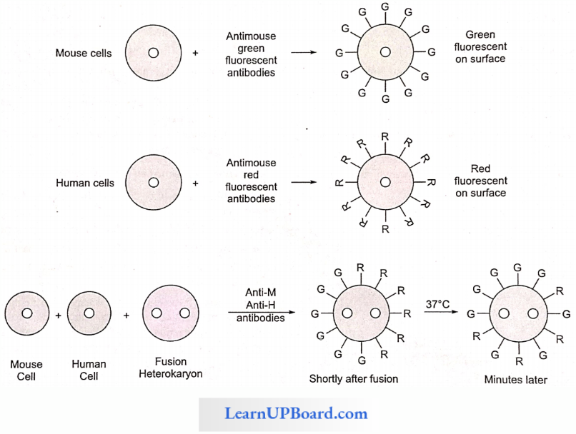
Experiment To Show Fluidity Of Cell Membrane: Larry Frye and Michael Edidin, at Johns Hopkins University, labeled the plasma membrane proteins of a mouse cell and a human cell with two different markers and fused the cells. Using a microscope, they observed the markers on the hybrid cell. The mixing of the mouse and human membrane proteins indicates that at least some membrane proteins move sideways within the plane of the plasma membrane.
Functions of Plasma Membrane: Transport
- Passive Transport: The transport of substances through a membrane along a concentration gradient is called passive transport it is downhill transport without the expenditure of energy.
- Active Transport: The transport of substance through the membrane against a concentration gradient with the expenditure of energy is called as active transport. Active transport is highly selective, and unidirectional.
- Bulk Transport: The transport of substances through the vesicle of the membrane is called as bulk transport.
Types Of Transport
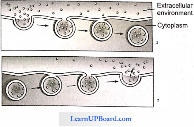
- Endocytosis: The intake of foreign substances through vesicles is known as endocytosis, The endocytosis of a solid particle is called phagocytosis (cell eating) and the endocytosis of liquid molecules is called pinocytosis (cell drinking).
- Phagocytosis results in the formation of phagosomes and pinocytosis results into the formation of pinosomes.
- Exocytosis: Exocytosis or endocytosis geophagy or cell vomiting is the outward movement of a substance from the cell.
Endomembrane System (GERL System): The term endomembrane system was coined by Navikoff The of cell is composed of a Golgi body, endomembrane system, and vacuole (called endoplasmic reticulum (ER), Iysosome, GERL system).
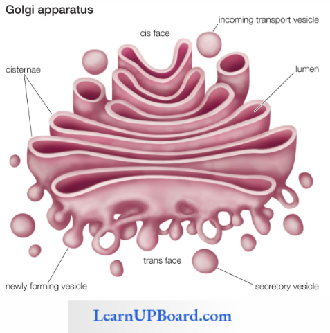
Golgi Body: The common names of the Golgi body are mitochondria, idioms, dictyosome, Dalton complex, traffic police, the middle man of the cell, apparato reticular, and Golgi complex. Golgi complex was discovered by Camillo Golgi in the nerve cell of an owl and cat and the term “Golgi body” was coined by Cajal.
Golgi Body Occurrence: Only found in the eukaryotic cell except for mature RBCs and sperm of mammals, mature sieve tube cells, mature sperm of bryophyte, and Pteridophyta. In the plant cell, the Golgi body is called a dictyosome. In fungi, the Golgi body is unicistemal, because its components are not packed.
Golgi Body Structure: It appears as a flattened sac-like structure bounded by a single membrane without any ribosome on its surface. It shows polarity due to differences in the structure of both ends. The convex end is called cis-face or forming (F-face) and the concave end is called transface or (maturing) M-face.
Golgi Body Is Composed Of Four Components:
- Cistenae,
- Tubule,
- Vesicle, and
- Golgian vacuole.
- Golgi Body Cistenae
- It is the most important component of the Golgi body.
- It is found in the form of stacks.
- Each stack contains 4-18 cistemae.
- It shows polarity due to the presence of two faces.
- Slightly curved and encloses a cavity inside
- Golgi Body Tubule: It looks like a branched network-like structure.
- Golgi Body Vesicle: Vesicles are formed by budding of the terminal end of tubular smooth ER. They are associated with the convex surface of cistemae.
- Golgi Body Golgian Vacuole: It is formed by the budding of the terminal end maturing face of Golgi apparatus or surrounding of cisternae. Golgi body is present in the cytoplasmic area and is free from other cell organelles. Hence, the area is called the zone of cell exclusion.
Function Of Golgi Body
- Processing and packaging of material: Vesicle enters into the Golgi body at cis- or F-face and the secretary vesicle comes out from the Golgi body from the trans- or M-face. Hence, called the traffic police.
- Formation of primary Iysosome
- Glycosidation is the formation of glycolipids from lipids.
- Glycosylation is the formation of glycoprotein from protein.
- Extracellular secretion: It secretes mucilage; hence, root caps are rich in the Golgi body.
- Intracellular secretion of pectin and other cellular materials.
- Formation of acrosome of sperm; hence, acrosome is also called modified Golgi body.
- Transport vesicle formed by endoplasmic reticulum.
- Lysosomes and secretory vesicles originate from the M-face of the Golgi body.
Endoplasmic Reticulum: Endoplasmic reticulum was discovered by Porter and Thomson and the name was given by Porter and Khallan. ER is present in all eukaryotic cells except mature RBCs, monocytes of WBC, undifferentiated cells eggs, etc. It is completely absent in prokaryotic cells.
- Endoplasmic Reticulum Structure: The endoplasmic reticulum forms a hollow tubular network such as the structure in cytoplasm and is composed of three components
- Endoplasmic Reticulum Cisternue: Simple unbranched tube-like structures; 40-50 pm in diameter; generally arranged parallel and interconnected to each other.
- Endoplasmic ReticulumTubule: Tubules are branched tube-like structures 50-200 pm in diameter.
- Endoplasmic Reticulum Vesicle: Small vacuole-like structures, 25-500 pm in diameter. Ribosomes are not attached. The endoplasmic reticulum is attached to a nuclear membrane at one end and a cell membrane at the other end.
Types Of Endoplasmic Reticulum
Rough ER (RER): The rough surface of the endoplasmic reticulum is due to the presence of ribosomes. Only a large unit (60S) of the ribosome is attached to the surface of RER with the help of two types of glycoprotein called Ribo- phorrin-1 and Ribophorrin-2.
- RER forms two-thirds of the total ER of cells.
- RER is only composed of cisternae and tubules.
- RER bears a pore at the point of attachment of the ribosome.
Smooth ER (SER): Smooth endoplasmic reticulum surface is smooth due to the absence of ribosomes.
- SER forms only one-third of the total ER of cells.
- SER is composed of only tubule and vesicle.
Endoplasmic Reticulum Note:
- The ER of muscle is called the sarcoplasmic reticulum, in the retina of the eye is called the myeloid body, and in the nerve cell is called NissTs granule.
- RER is found in cells involved in protein synthesis.
- RER is basophilic in nature due to the high content of RNA. Hence, also called ergastoplasm or basophilic body.
- RER originates from the nuclear membrane, but SER originates from RER.
- SER is found in cells that are involved in the synthesis of lipids, glycoproteins, and hormones.
- The liver cell has both types of RER and SER.
Common Functions Of RER And SER
- The endoplasmic reticulum gives mechanical support to the cytoplasm since it forms a network-like structure inside the cell.
- Transport of material within cell.
- It helps in the formation of the nuclear membrane.
- Both RER and SER increase surface area for enzymatic action.
Functions Of SER
- Synthesis of lipid and glycogen
- Detoxification (protection of cell by the toxic effect of chemicals by using cytochrome P450)
- Helps in muscle contraction by release and uptake of Ca
- Formation of organelles such as spherosomc and glyoxysome
Functions Of RER
- Synthesis and transport of protein
- Formation of SER
- Synthesis of lysosomal enzyme
Lysosome
- Lysosome Common Name: Suicidal bag, disposal unit, recycling center, scavenger of cell, and atom bomb of cell. Lysosomes originate from the Golgi body and they were discovered by Christian de Duve. The term was coined by Navikaff.
- Lysosome Occurrence: Mainly found in eukaryotic animal cells, but also found in fungi and root tip cell of maize and cotton.
- Lysosome Structure: They are the smallest single membrane-bound cell organelles and are mainly found in animal cells. Lysosomes are small, oval, or spherical structures filled with about 50 types of hydrolytic enzymes collectively called acid hydrolyses. The marker enzyme of lysosomes is acid phosphatase.
All enzymes work at acidic pH. The acidic condition is maintained by the pumping of proton inside lysosomes. The excess of lipid-soluble vitamins, steroidal sex hormones, bile salts, X-rays, and UV rays are called membrane destabilizers. Lysosome membrane has some stabilizers as cortisone, cholesterol, heparin, etc. Lysosomes show polymorphism found in four different forms
- Primary Lysosomes: Newly formed lysosomes containing inactive enzymes. They are produced from the transface of the Golgi body.
- Secondary Lysosomes Or Digestive Vacuoles Or Hetrophagosomes: Secondary lysosomes contain primary lysosomes and food vacuoles (pinosomes or phagosomes).
- Tertiary Lysosomes Or Residual Bodies: Lysosomes containing undigested food material.
- Autophagic Vacuoles: They are formed by the union of many primary lysosomes around old and dead organelles.
Enzymes produced by lysosomes are classified into four groups.
- Proteases,
- Acid phosphatases,
- Sulphatases, and
- Ribonucleases, deoxyribonucleases.
Lysosome Function
- Perform extracellular and intracellular digestion.
- Removal of dead cell organelles. Hence, act as scavengers.
- Digestion of harmful substances of cells.
- Autophagy: If a person is on prolonged last, then lysosomes provide energy by digesting the stored food of cell.
- Autolysis: Disappearance of the tail of a tadpole larva of a frog during metamorphosis is due to an enzyme cathepsin.
- In old age, the number of lysosomes increases, and the number of mitochondria decreases.
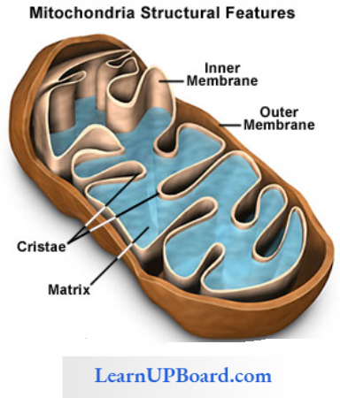
Mitochondria: Mitochondria are the largest cell organelles and the second largest cellular component of animal cells. They are the second largest cell organelles in plant cells and the third largest cellular component of plant cells.
- Mitochondria were discovered by Kollikar in the striated flight muscle of insects, and he called it as sarcosome. The term mitochondria was given by Benda. All mitochondria of a cell are called chondrites.
- A mitochondrion is also known as a sarcosome, bioblast, plasmosome, chondroplasty, plastochondria, and a powerhouse of the cell. It is also considered as a cell within cell.
- The vital stain for mitochondria is Ganus grcen-B which does not kill the mitochondria. The number of mitochondria is variable in different organisms. The number of mitochondria depends on the metabolic activities of a cell.
Mitochondria Number
- 1 in Chlorella, Trypanosoma, and Microasterias
- 25 in human sperm cell
- 300 -400 in human kidney cell
- 500-1000 in lever cell
- 50,000 in giant amoeba Chaos chaos
- 140,000-150,000 in the egg of sea urchin
- 500,000 in-flight muscle cells of insect
- Plant cells have less number of mitochondria than animal cells because plant cells have both chloroplast and mitochondria for ATP production
Mitochondria Size: The smallest mitochondria are found in yeast cells and the largest in the oocyte of Rana pipens.
Mitochondria Occurrence: Found in all eukaryotic cells except mature RBC.
Mitochondria Structure: The mitochondrion is a double membrane-bound sac-like structure. Each membrane is 60-70 A thick and composed of lipoprotein. Both membranes arc structurally and functionally quite differently from each other. The outer membrane is continuous, fully permeable, rich in lipids, and contains porin (protein). The outer membrane is poorer in protein.
- The lipid-protein ratio of the outer membrane is 40:60.
- The lipid-protein ratio of the inner membrane is 20:80.
- The inner membrane contains cardiolipin (having four fatty acids).
- The inner membrane is infolded as a finger-like structure called a crista.
- Cristae are meant to increase the physiological active area of the inner membrane.
- Cristae bear stalked particles called exosomes.
The cavity of mitochondria is divided into two chambers.
- Outer Chamber Or Perimitochondrial Space: It is the space between the outer and inner membranes. The thickness of the outer chamber is 60-80 A and it is filled with the homogenous fluid of phospholipids.
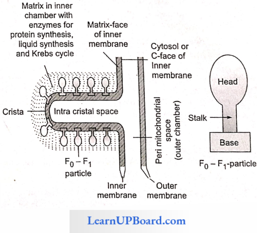
- Inner Chamber: It is the space enclosed by the inner membrane. It is filled with a proteinaceous liquid called matrix. The matrix contains DNA (with high GC ratio), RNA,70S ribosomes, and all enzymes of Kreb’s cycle except succinic dehydrogenase. (SDH). SDH is present in the inner membrane of mitochondria.
Mitochondria Chemical Composition
- Protein: 65-70%
- Lipid: 25-30%
- RNA: 5-6%
- Ribosome: 70S type
Two States Of Mitochondria
- Orthodox: It is the inactive state of mitochondria where ATP production is not involved. Hence, the matrix becomes broad and the perimitochondriai space becomes narrow.
- Condensed: The active state of mitochondria is involved in ATP production. Hence, the matrix becomes narrow, but the outer chamber becomes broad.
Mitochondrial DNA
- Mitochondrial DNA forms only 1% of the total DNA of the cell.
- It is much smaller than the nuclear DNA. Hence, contains few information.
- It is double-stranded, circular, and Naked (Histone absent)
- GC content is high. Hence, Tm (melting temperature) is high.
- Mitochondria have their own DNA polymerase enzyme. Hence, it replicates its own DNA.
- Mitochondrial DNA is responsible for cytoplasmic inheritance. For example, male sterility in maize, and petite character in yeast.
Mitochondria contain about 70 types of enzymes which is 70% of the total enzymes of the cell, which indicates the highest concentration of enzymes in cell mitochondria.
Oxysome: Oxysome is also called F0-F1 particle elementary particle or subunit of Fernandez and Moran or ATPase particle.
Structure Of Oxysome
- Head (F1 Particle): Site of ATP synthesis
- Stalk
- Base (F0 Particle): Proton tunnel
The space between two exosomes is 100Å. The number of exosomes is 104-105 per mitochondrion.
Oxysome Note: Mitochondria are called semi-autonomous cell organelles because they have their own DNA, RNA, and 70S type of ribosome, but they also depend on the nucleus and cytoplasm for most of the metabolic activity. Mitochondria are similar to bacteria cells or prokaryotic cells in the following points
- Presence of porin protein in the outer membrane
- Mitochondria show binary fission of division
- Presence of 70S type of ribosomes
- DNA double-stranded, circular, and naked
On these bases, mitochondria may be called as “cell within the cell” “intracellular prokaryotic parasite” or “bacterial endosymbiont.”
Functions Of Mitochondria
- Mitochondria act as the site of respiration except during glycolysis and anaerobic respiration.
- They act as the site of oxidative phosphorylation (formation of ATP by the oxidation of reduced co-enzyme).
- They are also called as the powerhouse of cells.
- Mitochondria form the sperm tail.
- The synthesis of many amino acids and fatty acids occurs in the mitochondria.
- Mitochondria also take part in lipid synthesis.
- Mitochondria may store and release calcium according to requirements.
Ribosome: In plant cells, ribosome was discovered by Robison and Brown, and in animal cells, by Palade. Ribosomes are the smallest nonmembranous and most abundant cell organelle that are negatively charged. The number of ribosomes is 104-105 per cell. Ribosomes are also called as palate particles or protein factories or engines of cell or ribonucleic acid proteinic particles (r-RNA + Protein).
Ribosome Occurrence: Ribosomes are found in both prokaryotic and eukaryotic cells except mature RBCs. In a prokaryotic cell, ribosomes are found freely in the cytoplasm or form polysomes (ribosomes + mRNA). The function of polysomes is the synthesis of multiple copies of a single protein. In eukaryotic cells, two types of ribosomes are found:
- Cytoplasmic Ribosomes: 80S type of ribosomes are found in cytoplasm freely or attached with ER or nuclear membrane.
- Organelle Ribosomes: 70S type of ribosomes are found in mitochondria and plastids.
Hence, the ribosome is also called organelles within organelles.
Ribosome Type: On the basis of sedimentation coefficient, ribosomes are two types
- 70S Type: It is found in both prokaryotic and eukaryotic cells. The ratio of r-RNA and protein is 60:40. The large subunit is 50S (34-protein + 5S r-RNA + 23S r-RNA)and the small subunit is 30S (23-protein + 16S r-RNA).
- 80S Type: It is reported only in eukaryotic cells. The ratio of r-RNA and protein is 40:60. The smaller unit is 40S (33-protein + 18S r-RNA) and the larger unit is 60S (40-protein + 5S r-RNA + 5.8S r-RNA + 28S r-RNA).
Ribosome Origin: In prokaryotes, ribosome synthesis takes place in the cytoplasm, but in eukaryotes, it is synthesized in nucleolus.
Association And Dissociation Of Ribosomes: Both subunits of ribosomes are found separately in the cytoplasm. The association and dissociation of ribosomes depend on the concentration of Mg+2 ions. In the high concentration of Mg+2 ions, both subunits associate to form a complete ribosome called dimmer but in low concentration, it dissociates. Both subunits are assembled at the time of protein synthesis.
Functions Of Ribosomes
- Site of protein synthesis, hence called protein factory.
- Free ribosomes synthesize non-secretary proteins, while ER-associated ribosomes synthesize secretory proteins.
- Proper folding of proteins requires chaperones.
Plastid: Plastids were discovered by Hackel. They are only found in plant cells. They are the largest cell organelle and the second largest cellular component. A plastid originates from a protoplastid (found in meristem). All plastids of a plant are collectively called plastidome, which was coined by Dangered. Schimper discovered different types of plastids. Plastids can be divided into three types on the basis of the presence or absence of the pigment.
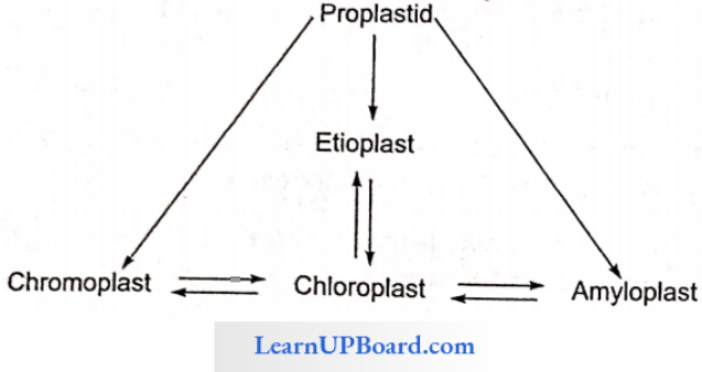
- Chloroplast: Green plastids (chlorophyll present); are found in the green parts of plants.
- Chromoplast: Colored plastids (chlorophyll absent but other pigments present); provide coloration to flowers and fruits.
- Leucoplast: Colorless plastids (pigments and grana absent). These are concerned with the storage of carbohydrate proteins and lipids in the underground parts of the plant. These are the largest plastids. Different types of leucoplasts are as follows
- Elaioplast Or Oleoplast: Stores fat
- Amylop Last: Stores starch
- Aleuroplast: Stores protein
Plastid Note: All three forms of plastid, chloroplast leucoplast, and chromoplast are interconvertible. For example, in tomatoes, chloroplasts change into leucoplasts, and then in chromoplasts. In potatoes, leucoplasts change into chloroplasts. In mango or chili, chloroplasts change into chromoplasts. In carrots, leucoplasts change into chloroplasts.
Chloroplast
- The number, shape, and size of the chloroplast are variable.
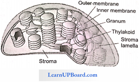
- In higher plants, the chloroplast is discoid in shape and bounded by a double membrane.
- Each membrane is 90—100 A thick and composed of lipoprotein.
- Both membranes are structurally and functionally different.
- The outer membrane is fully permeable, rich in lipid, and contains porin in protein.
- The inner membrane is selectively permeable and rich in protein.
- Chloroplast may be called “cell within the cell” “intra-cellular prokaryotic parasite” or “bacterial endosymbiont.”
- Chloroplast is also a semi-autonomous structure like mitochondria.
- The DNA of chloroplast is double-stranded, circular, and naked (without histone).
- The DNA has high GC content, hence high Tm. (melting point).
- Chloroplast is also responsible for cytoplasmic inheritance, for example, plastid inheritance in Mirabilis jalapa.
Structure Of Chloroplast: The cavity of chloroplast is divided into two chambers
- Outer Or Periplasmic Space: The space bounded by outer and inner membranes. It is 100-200 A thick.
- Inner Chamber: The space enclosed by the inner membrane. It is filled with a proteinaceous liquid called stroma. It contains DNA, RNA, 70S type of ribosomes, and all enzymes (RuBisCO) taking part in the Kelvin cycle.
RuBisCO: RuBisCO forms 50% part of the stroma and about 16% part of the total chloroplast protein. It is considered as the most abundant enzyme or protein on earth. It is also known as RuBP carboxylase, RuBP oxygenase, and carboxy dismutase.
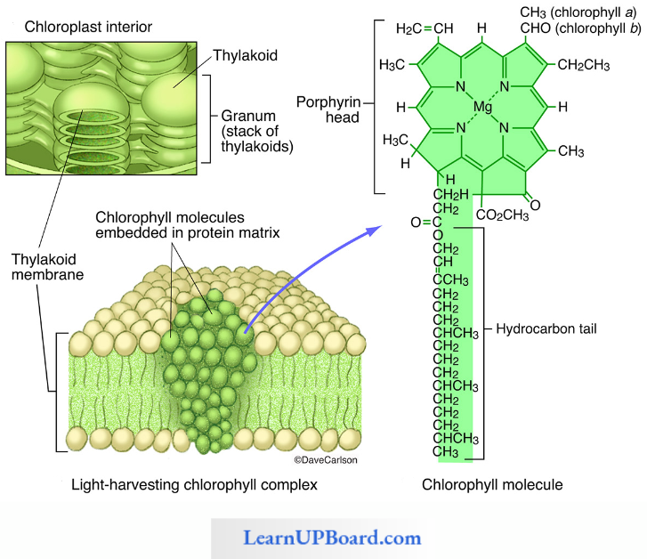
In the stroma of chloroplast, several double membrane-bound sac-like structures called baggy trousers or thylakoids are reported. The term “thylakoid” was coined by Menke. Thal-alkaloid is considered as the structural unit of the chloroplast.
- Thallakoid is an analogous structure to the cristae of mitochondria.
- Several thylakoids become stack-like structures and form granum.
- The number of thylakoids in a granum is 2-100.
- The number of grana in one chloroplast is 40-60.
- Grana are interlinked with each other by fret channel or stroma lamella which may lack pigments.
- In the membrane of thylakoid, about 200-300 quantasomes are present.
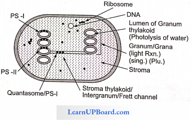
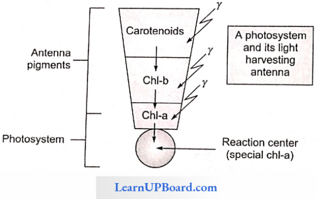
Quantasome: Quantasome was discovered by Parks and Biggin, but the term was coined by Park and Pon.
- It contains about 230 chlorophylls (160 chl-a, 70 chl- b) + 50 carotenes. Quantasome absorbs one quantum of light.
- Quantasome is called the functional unit of chloroplast, or the unit of photosynthesis.
- Pigments associated with quantasomc form two types of photosystems (PS). Photosystems are composed of two parts
- Reaction center and
- Light-harvesting complex (LHC) or antenna.
- The reaction center contains one molecule of chl-a and LHC contains other pigments.
- Each photosystem is composed of 250—400 pigments.
- LHC is concerned with the light-harvesting mechanism. Different wavelengths of light are absorbed by LHC.
- The reaction center (chl-a) is concerned with the conversion of light energy into chemical energy.
- On this basis, chl-a is called the main pigment and others are called necessary pigments.
Type Of Photosystems
- PS-1: Located at the non-appressed part of grana and stroma thylakoid reaction is P700 (fret channel).
- PS-2: Located at the appressed part of grana and the reaction center is P680. The reaction center of bacteria is B810.
Photosystems Note:
- Chloroplast is agranular in algae and bundle sheath cells C4 of plant
- If thylakoids are directly scattered in the cytoplasm, then they are called chromatophores.
- Chloroplast in which thylakoids are present but not stalked is called adrenal chloroplast
- In higher plants, grana contains pigments: chl-a, chl-b, and carotene.
- In algae, xanthophyll is the main pigment and the other pigment is phycobilin (phycocyanin and phycoerythrin).
Functions Of Chloroplast: Chloroplast is the site of photosynthesis. The light reaction takes place in grana and the dark reaction in the stroma. Hence, the chloroplast is called the glucose factory. The thylakoid membrane is the site of photophosphorylation (synthesis of ATP by using light)
Structure Of Chlorophyll: Chlorophyll without Mg is called pheophytin which acts as an electron acceptor. Chl-a contains -CH3 group present at the third carbon of the second pyrrole ring. Chl-b contains -CHO group present at the third carbon of the second pyrrole ring.
Microbodies: Microbodies are single membrane-bound cell organelles concerned with oxidation reactions other than respiration. Micro¬bodies include
- Peroxisomes,
- Glyoxysomes, and
- Spherosomes.
- Peroxisome
- Discovered by de Duve in the leaf cell of spinach.
- Peroxisome is mainly found in the mesophyll cells of C3 plant.
- It is concerned with photorespiration which involves glycolate metabolism.
- Catalase is the largest enzyme and peroxidase is the smallest enzyme of peroxisome.
- It is mainly concerned with the formation and destruction of H2O2. Hence, it protects cell from the toxic effect of H2O2.
- Peroxisomes originated from ER.
- Peroxisome converts glycolic acid into glycine by the help of glycolic acid oxidase enzyme.
- In animals peroxisomes found in lever and kidney cells and are concerned with the synthesis and degradation of lipids.
- Glyoxysome: Glyoxysomc was discovered by Beevers and Tolbert from the germinating seed of the castor. It is mainly found in the germinating fatty seeds (caster, ground nut, etc.) and also found in some fungi (Neurospora and yeast). It is concerned with the following activity
- β-oxidation of fatty acid.
- Conversion of fat into carbohydrate. This process is called glyoxylate metabolism which requires two enzymes that are found in glyoxysome.
- These enzymes are
- Isocitrate lyase,
- Malate synthase. These two enzymes convert acetyl CoA into glucose.
- Glyoxysomes originated from smooth ER. They are destroyed in cell if the germination of seeds is completed.
- In animal cell, β-oxidation of fatty acid takes place in mitochondria.
- Spherosome Or Oleosome: Spherosome was discovered by Pemer and Hanstein. Spherosome originates from smooth ER and is filled with several hydrolytic enzymes. The term was coined by Dangeard. Spherosomes are half-membrane-bound cell organelles filled with several hydrolytic enzymes. They are only found in plant cells, hence called plant lysosomes. The enzymes of spherosomes are different from those of lysosomes.
- Spherosome Or Oleosome Functions
- Synthesis, storage, and translocation of fat.
- Synthesis of the intermediate compound of suberin.
- Spherosome Or Oleosome Functions
Centriole: A Centriole is a cylindrical or rod-like structure that is not bounded by any membrane and is present close to the nucleus, mainly in the eukaryotic animal cell.
- Centriole is also found is some motile plant cells. For example, motile algae, zoospores of algae, antherozoids of bryophytes, and pteridophytes.
- Centrioles are absent in prokaryotic cells, amoeba, gymnosperm, and angiosperm.
- Centrioles are found in pairs situated at right angles to each other called diplosomes.
- Diplosome is found in the exclusive area of cytoplasm which is free from any other cell organelle. This inclusion area is called cytocentrum centrosphere kinoplasm or zone of cell exclusion.
- Diplosome and cytocentrum are collectively called Centrosomes.
- Centriole does not contain RNA or DNA but duplicates directly during the end of the S-phase of the cell cycle.

Duplication completes in G2 phase. Centriole is a cart wheel-like structure and shows 9 + 0 organization.
- It has nine peripheral triplet fibers but no central fiber.
- Each peripheral triplet fiber is composed of three microtubules which represent A, B, and C from inside to outside. Hence, the total number of microtubules in a centriole is 27 (9 x 3).
- The central part of centriole is called HUB which has no microtubule.
- HUB is attached to the peripheral fiber by pretentious spokes.
- Each peripheral triplet fiber is linked with each other by an A-C or C-A linker.
- Nine amorphous masses of protoplasm are present on the peripheral part of the centriole. These are called masses (MTG micro tubule generator), and pericentriolar satellite/nucleating center. It is concerned with the formation of new centriole.
Centriole Function:
- Centrioles polymerize microtubules for the formation of spindle fibers and astral rays during cell division.
- They form the basal body of cilia and flagella.
- They also form spindle fibers during cell division.
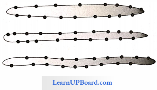
Cilia And Flagella: Both cilia and flagella are locomotory organs and both show more or less similar structural organization. Cilia are smaller in size, more in number, present on the entire surface of cell, and beat in a coordinated synchronous manner. Flagella are fewer in number, more elongated, generally pre¬sent at the anterior end of the cell, and beat independently.
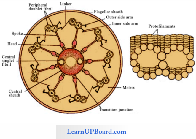
Eukaryotic Cilia/Flagella: The cilia and flagella have a microtubular composition. Both are bounded by a unit membrane. The bounded space is filled with matrix. In the matrix, there remain embedded nine doublets and two singlets of microtubules. Two singlets are located in the middle and called a central axoneme. Nine doublet fibrils are arranged peripherally around the central fibrils called peripheral axoneme.
Functions Of Cellia And Flagella
- Both flagella and cilia act as the organ of locomotion. Cilia may help in the aeration and circulation of food.
- Cilia may beat in a metachronous (beat one after one) or synchronous (beat simultaneously) rhythm.
- The cilia of the respiratory tract help in retaining the dust.
Cytoskeleton: An elaborate system of network-like structure, which is composed of proteins and filamentous structures present in the cytoplasm of eukaryotic cells to support the cytoplasm, is collectively called as cytoskeleton. It includes three structures
- Peripheral axonemes are linked to the central axoneme by radial spokes.
- Peripheral axonemes are linked with each other by A-B linker.
- A side arm develops from microtubule A of the peripheral axoneme.
- The central and peripheral axonemes are composed of tubulin protein.
- The side arm is composed of dynein protein which has ATP are activity.
- A-B linker is composed annexin type of protein.
- Energy required for the movement of cilia or flagella is provided by dynein protein. It has ATPase zyme which hydrolyzes ATP into ADP and releases energy.
- Microtubule
- Intermediate filament
- Microfilament
- Microtubule
- Discovered by de Robertis and Franchi. Microtubules are unbranched, hollow, non-contractile filaments of tubulin protein that form the basal structure of centriole, basal body of cilia and flagella, spindle fiber, and axoneme of cilia and flagella.
- The assembly and disassembly of microtubules require GTP and Ca+2 ion.
- Microtubules are responsible for cell motility and maintenance of the cell shape. They also show polarity.
- The boundary of the microtubule is composed of 13 parallel protofilaments.
- Microfilament
- Discovered by Paleviz.
- Microfilament is a branched or unbranched contractile solid filament composed of actin protein and found just below the cell membrane.
- Responsible for cyclosis, sol-gel interconversion, pseudopodia formation, and formation of cleavage furrow during cell division.
- Intermediate Filament: It is a hollow, branched non-contractile filament composed of three types of acidic protein
- Keratin (acidic in nature)
- Vimentin (acidic in nature)
- Desmin (acidic in nature)
- It forms a nuclear matrix or nuclear lamina present on the surface of the inner nuclear membrane
- It provides stability to the cell.
- It is involved in the formation of a scaffold for chromatin.
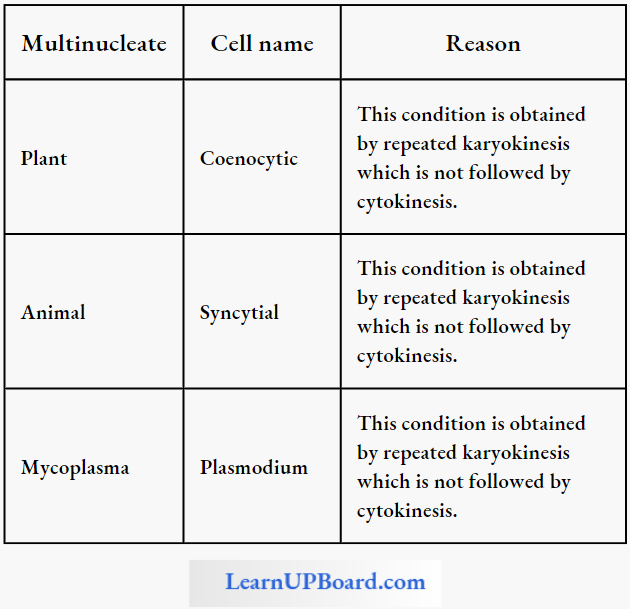
Nucleus: Nucleus is the largest cellular component in a cell. It is an extracytoplasmic component. The nucleus was discovered by Robert Brown in the root cell of an orchid. A cell may be uninucleate, binucleate, or multinucleate.
If the nucleus degenerates in a mature cell, then the cell is called enucleate, for example, a mature sieve tube, RBC of the animal. Hammerling called the nucleus as the brain of a cell or controlling the center of a cell by using a grafting experiment with two species of Acetabularia (unicellular largest marine green alga): A. crenulata and A. mediterranea.
Structure Of Nucleus: Nucleus can be easily distinguished into the following four parts nuclear membrane, nucleoplasm, nucleolus, and chromatin.
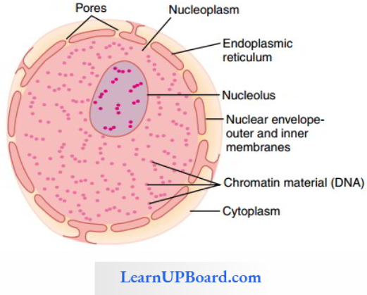
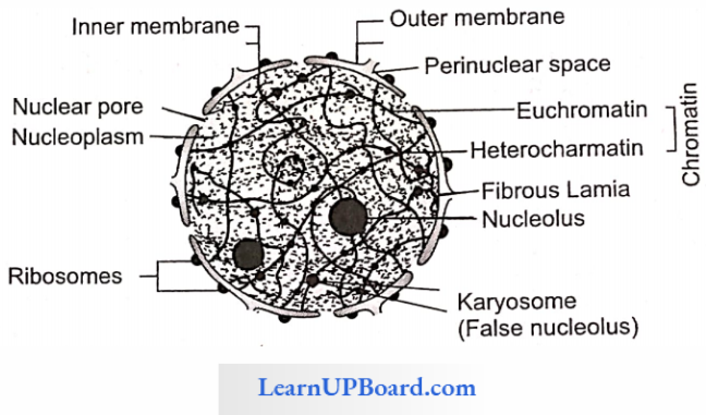
Nuclear Membrane
- The nuclear membrane is also called nucleolemma or karyotheca.
- The nucleus is also bounded by the double unit membrane of lipoprotein.
- Each membrane is 90-100 Å thick and composed of lipoprotein.
- The space, enclosed by two membranes (75 Å) is called perinuclear space and is 100-300 Å thick.
- The nucleus membrane is punctured by several pores called nuclear pores.
- Nuclear pores allow the movement of m-RNA.
- These pores may be circular or octagonal.
- Pore complex = Pore (1000 Å) + Annulus (a special type of protein that plugs the nucleus pore)
- The outer membrane is connected with the cell membrane through ER and also bears ribosomes (60S unit) on its surface.
- A network of intermediate filaments is present on the inner surface of the nucleus membrane called nuclear lamina or nucleus matrix.
- The nucleus lamina functions as the attachment point of the telomere during cell division (leptotene stage of meio- sis-I).
Nucleoplasm
- A part of protoplasm present within the nucleus is called nucleoplasm.
- It is more denser than cytoplasm. Hence, the nucleus can be easily observed under a microscope.
- Nucleoplasm is mainly made up of nucleic acids, his-tone proteins, enzymes (DNA, R.NA polymerase), li¬pids, minerals (P, K+, Na, Ca, Mg), NHC proteins, and NHC non-histone chromosomal proteins.
Nucleolus
- Nucleolus was discovered by Fontana and the term was given by Bowman.
- It is the largest part of the nucleus which occupies about 35% of the nucleus.
- It is the site of the synthesis of r-RNA (except 5S RNA) and subunit of RNA.
- It is a non-membranous structure composed of proteins, Ca+2 ions, and r-RNA but lacks DNA.
- The nucleolus stores RNA and synthesizes both subunits of ribosomes. DNA which synthesizes RNA is known as r-DNA of nucleolus.
- The nucleolus is composed of four parts:
- Pars Granulosa: Granular part of nucleolus
- Pars Amorpha: Amorphous part
- Pars Fibrosa: Fibrous part
- Pars Chromosoma: Chromosomal part
- The nucleolus is found at the secondary constriction (nucleolus-organizing region, NOR) of the chromosome.
- In haploid cells, generally, it is 1 and in diploid cells, it is 1-4.
- A maximum number of nucleoli is found in the oocyte of Xenopus (1600 nucleoli).
Chromatin Network
- During the interphase stage of cell division, nucleoplasm contains a fine network-like structure called a chromatin network.
- The term “chromatin” was coined by W. Flemming.
- Chromatin is fine fiber composed of DNA, RNA, and protein that takes differential stains in basic dye (Feulgen, acetocarmine, hematoxylin). This staining technique is called heterozygosis.
- The chromatin network becomes condensed during cell division and forms rod-like structures called chromosomes.
Types Of Chromatin
- Heterochromatin: The dark stain part of chromatin which is genetically inactive and has highly coiled or tightly packed DNA with histone is called heterochromatin.
- Euchromatin: The light stain part of chromatin which is genetically active and DNA is loosely packed with histone transcription occurs in euchromatin.
Types Of Heterochromatin
- Facultative: It is the inactive and condensed part of one of the two homologous chromosomes that take part in the formation of bar body, Y-spot, and drum stick.
- Constitutive: It is rich in repetitive DNA and present in the same region of both chromosomes of a homologous pair.
Chromosome: The word chromosome has two words: chroma and soma. Chroma means color and soma means body.
- A chromosome is a rod-like or thread-like structure present in the nucleus of the eukaryotic cell and only visible during cell division.
- The chromosomal shape is observed during anaphase, but the size is measured during metaphase due to maxi¬mum coiling.
- Chromosomes were discovered by Hofmeister in the pollen mother cell of Tradescantia.
- It was named as chromosome by Waldeyer for its deeply straining nature with basic dye.
- The number of chromosomes in a particular species is fixed.
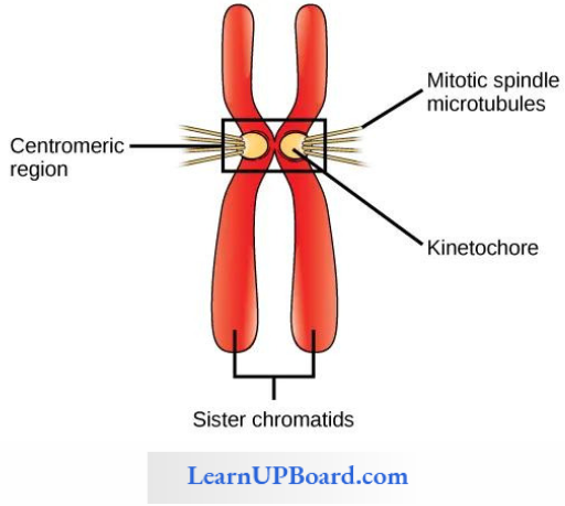
- Chromosomes are composed of two chromatids which are held together at a point called centromere.
- Each chromatid is composed of an elongated fiber called chromonemata. Chromonemata bear several beaded structures called chromomere. The outer covering of the chromosome is called a pellicle.
Maximum Number Of Chromosomes: In higher plants, Poa literosa (Poaceac family) has 266 chromosomes.
- In lower plants, Ophioglossum (pteridophyte; Adder’s tangus fern) has 1262 chromosomes.
- In animals, Aulacantha (radiolarians) has 1600 chromosomes.
Minimum Number Of Chromosomes
- In higher plants, Haplopappus gracilis (Asteraceae) has four chromosomes.
- In lower plants, Mucor has two chromosomes.
- In animals, Ascaris has two chromosomes.
Centromere: The Centromere is technically the non-chromatid part of the chromosome. It is the attachment point of spindle fiber during cell division. The attachment point of the centromere is called the kinetochore. A chromosome can fold at the centromere, hence this point is called primary constriction.
Types Of Chromosomes: On the basis of the position of centromere, chromosomes are of four types
- Metacentric: Centromere in the central position (V shape).
- Submetacentric: Centromere in the subcentral position (L shape).
- Acrocentric: Centromere in the subterminal position (J shape).
- Telocentric: Centromere in the terminal position (I shape).
- Acrocentric Or Holocentric: Centromere absent (I shape).
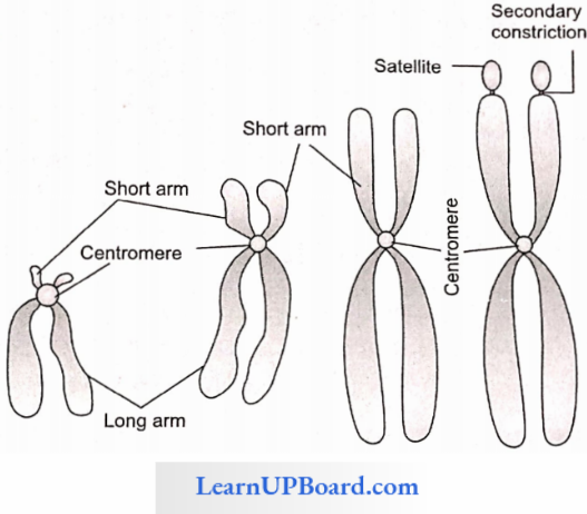
Salient Features Of Chromosomes
- The part of the chromosome after the secondary constriction is called a satellite.
- Chromosome-bearing satellite is called SAT-chromo-some (marker chromosome).
- SAT stands for sine acid thymonucleinico or satellite.
- Secondary constriction is the site of the formation of nucleolus, hence called nucleolus-organizing region (NOR).
- The terminal end of the chromosome is called the telomere which is the point of chromosome sealing.
- The telomere is the sealing end of a chromosome.
- Nucleosome is the fundamental stage of the packaging of the eukaryotic chromosomal DNA.
- The term “nucleosome” was coined by Oudet. According to this model, the nucleosome represents the bead-on-string organization.
- Metaphasic chromosomes are composed of two DNA molecules.
- Anaphasic chromosomes are composed of one DNA molecule.
- The chromosomes present in the plastids are called as plastogenes.
The chromosomes present in the mitochondria are called chondriogenes.
- Q-banding: It is the AT-rich region of the chromosome.
- C-banding: It is the constitutive heterochromatin part.
- R-banding: It is the sulfur-deficient region.
- G-banding: It is the sulfur-rich region and low GC content of chromosomes.
Functions Of Chromosomes
- Since the number of chromosomes in a particular species is constant, hence it is used to determine species.
- It plays an important role in the cell division of eukaryotic cell.
Special Types Of Chromosomes: Some special types of chromosomes or supplementary chromosomes are:
- Giant chromosomes
- Lampbrush chromosome
- Salivary chromosome
- Small-sized chromosomes
- β-chromosome
Lampbrush Chromosome Or Diplotene Chromosome: The Lampbrush chromosome was discovered by Ruckert in the oocyte of a shark, although first observed by W. Flemming in an amphibian oocyte. It was also found in the oocytes of several vertebrates and invertebrates which produce large-sized and yolky eggs.
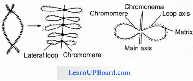
- It is suggested that the loop represents an operon composed of several cistrons which are composed of spacer DNA.
- It is the largest and reproductive chromosome which is three times larger than polytene or salivary chromosome.
- It is a bivalent chromosome found in homologous pair and present permanently in the diplotene stage of prophase 2 of mitosis, hence called diplotene chromo¬some which does not undergo cell cycle.
- Each chromosome is composed of two parts: the main axis and loop.
- The main axis is composed of two chromatids that run parallel to each other and each chromatid is composed of DNA coated with RNA and protein.
- The loop axis is composed of DNA and the matrix is composed of RNA and NH-protein.
- Each loop is the active site of RNA synthesis (m- RNA), hence also the site of the formation of yolk (protein).
- If the loop is treated with DNAase enzyme, then the loop axis dissolves. It indicated that the loop is composed of DNA.
- If the loop is treated with RNAase enzyme, then the matrix gets dissolved. It means the matrix is composed of RNA and protein.
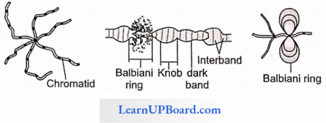
Salivary Or Polytene Chromosome: The salivary chromosome was discovered by Balbini from the salivary gland of the Chironomus larva (insect). It is also found in the salivary gland and mal-pighian tubule of Drosophila as well as the endosperm and antipodal cells of plants.It is a multithreaded chromosome, composed of several chromonemata, hence called polytene chromosome.
- Number Of strands In Drosophila: 1024
- Number Of Strands In Chironomus: 4096
- The term “polytene chromosome” was coined by Roller and Railing.
- It is the second largest somatic chromosome which is 2000 times larger than the mitotic metaphase chromosome of Drosophila.
- Each polytene chromosome represents a pair of homologous chromosomes which after repetitive endomitosis form multi-stranded structures, but all strands remain attached to a common centromere called chromocenter.
- After staining, the polytene chromosome transverse band appears which is composed of dark and light bands alternating each other.
- It also shows swelling at several intervals.
- A dark band rich in DNA is considered a chromomere where DNA is supercoiled.
- The inter or light band represents the inactive part of the chromonemata.
- Swelling is composed of several loops called puffs or Balbiani rings which represent the active site of RNA synthesis
β-chromosome: γ-chromosome is much smaller than the normal chromosome found in maize
- It is heterochromatin and genetically inert.
- It has a slow rate of replication and get destroyed later on.
- It affects the viability of seeds and the fertility of chromosomes.
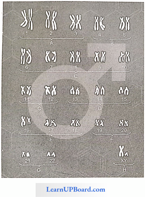
Karyotype And Idiogram: A characteristic pattern of a set of chromosomes present in a diploid somatic cell is called karyotype. The diagrammatic representation of a karyotype is called an ideogram. All chromosomes of the cell are present at the mitotic metaphase stage and arranged in the order of decreasing size.
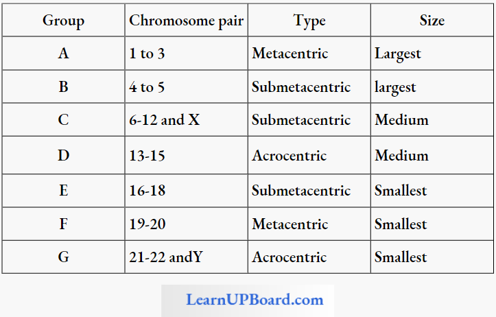
- SAT (Chromosome): 13, 14, 15, 21, 22
- NOR (Chromosome): 13, 14, 15,20, 21
- Smallest Autosomal Chromosome: 21st pair
- Smallest Sex Chromosome: Y chromosome
Types Of Karyotype
- Asymmetric: Karyotype having few metacentric and few other telocentric chromosomes, and there is much difference between the smallest and the largest chromosomes. It indicates advanced features of the organism.
- Symmetric: Karyotypes with several metacentric chromosomes, and there is a gradual change in the size of chromosomes. It indicates a primitive type of karyotype.
Significance Of Karyotype
- With the help of karyotype, any change in the chromosome and any abnormality can be detected.
- It indicates the primitiveness and advancement of the organism.
Human Karyotype
- In humans, WBC is used in the preparation of karyotypes.
- WBC is first treated with colchicine to arrest cell inducting division.
- It is further stained with fuelgen and an idiogram is prepared.
- Tejo and Levan proposed the diploid number of chromosomes in humans for example 23 pairs= 46 chromosomes.
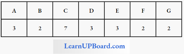
- Denver classified these chromosomes into seven groups represented by the karyotype formula.
Types Of Chromosomes Based On Karyotype Formula
Significance Of Karyotype
- Evolutionary relationship between different species by karyotype analysis.
- Various types of abnormalities present in chromosomes can be easily identified.
- Indicates primitiveness or advancement of the organism.
Genome: A haploid set of chromosomes is called genome.
Plasma: Hereditary factors or genes present in the cytoplasm are called as plasma.
NEET Biology Notes Cell The Unit Of Life Assertion Reasoning Type Questions And Answers
In the following questions, an Assertion (A) is followed by a corresponding Reason (R). Mark the correct answer.
- If both Assertion and Reason are true and the Reason is the correct explanation of the Assertion.
- If both Assertion and Reason are true, but the Reason is not the correct explanation of the Assertion.
- If Assertion is true, but Reason is false.
- If both Assertion and Reason are false.
Question 1. Assertion: RBC membrane is highly flexible.
Reason: The amount of external protein in the cytoplasmic face of the membrane is more.
Answer: 1. If both Assertion and Reason are true and the Reason is the correct explanation of the Assertion.
Question 2. Assertion: Cells of Zona reticularis contain a large number of SER.
Reason: They are present in the adrenal cortex.
Answer: 2. If both Assertion and Reason are true, but the Reason is not the correct explanation of the Assertion.
Question 3. Assertion: Centriole does not form any compartment in a cell.
Reason: Centriole is a non-membranous cell organelle.
Answer: 1. If both Assertion and Reason are true and the Reason is the correct explanation of the Assertion.
Question 4. Assertion: Janus green B is a vital stain for locating mitochondria.
Reason: Janus green is oxidized by cytochrome a-, present in mitochondria.
Answer: 1. If both Assertion and Reason are true and the Reason is the correct explanation of the Assertion.
Question 5. Assertion: Lysosomes help in the digestion of foreign particles in the animal cells.
Reason: They have respiratory enzymes.
Answer: 3. If Assertion is true, but Reason is false.