
NEET Biology Notes Digestion And Absorption Nutrition
The sum total of the processes starting from taking the food up to its utilization is called nutrition. Depending upon the mode of nutrition, organisms can be classified as autotrophs and heterotrophs. Euglena is mixotrophic (both autotrophic and saprophytic).
Animals Nutrients
- Nutrients may be organic or inorganic in nature.
- The organic constituents of nutrients are carbohydrates, lipids, proteins, and vitamins, and inorganic constituents are minerals and water.
- Carbohydrates, lipids, and proteins are macronutrients or proximate principles of food because these constitute energy sources for the production of heat and different organic functions.
- Minerals, vitamins, and water are micronutrients or protective principles of food because although these do not provide energy, yet their deficiencies are related to specific abnormalities in man.
- About 21 minerals (for example, sodium, potassium, calcium, sulfur, phosphorus, magnesium, and chlorine) or macroelements are known to be essential for human nutrition; they are required as 100 mg per day.
- Trace elements or microelements (for example, iron, iodine, zinc, manganese, cobalt, copper, molybdenum, etc.) are required in very small amounts.
- Altogether 20 vitamins are thought to be required in small amounts in human nutrition.
“digestion and absorption neet notes “
Types of Nutrition in Animals: Based on the methods of procurement or collection of food, heterotrophic nutrition is of three major types:
1. Holozoic Nutrition: When whole plants (or their parts) or whole animals (or their parts) or both are consumed either in solid or liquid form, through the mouth.
It can be classified in the following groups
- Herbivores: Feeding on plants, for example, rabbit, goat, cow, etc.
- Carnivores: Feeding on other animals, for example, tigers, lions, etc.
- Omnivores: Feeding on all types of food, for example, humans, cockroaches, etc.
- Detrivores: Feeding on detritus or organic remains, for example., earthworms, etc.
- Scavengers: Feeding on carrions (carrion eaters), for example., hawks, vultures, etc.
- Insectivores: Eating insects, for example, common bats, etc. Cannibals: Eating other members of one’s own species, for example, many snakes, etc.
- Frugivorcs: Feeding on fruits, for example, parrots, etc. Sanguivores: Taking meals of blood, for example, leech, female mosquito, etc.
- Coprophagous: Eats its own fecal matter, for example, rabbit.
2. Saprozoic Nutrition: When decaying organic materials of plant or animal origin are consumed.
The organisms following such nutrition secrete digestive enzymes directly onto their food and therefore the food is digested outside the body, for example, fungi, bacteria some protozoans, etc.
3. Parasitic Nutrition: When a living organism feeds on another living organism and causes harm to it, for example., tapeworm, malarial parasite, etc.
Read and Learn More NEET Biology Notes
NEET Biology Notes Digestion And Absorption Digestive System Of Mammals
Digestion is the catabolic process in which the non-diffusible complex of organic compounds converts into diffusible simple compounds by hydrolytic enzymes. The digestive system comprises (1) an alimentary canal and (2) acessary digestive glands.
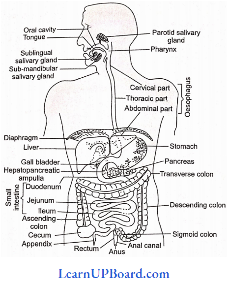
Alimentary Canal: It is a coiled muscular tube about 6-9 m long, extending from mouth to anus. It is divided into three parts:
- Foregut Or Stomodeum: Ectodermal in origin, it includes a mouth and buccal cavity.
- Midgut Or Mesenteron: Endodermal in origin, it includes pharynx to rectum.
- Hindgut Or Proctodeum: Ectodermal in origin, it includes the anal canal and anus.
Path Of Food From Mouth To Anus: Mouth → Buccal cavity → Oropharynx → Oesophagus → Stomach → Small intestine → Large intestine → Anal canal → Anus.
Parts Of The Alimentary Canal
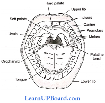
Mouth: The mouth is an opening bounded by upper and lower lips. Lips are attached on the inner side with gums by a thin transparent fold called the labial frenulum of the mesentery.
- The motility of lips is due to orbicular oris muscles.
- Whales and platypuses have immovable lips.
- In men, the middle part of the upper lip is depressed and is called filtrum (a beauty sign).
- The lip of a rabbit has a cleft called a hare lip.
Buccal Cavity
- The buccal cavity is divided into (1) vestibule and (2) oral cavity. The space between lips and teeth is called a vestibule.
- The roof of the oral cavity is made up of a palate. On the anterior side, the hard palate (maxilla, premaxilla, and palatine bone), and on the posterior side, the soft palate is present.
- The mucus epithelium has thick transverse folds called palatine rugae.
- The terminal part of the soft palate hangs in the throat called the uvula.
- On the sides of the uvula, tonsils are present which are made up of lymphatic tissue.
- The floor of the oral cavity is occupied by a muscular tongue.
” class 11th digestion and absorption notes”
Tongue
- The tongue is a voluntary muscular and glandular structure.
- It is attached to the floor of the buccal cavity by a fold called the lingual frenulum of the tongue.
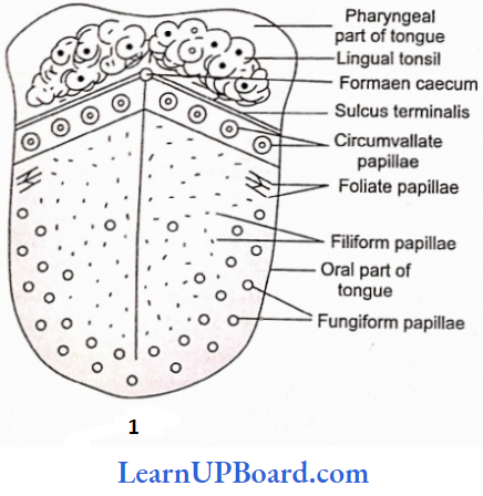
- An inverted V-shaped furrow termed sulcus terminalis divides the upper surface of the tongue into the anterior oral part and posterior pharyngeal part.
- The apex of the sulcus terminalis projects backward and is marked by a small median pit, named foramen cecum.
- Foramen cecum is an embryological remnant and marks the site of the upper end of the thyroglossal duct.
- The upper surface of the oral part of the tongue has small projections called papillae on its surface. These are:
Filiform Papillae: Smallest, most abundant, and have no taste buds.
Fungiform Papillae: These appear as red dots on the tongue and contain taste buds.
Foliate Papillae: Leaf-like, absent in humans.
Circumvallate Papillae: Largest in size, knob-like, and contain taste buds.
Functions Of Tongue
- It helps in the mastication of food.
- It helps in the cleaning of the buccal cavity.
- Helps in speech articulation.
- The sweat glands of dogs are present on the tongue (panting of dog); so it helps in thermoregulation.
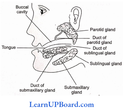
Salivary Glands: Four pairs of salivary glands open in the buccal cavity:
Parotid: Largest—present below and in front of ears—Stenson’s duct.
Submaxillary: Medium-sized —present at the angles of the lower jaw—Wharton’s duct.
Sublingual: Smallest—located below the tongue__ Rivinus duct.
Infraorbital: Absent in man, otherwise present below eyes, for example., rabbit.
- Daily secretion of saliva is 1.5 L (pH of saliva is 6.7) and has salivary amylase (ptyalin), maltase, and lysozyme.
- Salivary glands are stimulated to secrete saliva by parasympathetic innervation; on the other hand, sympathetic nerves cause reduced secretion of saliva leading to the drying of the mouth.
- Cl- ions are required for the activation of salivary amylase.
- Viral infection of salivary glands (mainly parotid) causes mumps disease.
- Food mixed with saliva in the buccal cavity is called bolus.
Teeth: Teeth of human beings are of the following types:
1. Thecodont: Part of teeth present in the bony socket, called alveoli.
2. Heterodont: Different sizes and shapes of teeth are called heterodonts.
Teeth present on the upper and lower jaws are
- Incisors: For cutting; have a single root.
- Canines: For tearing; has a single root.
- Premolars: Upper premolars have two roots; lower premolars have one root.
- Molars: For chewing, the upper molars have three roots and the lower molars have two roots.
- Canines: Well-developed in carnivores. In rabbits, canines are absent and this space is called diastema.
3. Diphyodont: Teeth appear twice in life.
- Milk teeth (deciduous teeth/lacteal teeth)
- Permanent teeth (remains throughout life)
Classification Of Teeth According To Position
- Acrodont: These teeth arise from jaw bones, for example, reptiles, amphibians, and fishes.
- Pleurodont: Teeth fixed to the lateral surface of the jaw ridge, for example, fangs of snakes.
- Thecodont: Embedded in sockets, for example, mammals and crocodiles.
Classification Of Teeth According To The Arrangement Of Enamel And Dentine
- Bunodont: Small cusps, for example, human.
- Lophodont: Long transverse dentine covered by enamel, for example., elephants.
- Selenodont: Crescent-shaped cusps, for example, sheep/ cattle.
- Secodont: Pointed cusps, for example, carnivores.
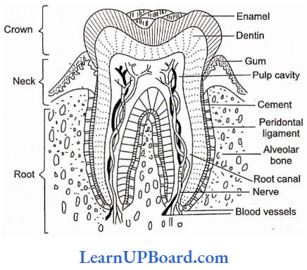
Structure Of Teeth: A tooth is made up of three parts:
- Crown: It is the outer part of the tooth, exposed outside gums.
- Neck: It is the middle part of the tooth which is embedded inside the gums.
- Root: It is the part of the tooth that is inserted inside the socket of the jaw bone (alveoli).
Dental Formulae:
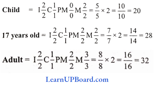
In a human being, 20 teeth grow twice during a lifetime, i.e., diphyodont \(\frac{2102}{2102}\) (premolars and last molars absent), and 12 teeth appear only once in life, i.e., monophyodont.
![]()
Additional Points Regarding Teeth
- Enamel (secreted by ameloblast): The hardest substance of the body—ectodermal in origin.
- Dentine (secreted by odontoblasts): Main part of tooth—mesodermal in origin.
- Caries: Decay of teeth due to degeneration of the enamel and formation of cavities.
- Pyorrhea: Infected gums and tooth sockets.
Pharynx: The pharynx is a common passage in swallowing food and breathing.
- The gullet is the aperture which leads into the esophagus air.
- The glottis is the structure that allows air to enter into the trachea.
- Epiglottis is the structure that prevents entry of food into the windpipe during swallowing in mammals.
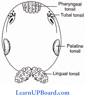
Tonsils: The lymphoid tissues of the pharynx are called tonsils.
- Tonsils are of the following types:
- Nasopharyngeal/pharyngeal tonsil/adenoids
- Palatine/faucial tonsils
- Lingual tonsils
These are arranged in a ring-like manner called Waldeyer’s ring.
Oesophagus: It is a long (25cm) and thin tube that pierces the diaphragm and enters the abdominal cavity. Oesophagus is characterized by:
- Absence of visceral peritoneum. Its outermost fibrous (non-coelomic) covering is called tunica adventitia.
- Absence of digestive glands.
- It has mucus-secreting goblet cells.
- Presence of mucous membrane formed of non-keratinized stratified squamous epithelium.
- Presence of voluntary (anterior one-third) and involun¬ tary (posterior two-thirds) muscle fibers.
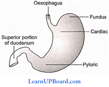
Stomach: The stomach is oval and pouch-like, divisible into cardiac,fundic, and pyloric parts
digestive system class 11th
- The cardiac sphincter is present at the opening of the esophagus into the cardiac stomach and prevents the regurgitation of food into the esophagus.
- The pyloric part opens into the small intestine and the opening is guarded by the pyloric sphincter.
- The wall of the stomach has three layers of muscles, the outermost longitudinal layer, the middle circular layer, and the innermost oblique layer.
- Mucosa has folds called gastric rugae and cardiac, fundic, and pyloric glands.
- Only fundic glands secrete gastric juice. These contain neck cells (secrete mucus and are present in all three types of glands).
- Oxyntic or parietal cells secrete HCl and Castle’s intrinsic factor for absorption of B12.
- HCl of gastric juice converts Fe3+ into Fe2+ which makes the absorption of iron possible.
- Non-secretion of HCl (achlorhydria) or gastrectomy can lead to iron deficiency (anemia).
- Peptic cells chief cells or zymogenic cells release large quantities of pepsinogen.
Mucus cells: A large amount of mucus is secreted, Semisolid food, mixed with gastric juices in the stomach, is known as chyme (it is highly acidic).
Compound stomach
- The stomach of ruminants is known as the compound stomach.
- It has four well-defined chambers or compartments, viz., rumen, reticulum, omasum, and abomasum.
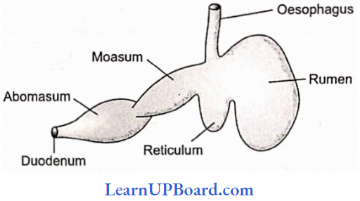
- Rumen is the first and the largest chamber mainly meant for the storage of food.
- In camel and deer, omasum is absent, and water cells project from rumen. Digestion of cellulose takes place by fermentation, with the help of symbiont bacteria and protozoans.
- The abomasum is the true stomach, which secretes gastric juices.
- Rabbits eat their own feces (coprophagy) to complete the digestion of cellulose. It is taken as pseudo-rumination.
Small Intestine: It consists of three parts: (1) duodenum, (2) jejunum, and (3) ileum.
- The first part is the duodenum, 25 cm long, C-shaped in humans, and has an opening of the hepatopancreatic duct (bile duct + pancreatic duct).
- A small swelling is present at the opening of the hepatopancreatic duct and is called the ampulla of Vater or hepatopancreatic ampulla and the opening is regulated by the sphincter of Oddi.
- The next part ofsmall intestine is jejunum and ileum.
- The wall of the intestine has thin layers of longitudinal and circular muscles.
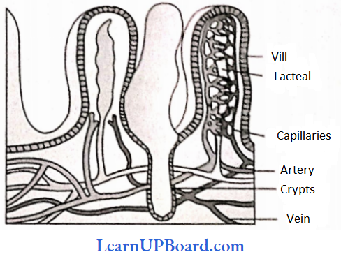
- Mucosa has folds plicae circulars (folds of Kerkrings or valvulae conniventes) and villi toward the lumen of the intestine.
- Epithelial cells lining the villi have microvilli which further increase the absorptive area.
- Intestinal glands or crypts of Leiberkuhn have epithelial cells (secrete mucus), path cells (secrete digestive enzymes), and argentaffin cells (probably secrete hormones).
” difference between digestion and absorption”
- In the duodenum, Brunner’s glands are also present (located in the submucosa) which secrete mucus.
- Diffused patches of lymphoid tissues are present throughout the small intestine and are aggregated in the ileum to form Peyer’s patches.
- Intestinal villi are mainly concerned with absorption.
- The main function of intestinal villi is to provide a large surface area for absorption.
- The absorption of digested food mainly occurs in the small intestine mainly the ileum.
- The lymph capillary present in a villus is called a lacteal. It is concerned with the absorption of digested fat.
- The distal end of the ileum is expanded to form a small dilated spherical sac called sacculus rotundus in rabbits. The ileum opens into the cecum through an ileocecal valve.
- Half liquid food mixed with bile, pancreatic juice, and succus entericus in the intestine is called chyle (highly alkaline).
Food + Bile + Pancreatic + Intestinal juice = Chyle
Large Intestine
- It is about 1.5 m long and consists of three parts: (1) caecum, (2) colon, and (3) rectum.
- A blind pouch of the cecum is a vermiform appendix.
- These parts help in the digestion of cellulose in herbivores (rabbit).
- The wall of the colon has sac-like haustra. Histologically, the wall of the colon has three bands of longitudinal muscles called taenia coli.
- Another characteristic of the colon surface is the presence of small fat-filled projections called epiploic appendages.
- The colon part is divisible into ascending, transverse, descending, and sigmoid colon.
- The ascending colon is the smallest and is without mesentery.
- The last part of the rectum is the anal canal having a strong sphincter. It opens outside by the anus.
- In certain conditions (such as persistent constipation), rectal veins can get distended or enlarged due to weakening valves (varicosity). It leads to swollen areas called hemorrhoids (piles).
Histology Of Alimentary Canal: The alimentary canal consists of four basic layers. From the outer surface toward the inner side of the tire lumen (cavity), the layers are as follows:
Visceral Peritoneum (serous membrane or serosa): It is the outermost layer made up of squamous epithelium. It is continuous with the mesentery.
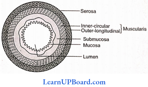
Muscular Coat: It is composed of outer longitudinal and inner circular muscle fibers.
- In the stomach, an additional layer of oblique muscle fibers is found in the circular muscle fibers.
- These muscle fibers are unstriped (smooth). The muscular coat also contains the major nerve supply to the gastrointestinal tract—the mesenteric plexus (plexus of Auerbach), which consists of fibers from both au autonomic divisions and mostly controls the peristaltic movements in the alimentary canal.
Submucosa: It consists of loose connective tissues richly supplied with blood and lymphatic vessels and in some areas with glands.
- Meissner’s plexus (submucosal plexus) is present between the muscular coat and the mucosa which is part of the autonomic nerve supply to the smooth muscles and secretory cells of mucosa glands.
- It controls various secretions of the alimentary canal and movements of the mucosa.
Mucosa (mucous membrane): It is so named because it secretes mucus to lubricate the inner lining of the gut. It is composed of three layers:
- The thin muscularis mucosa lies next to the submucosa. It consists of outer longitudinal and inner circular muscle fibers, both arc unstriped.
- Lamina propria, the middle layer of mucosa, consists of loose connective tissues, blood vessels, glands, and some lymphoid tissues.
- The innermost layer is the epithelium, which forms gastric glands in the stomach, and villi and intestinal glands in the small intestine. In the upper one-third of the esophagus, both Auerbach and Meissner’s plexuses are absent.
Digestive Glands
Liver
- Largest digestive gland. It lies in the upper right side of the abdominal cavity just below the diaphragm.
- The liver is divided into two main lobes: right and left.
- The right lobe is differentiated further into the right lobe proper, a quadrate lobe, and a caudate lobe on the posterior surface.
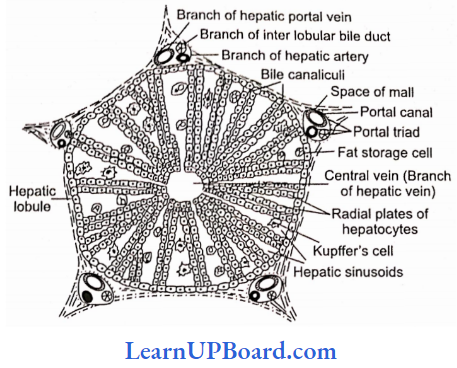
- The liver is surrounded by Glisson’s capsule’, its trabeculae divide liver lobes into hexagonal lobules.
- Polyhedral hepatocytes arc arranged in cords around a central venule.
- Portal triads contain the hepatic artery, portal venule, bile ductule, and lymphatics.
- Blood sinusoids are present. Kupffer cells are present in sinusoids and are phagocytic in nature.
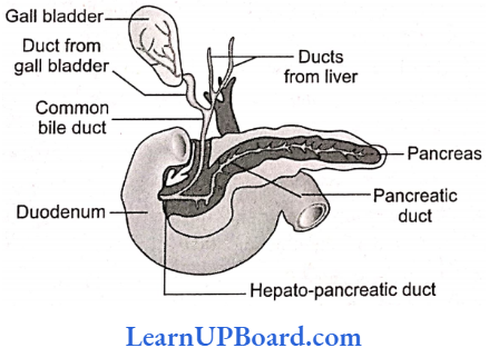
- Gail’s bladder is situated on the inferior surface of the right lobe. It is 8 cm long and 2 cm wide.
- Bile is secreted by hepatocytes into the bile canaliculi, a series of narrow spaces between adjacent liver cells.
- The canaliculi drain via bile ductules into bile ducts, which run in portal tracts; the bile ducts themselves discharge into the right and left hepatic ducts which unite to form the common hepatic duct at the hilum of the liver.
- Gall bladder has the capacity of storing 30 to 50 mL of bile. It fills and empties via the cystic duct which joins the common hepatic duct to form the bile duct; this in turn empties into the duodenum through the ampulla of Vater (hepatopancreatic ampulla).
- At the point of its entry into the duodenum, the bile duct and adjacent pancreatic duct join each other.
- The sphincter of Boyden surrounds the opening of the bile duct into the pancreatic duct. The sphincter of Oddi surrounds the ampulla of Vater.
Functions Of Liver
- Secretion of bile: Daily secretes 700-1000 mL. In the gall bladder, electrolytes and water are reabsorbed concentrating bile approximately 10 to 15 times.
- The bile contains bile pigments bilirubin and biliverdin, bile salts sodium glycocholate, sodium taurocholate, and sodium bicarbonate.
- Sodium bicarbonate is mainly responsible for the alkaline nature of bile (pH = 8.0).
- Cholesterol is insoluble in water but its association with bile acid and phospholipid makes it soluble.
- In the subjects, who regularly produce bile, supersaturated with cholesterol, the cholesterol may be deposited in the gall bladder as gallstones (cholelithiasis).
- Inflammation of the gall bladder is called cholecystitis.
- Storage of fat.
- Urea synthesis.
- Erythropoiesis (during embryonic period only).
- Breakdown of RBCs.
- Iron is stored as ferritin.
Pancreas (Heterocrine Gland)
- A racemosely branched gland situated between the stomach and duodenum.
- The pancreas consists of acini (which secretes digestive enzymes) and islets of Langerhans (which secretes insulin and glucagon hormones).
- The pancreas has two ducts with it. The first is the duct of Santorini which is accessory or non-functional, opening directly into the duodenum and the other is the duct of Wirsung which is functional and combines with the bile duct to form a common hepatopancreatic duct.
Mobility Of Alimentary Canal
- The alimentary canal undergoes regular contraction for proper digestion and absorption of food.
- Food enters into buccal cavity where it is mixed with saliva.
- Food is masticated with the help of teeth. The salivary enzyme ptyalin (salivary amylase) causes chemical breakdown of food (i.e., carbohydrates).
- Smaller food particles are held together by the mucin of saliva forming the food bolus which is then swallowed.
- The food bolus gets swallowed, i.e., enters the esophagus with the coordinated activity of the tongue, soft palate, and pharynx.
- Waves of contraction or peristaltic waves in the esophagus push it downward.
- As the food reaches the end of the esophagus, the cardiac sphincter, regulating the opening of the esophagus into the stomach, relaxes to allow the entry of food into the stomach.
- If the sphincter fails to open up properly, it leads to the accumulation of food in the lower part of the esophagus called cardia achalasia.
- In the stomach, mechanical churning of the food occurs by waves of contractions passing from the cardiac to the pyloric end, and mixing of food with gastric juice also occurs.
- During this activity, cardiac and pyloric sphincter remains closed.
- If the cardiac sphincter remains open, acidic gastric contents may escape into the esophagus, leading to a heart bum condition due to a burning sensation in the esophagus.
- From the stomach, acidic chyme enters the small intestine where digestion is completed followed by the absorption of digested food.
- From the small intestine, the chyle enters the large intestine and is finally egested out.
- Movements in the alimentary canal are caused by myenteric plexus, as well as hormones such as motilin, villikinin, gastrin, etc. It shows peristaltic movements.
- The peristaltic movements involve contraction and relaxation resulting in wave-like movement.
- Contraction is due to the contraction of circular muscles and relaxation of longitudinal muscles. Relaxation is caused by the simultaneous contraction of longitudinal muscles and the relaxation of circular muscles.
- Peristaltic movements start from the esophagus.
- The churning movements of the stomach are also peristaltic movements which become powerful as they proceed toward pylorus.
- In the large intestine, peristaltic movements are moderately weak.
Digestion And Gastrointestinal Secretions
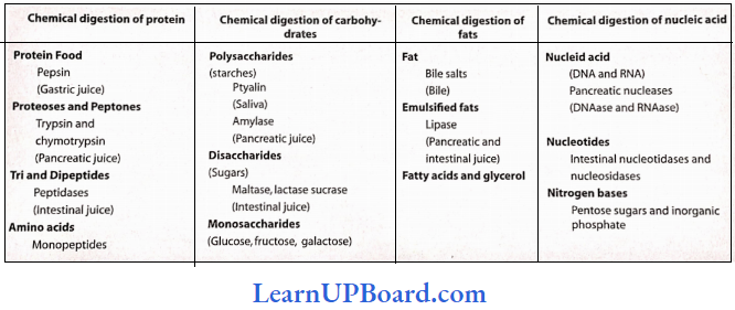
Digestion Of Carbohydrates
- The diet of most of the animals including man consists of carbohydrates.
- Depending upon the complexity, carbohydrates are of three types: polysaccharides, disaccharides, and monosaccharides.
- During the process of digestion, both poly-and disaccharides are broken down into monosaccharides, and in this form, they can be absorbed into the body.
- Some of these complex carbohydrates are starch and cellulose present in cereal grains, potatoes, fruits, and tubers, sucrose present in cane sugar, lactose present in milk, etc.
- Enzymes that act- on carbohydrates are collectively known as carbohydrases. In the mouth cavity, the food is mixed with saliva.
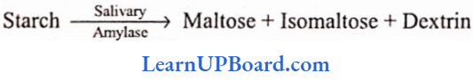
- It contains an enzyme called salivary amylase or ptyalin.
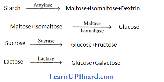
- Salivary amylase acts on starch and converts it into maltose, isomaltose, and small dextrins or limit dextrin’ (disaccharides).
- Chewing and mastication of food increase the action of salivary amylase on starch by increasing the surface area of food on which the enzyme acts.
- Cooking of food also helps the action of salivary amylase by causing breaches in the cellulosic cell walls.
- The amylase can, thus, enter into the food and digest starch.
- About 30 % of the starch present in food is hydrolyzed in the mouth itself.
- The action of salivary amylase continues for sometimes even in the stomach but soon. HCl present in the gastric juice destroys all the enzymes.
- Salivary amylase is absent from the saliva of many mammals such as cows and buffaloes; and predatory carnivores such as lions and tigers. However, pigs have got amylase in their saliva.
- Pancreatic juice and intestinal juice also contain carbohydrate-digesting enzymes.
- Pancreatic juice contains pancreatic amylase that acts on starch to digest it into maltose, isomaltose, and dextrin.
- Intestinal juice contains a number of carbohydrates such as maltase, isomaltase, sucrase, and lactase.
- Maltase and isomaltase act on maltose, isomaltose, and dextrin and convert them into glucose; sucrase acts on sucrose to convert it into glucose and fructose, and lactase acts on lactose to convert it into glucose and galactose.
- Human beings can digest lactose present in the milk. But with advancing age, they also cannot digest milk. This is because less lactase is produced. In them, lactose remains undigested and gets fermented in the intestine producing gases and acids. This results in flatulence, intestinal cramps, and diarrhea. So these persons must consume curd or yogurt (sweetened curd) as lactose is fermented to lactic acid in them.
- Galactosemia: Galactosemia is a metabolic disorder due to the absence of the enzyme uridyl transferase. As a result, galactose will accumulate leading to mental retardation.
- It can be prevented in galactosemic children by giving them a milk-free diet.
Digestion Of Proteins
- Proteins are complex organic compounds made up of single units called amino acids.
- So in the process of digestion, all proteins are broken down into amino acids.
- Enzymes that hydrolyze proteins are collectively known as proteases or peptidases.
- Many of these enzymes are secreted in their inactive font or proenzymes as their active form would hydrolyze cellular and extracellular proteins of the organism itself.
- Inactive enzymes are converted to their active form only at the site of action.
- Saliva as such does not contain any protein-digesting enzyme, but it can denature the uncooked natural proteins, like the ones present in raw egg, unboiled milk, or uncooked germinating seeds.
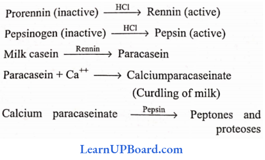
- The action of gastric juice: The gastric glands of the stomach produce light-colored, thin, and transparent gastric juice.
- It contains water, hydrochloric acid (0.3%), and inactivated enzymes, prorennin and pepsinogen.
- The presence of MCI makes the medium highly acidic (pH = 1 or 2) so that pepsin can act on proteins to convert them into peptones and proteoses. However, there is no pepsin in invertebrates.
- Both prorennin and pepsinogen are converted to their active forms in the presence of HCl.
- Pepsin and rennin can also perform the same function once they are formed. HCl helps to kill bacteria and other harmful organisms that may be present along with the food.
- Rennin acts on the casein protein of milk and converts it into paracasein which in the presence of calcium ions forms calcium paracaseinate (curdling of milk).
- Adult humans do not produce renin.
- The function of renin is then taken over by pepsin and other milk-coagulating enzymes in them. These reactions are summarized below:
- Pepsin can even digest collagens of connective tissue fibers, but it cannot act on the keratins of horns, hair, skin, or nails.
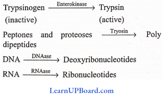
- The action of pancreatic and intestinal juice: Both pancreatic juice and intestinal juice (succus entericus) are poured into the small intestine.
- Pancreatic juice contains trypsinogen, chymotrypsinogen, procarboxypeptidases, lipases, amylases or amylopsin, DNAases, and RNAases.
- All these enzymes of pancreatic juice can act only in the alkaline medium.
- This change in the medium of food, from acidic to alkaline, is done by the bile juice. Therefore, bile juice acts on the food before the action of pancreatic juice. All these actions are given below:
- In predatory animals, trypsin can hydrolyze fibrinogen of blood into fibrin leading to blood coagulation. However, it is unable to bring about coagulation of milk. Also, trypsin cannot hydrolyze keratins.
- Chymotrypsinogen (inactive) is activated to chymotrypsin by trypsin itself. Chymotrypsin is another important milk-coagulating enzyme and can hydrolyze casein into paracasein which then coagulates to form calcium paracaseinate. However, unlike renin, it acts in an alkaline medium. Chymotrypsin can act on other proteins also.
- Carboxypeptidase hydrolyzes the terminal carboxyl group from peptide bonds to release the last amino acid from the peptides thus making the peptide shorter.
- Intestinal juice or succus entericus contains two protein-digesting enzymes aminopeptidases and carboxypeptidases which hydrolyze the terminal amino group from peptide bonds to release the last amino acid from the peptides thus making the peptide chain shorter.
- Dipeptidase acts on dipeptides to release individual amino acids.
- Enterokinase is also released which activates trypsinogen to trypsin.
Digestion Of Fats
- Fat digestion starts only when the food reaches the small intestine.
- It starts with the action of bile juice from the liver.
- Bile juice contains bile salts that break down the bigger molecules of fat globules into smaller droplets by reducing the surface tension of fat droplets. This process is known as emulsification of fats.
- Lipase is the chief enzyme that acts on emulsified fats.
- It is present both in pancreatic juice and intestinal juice.
- Pancreatic lipase (steapsin) is the strongest lipase.
- Lipase converts emulsified fats into diglycerides and monoglycerides releasing fatty acids at each step.
- At the end of digestion, all fats are converted into fatty acids, glycerol, and monoglycerides.
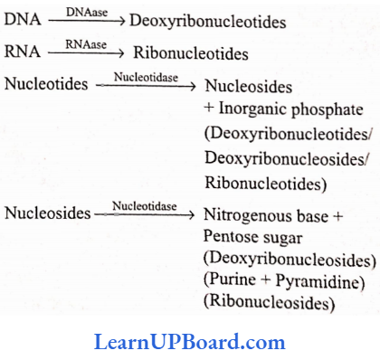
Digestion Of Nucleic Acids
- Nucleic acids are digested in the small intestine with the help of pancreatic and intestinal juices.
- Pancreatic juice contains two nucleases: DNAase and RNAase. Intestinal juice contains nucleotidase and nucleosidase.
NEET Biology Notes Digestion And Absorption
Almost no absorption takes place in the mouth and esophagus. Water, alcohol, simple salts, and glucose are absorbed in the stomach. In the small intestine, absorption of all digested materials takes place by active, passive, and facilitated transport.
- Glucose, sodium, and amino acids are absorbed actively.
- The absorption of glucose or amino acids involves carrier-mediated transport which binds glucose/amino acid at one site and Na+ at the other site. Therefore, the movement of glucose/amino acid is coupled to the concentration gradient of Na+ (Co transport).
- Na+ moves along the concentration gradient while glucose/amino acids are moving against a concentration gradient.
- The rate of absorption of galactose is the highest.
- Fructose is absorbed by facilitated diffusion.
- The products of fat digestion, monoglycerides, fatty acids, and glycerol are first incorporated into water-soluble droplets called micelles (a combination of fatty acids, monoacylglycerols, and bile salts); reconstructed to triglycerides in the absorptive cells; and released into lymph in the form of protein-coated water-soluble fat droplets called chylomicrons.
- In the large intestine, only water is absorbed. Absorption of vitamin B12 (cobalamine) in man requires a glycoprotein, called intrinsic factor (IF) secreted by the parietal cells of the stomach. Failure to absorb cobalamine causes pernicious anemia associated with a failure of RBC maturation (megaloblastic anemia) and neurological abnormalities.
NEET Biology Notes Digestion And Absorption Assimilation Of Food
The absorbed food materials are transported by blood and lymph. Lymph is finally transferred to the blood circulation. The blood transports absorbed food materials to different body cells where food materials become integral components of the living protoplasm and are used for energy, growth, and repair. This is called the assimilation of food.
- Amino acids are not stored but are taken up by the cells in connection with the synthesis of proteins.
- Proteins are used for growth, repair, etc. Excess amino acids can be converted into glucose and then to fat and are thus stored. This is an irreversible reaction.
- Amino acids can also be converted to glucose and used as fuel for the cell.
- During their conversion to glucose, the amino acids are deaminated (removal of amino groups —NH2).
- The liver is the chief site for deamination, i.e., a process by which the amino group is removed from amino acids resulting in the production of ammonia.
- Ammonia is soon converted into urea, which is filtered from the blood in the kidney.
- The excess of monosaccharides (glucose, fructose, and galactose) is usually stored in the liver and muscle cells in the form of glycogen (glycogenesis).
- Whenever there is a deficiency of glucose in the blood, glycogen is converted into glucose (glycogenolysis).
- Muscle glycogen is utilized during muscle contraction. Glucose is utilized in the production of energy for various body activities.
- A considerable amount of glucose is converted into fat and stored as such.
- The fat is stored in the fat deposits of the body, such as subcutaneous layers, mesenteries, etc.
- The stored fat is a readily available source of fuel for the cells.
- Fat has important insulating properties in connection with the conservation of heat and maintenance of body temperature.
- Fat also plays a protective role as filling or packing material, between and around the organs.
- In the liver, phospholipids are formed which are returned to the blood, to be used by all the cells. In liver cells, fats are converted into amino acids and carbohydrates.
- Vitamins, salts, and water are also useful for various metabolic processes.
NEET Biology Notes Digestion And Absorption Egestion
Peristalsis gradually pushes the slurry of indigestible materials of the small intestine into the large intestine or colon. Approximately, 1500 mL of chyme normally passes through the large intestine each day. The colon absorbs most of the water, electrolytes, and ions from these contents. This is accomplished by the active pumping of sodium and water by osmosis from die chyme.
- The other function of the colon is to help in the excretion of excess salts from the blood.
- The population of Escherichia coli (bacterium), which is a resident species of the colon, lives on this undigested matter. This bacterium, in turn, produces vitamin B12, vitamin K, thiamin, and riboflavin which are absorbed by the wall of the colon.
- Later on, the chyme is slowly solidified into coherent feces, which are about three-fourths water and one-fourth solid matter consisting of about 10-20% inorganic substance 30% dead bacteria, 10-20% fat, 2-3% protein, and 30% undigested roughage and dry constituents of digestive juices.
- Feces are given out through the anus by the process of defecation or egestion.
- The breakdown of bile pigments occurs, forming stercobilin pigment, which provides a brownish color to the feces.
- The foul smell of the fecal matter is due to skatole (3-methyl indole).
- Dark green mucilaginous material in the intestine of the full-term fetus is called meconium (includes residue from the swallowed amniotic fluid by the fetus and the residues of excretory products from intestinal mucosa and glands).
NEET Biology Notes Digestion And Absorption Cellulose Digestion In Ruminants
The minants ingest large quantities of fodder-rich food in cellulose or lignin. Neither substance can be digested directly by mammals, but the cellulose is attacked in the rumen by the enzymes produced by symbiotic bacteria. Sheep and cattle initially swallow food without masticating. In the rumen, food is mixed with water.
- The rumen has millions of bacteria of several species, some of which attack the cellulose walls of the fodder liberating the cell contents of the food.
- These bacteria also break down carbohydrates and proteins into simple substances.
- In the rumen, the main products of fermentation are acetic, propionic, butyric, and other acids, along with small quantities of other substances such as ethanol.
- Besides this, the symbiotic organisms in the rumen synthesize much more riboflavin and pantothenic acid than is normally procured through diet.
- In addition to bacteria, a number of protozoan ciliates have also been observed in the rumen. Some of these can break down cellulose.
- The stored carbohydrates and proteins within these ciliates are believed to serve as reserves for some time when fodder is not available.
- Cellulose breakdown by bacteria also takes place in the stomach of kangaroo (Setonix brachytirus), and sloth (Choloepus). In kangaroos, the stomach is larger, and in the sloths, there is a compound stomach.
- Cellulose-splitting bacteria are present mostly in the cecum and ventral colon of horses and in the cecum of rabbits. The rabbits, like other lagomorphs and many rodents, have developed a habit of ingestion or pseu domination.
- When a rabbit eats fresh food, it directly reaches the cecum, remains there for one or two days undergoing fermentation, and is then expelled as soft feces.
- These are then again eaten and this time they reach the cardiac stomach, but the freshly eaten food goes straight into the cecum as usual.
- After digestion and absorption, the twice-swallowed food is excreted as hard fecal pellets without letting it in through the cecum.
NEET Biology Notes Digestion And Absorption Nutritional Requirements Of Humans
Carbohydrates are used primarily as sources of chemical energy, to be either metabolized immediately as glucose or stored as glycogen. The synthesis of glycogen is called glycogenesis. The liver can store enough glycogen to maintain blood glucose levels for several hours. Under acute starved conditions, the liver cells begin to convert amino acids and glycerol (digestive products of fat molecules) into glucose. Such production of new glucose is known as gluconeogenesis.
- Proteins are used as structural components of tissues as channels, transporters, regulatory molecules, and enzymes.
- Proteins can also be utilized as energy sources when broken down into amino acids.
- Out of the 22 amino acids identified so far as the constituents of proteins, eight (10 in children) cannot be synthesized in the human body.
- These must be provided in the diet from outside and are designated as essential amino acids.
- Lipid (fat) molecules are especially suitable as concentrated energy reserves. The fat cells of adipose tissues are often called the fat depot of the body.
- Triglycerides are used as fuel. The human body is able to synthesize most of the lipids in enough quantity, except three polyunsaturated fats, such as linoleic, linolenic, and arachidic acids.
- These essential fatty acids must be provided to the human body through diets.
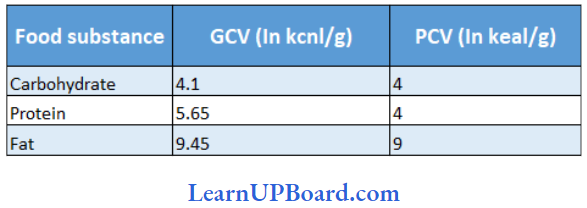
NEET Biology Notes Digestion And Absorption Calorific Value
The amount of heat liberated from the complete combination of 1g of food in a bomb calorimeter (a dosed metal chamber filled with O2) is its gross calorific value GCV) or gross energy value. The actual amount of energy liberated in the human body due to the combustion of 1g of food is the physiologic calorific value (PCV) of food.
- The energy requirements of animals and the energy content of food are expressed in terms of a measure of heat energy because heat is the ultimate form of all energies. This is often referred to as calorie (cal) or joule (J). which is the amount of heat energy required to raise the temperature of 1g of water through 1°C.
- Since this value is a tiny amount of energy, physiologists commonly use kilocalories (kcal) as a unit of measure (1 keal = 1000 eal) or kilojoule (kJ). One kilocalorie is the amount of energy required to raise the temperature of 1 kg of water through 1°C.
- Nutritionists traditionally refer to kcal as Calorie or Joule (always capitalized).
NEET Biology Notes Digestion And Absorption Minerals And Vitamins
Both minerals and vitamins occur as small molecules and mostly do not require digestion. Minerals are ingested as salts dissolved in water or as part of organic compounds (food). Still, a few of the minerals are absorbed with the aid of digestive juices (like bile) and gastric juices. Of the 21 essential minerals required by man, some are important for maintaining fluid balance, whereas others help to regulate metabolism by acting as a component of enzymes.
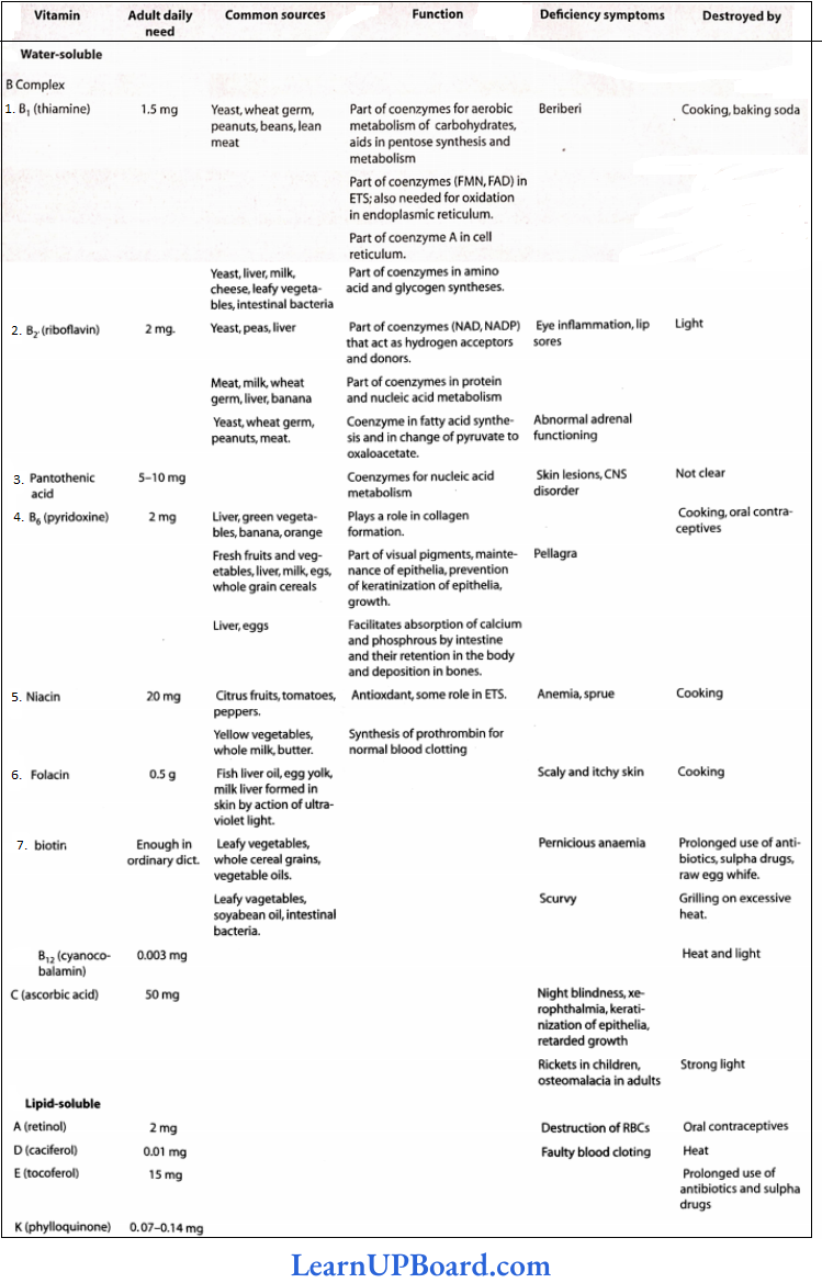
- Vitamins are essential for normal metabolism, growth, and sound health.
- Humans can synthesize vitamin A (retinol) with the help of plant pigment, carotene, which is available in yellow and green leafy vegetables.
- Vitamin A forms the retinal pigment of human eyes such as rhodopsin of rod cells and iodopsin of core cells.
- Humans can also synthesize vitamin D (calciferol) in their skin in the presence of ultraviolet rays of light. Although most animals can synthesize vitamin C from glucose, humans cannot; hence, they require it in their diet. Vitamin Bl7 is a newly discovered vitamin with anticancerous properties.
- Chemical nature: Vitamins differ greatly in their chemical nature. Generally, they have nothing in common. Some vitamins are alcohol, some arc proteins, some arc quinone, and some are sterol.
- Provitamins: These are compounds that are changed into vitamins in our body. Examples arc:
- Ergosterol present in food is changed in the skin into vitamin D in the presence of sunlight.
- Carotene, a pigment present in carrots is changed in the liver and in the small intestine into vitamin A.
- Vitamers: This term is used for different forms of the same vitamin. Vitamers are also called isotopic vitamins. Examples are:
- Vitamin A has vitamins A1 and A2 (vitamins).
- Vitamin D, has vitamin C, D2, and D3 (vitamins).
- Vitamin poisons: These are compounds that replace vitamins in our body, for example, antibiotics. Sulfa drugs replace vitamin C; tetracycline or ampicillin replace vitamin B complex.
NEET Biology Notes Digestion And Absorption Nutritional Deficiencies And Disorders
Deficiencies of nutrients such as vitamins, minerals, and proteins, in the food are related to specific disorders, diseases, and abnormalities in humans. Impairment of health due to improper intake of food or nutrients results in the effect recognized as malnutrition. Malnutrition is a term that covers problems of both undernutrition and overnutrition. Two types of diseases due to inadequate nutrition are described.
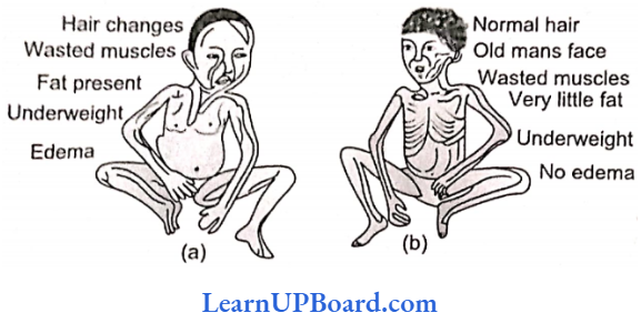
Differences between kwashiorkor and marasmus:
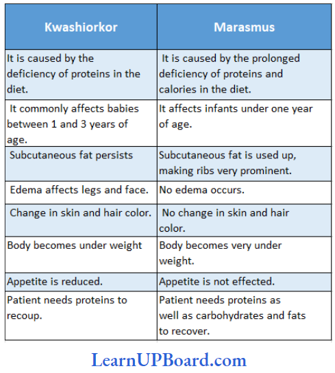
- An individual or a group of individuals may be undernourished due to non-availability of food, and hence, deficiency of minimum required food and nutrients.
- In this situation of undernutrition, the intake of food is too insufficient to meet the needs for metabolic energy. Consequently, the individual shall have to make up the shortfall by metabolizing some molecules of its own body.
- Excess intake of food and nutrients may cause a great deal of harm to the body. The excess nutrients are stored as increased body mass. Such a situation is attributed to overnutrition. Excess intake of saturated fats such as butter, ghee, vegetable oils, red meat, eggs, etc., often leads to hypercholesterolemia, a condition in which blood cholesterol content becomes abnormally high, ultimately leading to cardiac disorder.
- Deposition of cholesterol on the walls of blood vessels stiffens the blood vessels and increases blood pressure. Besides, excessive intake of calories (sugar, honey, ghee, etc.) may produce overweight and obesity (excessive accumulation of fat in tissues), which is the most common form of overnutrition.
- Very high intakes of minerals and fat-soluble vitamins (obtained from food sources alone) can be toxic. This is because they are stored in the body. With the exception of folic acid (women of childbearing age), people who have well-balanced diets that supply enough energy do not usually need to take dietary supplements.
- But if they do decide to take supplements, then they should follow the advice on the label to reduce the risk of an overdose.
NEET Biology Notes Digestion And Absorption Disorders Of The Digestive System
- Inflammation Of The Intestinal Tract: The most common bacterial viral infection may be caused by intestinal parasites such as tapeworms, roundworms, threadworms, hookworms, and pinworms.
- Jaundice: The Liver is affected, and skin and eyes turn yellow due to the deposition of bile pigments.
- Vomiting: This reflex action is controlled by the vomiting center in the medulla. A feeling of nausea precedes vomiting.
- Diarrhea: It reduces the absorption of food, due to abnormal frequency of bowel movement.
- Constipation: Due to irregular bowel movement, feces are retained within the rectum.
- Indigestion: Feeling of fullness as the food is improperly digested. It is due to inadequate enzyme secretion, anxiety, food poisoning, overeating, and spicy food.
- Belching: It occurs usually when the stomach is over-dilated, air rises, and is expelled through the mouth producing a burping sound.
- Obesity: It is a condition when fat storage in the body increases, leading to its abnormal deposition in the subcutaneous layer. It is caused when energy input exceeds energy output in the body. 8.3 calories of energy from surplus food increases 1g of fat deposition in the body causing obesity.
- Flatus: It is an accumulation of gases in the gastrointestinal tract that are expelled through the anus producing a characteristic sound.
- Hepatitis: It is a condition of inflammation of the liver caused by infection of bacteria, viruses, or protozoa. It may cause cirrhosis.
- Colitis: It is a disorder in which inflammation of the colon and rectum, loose motions, bloody feces, and dehydration occur. It is caused by the infection of protozoans such as Entamoeba.
- Appendicitis: The wall of the vermiform appendix ruptures and bacteria are released in the coelomic cavity. Severe infection may cause death. By surgery, the appendix is removed.
- Hernia: It is a protrusion of the intestine into the inguinal canal and may extend into the scrotal sac.
- Mumps: It is a viral infection in parotid salivary glands. The throat region swells and fever develops.
- Nausea: It refers to the discomfort which leads to vomiting. It may be caused by distension of the stomach or of the gastrointestinal tract.
- Vomiting: It is a condition developed by reverse peristalsis caused by harmful substances in the stomach irritation in the pharynx or disturbance in semicircular canals. Food substance comes out through the mouth. Vomiting center is located in the medulla oblongata.
- Tonsillitis: The pharyngeal tonsils (lymphoid tissues) become infected with microorganisms. It causes pain in the throat followed by fever.
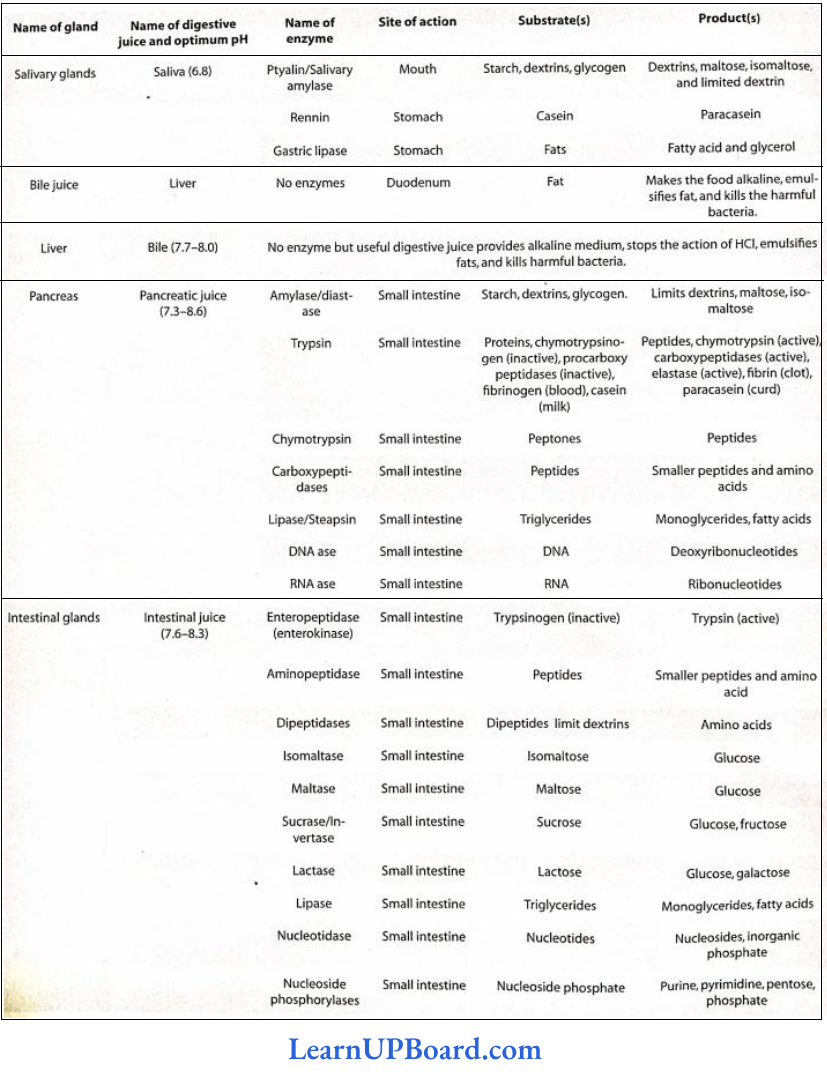
Summary Of Digestion:
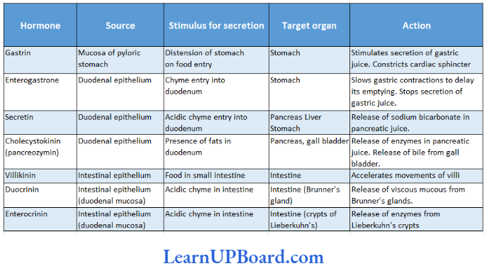
Gastrointestinal Hormones In Mammals:
Summary Of Chemical Digestion Of Food:
Summary Of Human Vitamins:
NEET Biology Notes Digestion And Absorption Points To Remember
- Assimilation: Utilization of absorbed material by the cell.
- The hunger center is in the hypothalamus.
- The Satiety center is also in the hypothalamus.
- Heartburn has nothing to do with the heart. It is caused by the regurgitation of acid from the stomach into the esophagus.
- Splanchnology is the study of the viscera.
- NIN: National Institute of Nutrition, Hyderabad.
- Anorexia: Loss of appetite.
- Spoilt hay of sweet clover Melilotus indica (fodder and green manure) contains a substance called dicumarol. Dicumarol prevents the action of vitamin K as it is antagonistic to it.
- What destroys the vitamins? Overcooking and excessive boiling, medicines such as aspirin, antacids, and diuretics lead to iron deficiency—anemia.
- Tea/coffee inhibits the absorption of iron from the diet. Prolonged consumption of tea or coffee after meals can lead to iron deficiency—anemia.
- In the upper one-third of the esophagus, only skeletal muscles are found.
- The chief seat of water absorption is the small intestine.
- The liver produces proteins such as albumin, fibrinogen, and prothrombin, but does not produce globulin.
- Poison glands of a snake are modified labial glands, homologous to parotid salivary glands.
- Vomerine teeth of frogs kill the prey.
- The tongue of a whale is not movable.
- The gall bladder is absent in adult lamprey (jawless vertebrate), grain-eating birds, rats, whales, all the perissodactyla (odd-toed hoofed mammals such as horses), and some artiodactyla (even-toed hoofed mammals).
- Alcoholics are short of vitamin C.
- C-shaped duodenum is a characteristic of man.
- During a high fever, one does not feel like taking meals because a high temperature shuts off the appetite center.
- Bile is alkaline in humans, but acidic in cats and dogs.
- Basal metabolic rate (BMR) is the minimum energy requirement for the maintenance of the body during rest or sleep. For a normal human adult, it is 1600 keal/day.
- Routine metabolic Rate (RMR): It is the energy requirement of a moderately active person. RMR is 2800 keal/day for adult males and 2200 keal/day for adult females.
- Entero-hepatic circulation: Of the total bile salts that enter the duodenum. 90-95% are reabsorbed actively from the terminal ileum in the portal vein and return to the liver, to be excreted again This is enterohepatic circulation. Approximately 93% of the cellular material is composed of C, H, and 0, and 2% is composed of N, P, Cl and S, I, F, B, are present in traces.
- Choloretics are the substances that increase bile secretion from the liver for example, bile salts.
- Cholagogues are the substances that cause the contraction of the gall bladder.
- Achalasia cardia condition is characterized by failure of the cardiac sphincter to relax completely on swallowing, causing food accumulation in the esophagus and proximal esophagus to dilate.
- Achloroydria means a lack of HCl secretion in the stomach. The capacity of the human stomach is 1.5-1.7 L.
- Elephant tusks are modified incisor teeth.
- Tusks of walrus are modified canines.
- The teeth of sloths and armadillos have no enamel.
- Spiny anteaters, scaly ant eaters, and some whales are toothless.
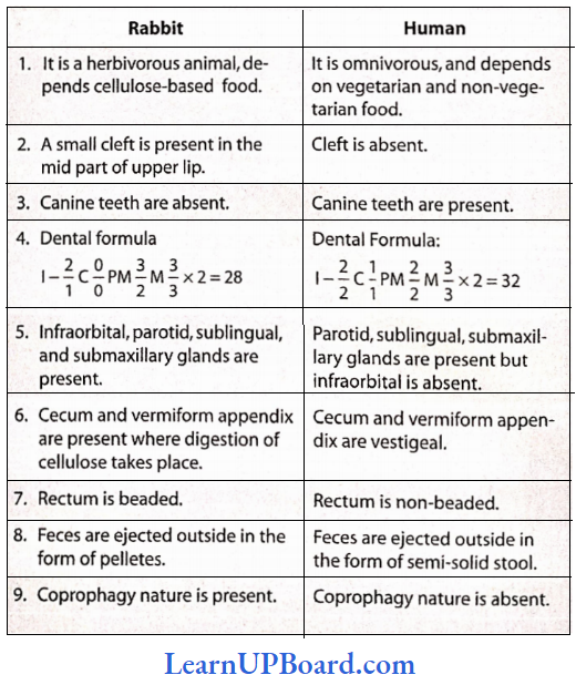
Comparative Study Between A Rabbit And A Human:
NEET Biology Notes Digestion And Absorption Assertion Reasoning Question And Answers
In the following questions, an Assertion (A) is followed by a corresponding Reason (R). Mark the correct answer.
- If both Assertion and Reason are true and the Reason is the correct explanation of the Assertion.
- If both Assertion and Reason are true, but the Reason is not the correct explanation of the Assertion.
- If Assertion is true, but Reason is false.
- If both Assertion and Reason are false.
Question 1. Assertion: Gastrectomy causes iron deficiency anemia.
Reason: Hydrochloric acid secreted by oxyntic cells converts ferric into ferrous and iron is absorbed as ferrous ions.
Answer: 1. If both Assertion and Reason are true and the Reason is the correct explanation of tire Assertion.
Question 2. Assertion: Cholagogues are substances that cause the contraction of the gall bladder.
Reason: These substances cause a release of CCK-PZ from the duodenum
Answer: 1. If both Assertion and Reason are true and the Reason is the correct explanation of tire Assertion.
Question 3. Assertion: Aptyalism patients have higher than normal incidences of dental caries.
Reason: Aptyalism is caused by the action of the parasympathetic nervous system.
Answer: 3. If Assertion is true, but Reason is false.
Question 4. Assertion: In humans, the duct of Wirsung from the pancreas combines with the bile duct before opening into the duodenum.
Reason: Blockage in the duct of Wirsung will hamper the endocrine function of the pancreas.
Answer: 3. If Assertion is true, but Reason is false.
Question 5. Assertion: In acute constipation, purgatives containing magnesium salts are generally used.
Reason: The osmotic effect of Mg2+ in the intestinal lumen prevents water reabsorption from the intestine. Mg2+ increases the solute concentration in the intestinal lumen because Mg2+ is absorbed very slowly.
Answer: 1. If both Assertion and Reason are true and the Reason is the correct explanation of tire Assertion.