
NEET Biology NotesLocomotion And Movement Locomotion
The act of changing place or position by the entire body or by parts of the body is called movement. Locomotion is the change of place for various purposes such as searching for food, shelter, protection from predators, and many other life activities.
NEET Biology NotesLocomotion And Movement Types Of Movements
Movements can be mainly of two categories: non-muscular and muscular movements.
Non-muscular movements: These movements persist in animals in some of their cells.
- Protoplasmic streaming: Streaming of protoplasm, called cyclosis, has been seen in most of the cells such as leucocytes, Amoeba, and other unicellular organisms.
- Pseudopodial movements: Leucocytes and macrophages move about in the tissues with the help of pseudopodia in the same manner as of amoeba.
- Flagellar movements: The flagella of certain cells (for example, choanocytes of poriferans) are maintained by their ceaseless vibrations a regular current of water through the canal system of sponges. The flagella of certain gastrodermal cells circulate fluid in the coelenteron of Hydra by regular beating. Sperms swim in water or in the female genital tract by flagellar movements.
- Ciliary movements: The cilia of cells lining the trachea, oviducts, and vasa efferentia propel dust particles, eggs, and sperm in a specific direction. The cilia of flame cells push waste material in excretory canals in flatworms.
- The non-muscular ciliary locomotion is retained by some animal larvae such as the planula larva of coelenterates and the trochophore of annelids, and even some adults such as planarians.
- Muscular movements: This depends upon the use of muscle fibers which have the ability to exert force by alternate contraction and relaxation. Most multicellular organisms have muscle fibers for moving different body parts or locomotion. A muscle contraction does not always result in movement. It may at times maintain the status quo, as in freshwater mussels (mollusks), muscle contract to keep the shell closed for safety.
locomotion biology
NEET Biology NotesLocomotion And Movement Locomotion In Humans
In humans, muscular movements are involved. There are three types of muscle tissues: striated or striped, non-striated or unstriped or smooth, and cardiac, according to their location, structure, and function.
Read and Learn More NEET Biology Notes
NEET Biology NotesLocomotion And Movement Structure Of Skeletal Muscle
The striated muscle forms 80% or more of the mass of soft tissues in a vertebrate body and is found in the body wall and limbs. It also occurs in the tongue, pharynx, and beginning of the esophagus.
- Skeletal muscles have a connective tissue sheath on the outer side called epimysium.
- A transverse section of it shows a number of bundles or fasciculi.
- Each fasciculus is surrounded by a connective tissue cover called perimysium.
- Within a fasciculus arc present a large number of muscle fibers, each surrounded by a connective tissue covering the endomysium.
- There is a broadband of fibrous connective tissues beneath the skin or around muscles called fascia.
- Each muscle fiber is cylindrical, and uniform in diameter. Sarcolemma is present on the outer side and at places is invaginated to form T or transverse tubules. A skeletal or striated muscle fiber is multinucleated or syncytial.
Ultrastructure of Skeletal Muscle Fiber:
A striated muscle consists of long, narrow, cylindrical, unbranched fibers with blunt ends.
- Each fiber is bounded by an elastic sarcolemma and contains many elongated, flattened nuclei characteristically located near the sarcolemma.
- The multinucleate condition results from cell fusion. Hence, a striated muscle fiber is a syncytium.
- The striated muscle fibers contain numerous mitochondria and glycogen granules for the supply of adequate energy.
- The myofibrils of a striated muscle fiber show alternating dark and light cross bands, striations, or stripes. Hence, the name of the muscle.
- The dark bands are called anisotropic or A bands. Each A band has at its middle a light zone termed Henson’s line or H zone. The light bands are isotropic and are known as the isotropic or I band. Each 1 band is crossed through its center by a dark membrane, the membrane of Krause or Z line.
This membrane continues right across the whole fiber and joins the sarcolemma surrounding the fiber. It seems to hold the myofibrils together and carry the signals for the contraction of fibrils inward from T-tubules (transverse tubules). The latter are invaginations of sarcolemma into the fiber adjacent to Z lines.
” locomotion in biology”
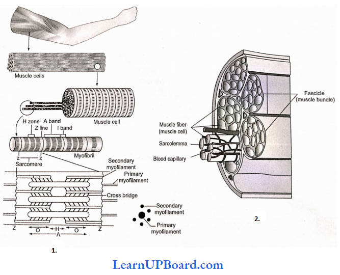
- The part of the myofibril between two successive Z lines functions as a contractile unit termed the sarcomere.
- The sarcoplasm also contains a protein pigment myoglobin, which can take up, store, or give up oxygen. For example, hemoglobin.
- An Electron microscope reveals that each sarcomere is a bundle of fine longitudinal myofilaments of two types: primary and secondary.
Primary myofilaments: The primary myofilaments are thicker and confined to the A band only. They are composed of myosin protein and bear minute projections called cross-bridges of the protein meromyosin and are free at both ends.
Secondary myofilaments: The secondary myofilaments are thinner and occur in I bands, but extend for some distance into A band between the primary myofilaments. This partial overlapping of primary myofilaments by secondary myofilaments imparts a dark appearance to A bands. The secondary myofilaments are composed of the proteins actin, tropomyosin, and troponin; have a smooth surface; and are attached to Z lines by one end, being free at the other end.
The secondary (actin) myofilaments are more numerous than the primary (myosin) myofilaments. Six actin myofilaments surround each myosin myofilament, and each actin myofilament is surrounded by three myosin myofilaments.
NEET Biology NotesLocomotion And Movement Muscle Contraction
When a nerve impulse (nerve action potential) reaches the synaptic end bulbs, it triggers the exocytosis of synaptic vesicles. In this process, the synaptic vesicles fuse with the plasma membrane and liberate ACh, which diffuses into the synaptic cleft between the motor neuron and motor end plate.
- When ACh binds to its receptor, a channel that passes small cations, most importantly Na+ opens.
- The inrush of Na+ changes the resting membrane potential, which triggers a muscle action potential that travels along the muscle cell plasma membrane and initiates the events leading to muscle contraction.
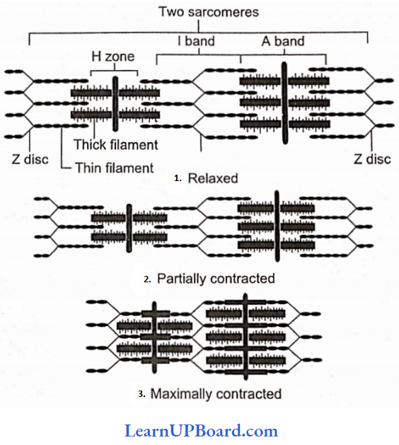
- Hanson and Huxley proposed that skeletal muscle shortens during contraction because thin filaments slide over thick filaments. Their model is known as the sliding filament mechanism of muscle contraction.
what is locomotion in biology
Sliding Filament Mechanism: During muscle contraction, myosin heads pull on the thin filaments, causing them to slide inward toward the H zone at the center of the sarcomere.
- The myosin cross bridges may even pull the thin filaments of each sarcomere so far inward that their ends overlap in the center of the sarcomere.
- As the thin filaments slide inward, Z discs come toward each other, and the sarcomere shortens, but the lengths of the thick and thin filaments do not change.
- The sliding of the filaments and shortening of the sarcomeres cause the shortening of the whole muscle fiber and ultimately the entire muscle.
Role of Calcium and Regulator Proteins: An increase in Ca2+ concentration in the sarcoplasm starts filament sliding, while a decrease turns off the sliding process.
- When a muscle fiber is relaxed (not contracting), the concentration of Ca2+ in its sarcoplasm is low.
- This is because the sarcoplasmic reticulum (SR) membrane contains Ca2+ active transport pumps that move Ca2+ from the sarcoplasm into the SR.
- Ca2+ is stored or sequestered inside the SR.
- As a muscle action potential travels along the sarcolemma and into the transverse tubule system, Ca2+ releases channels open in the SR membrane. As a result, Ca2+ floods into the sarcoplasm around the thick and thin filaments.
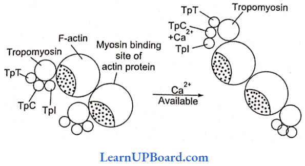
- Ca2+ released from the sarcoplasmic reticulum combines with troponin, causing it to change shape. This shape change moves the troponin-tropo-myosin complex away from the myosin-binding sites on actin. Troponin has three units (tri-unit structure): TpT (tropomyosin-binding troponin), TpC (calcium-binding protein), and Tpl (inhibitor, i.c., blocks the myosin-binding site of actin proteins).
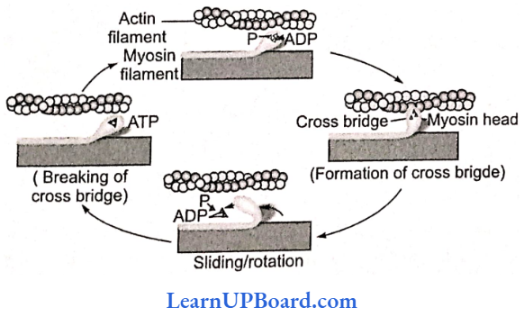
Power Stroke And The Role Of ATP: As we have seen, muscle contraction requires Ca2+ ions and energy in the form of ATP. The sequence of events during the sliding of the filaments is as follows:
While the muscle is relaxed, ATP attaches to ATP-binding sites on the myosin cross-bridges (heads). A portion of each myosin head acts as an ATPase, an enzyme that splits ATP into ADP+ P (phosphate group) through a hydrolysis reaction. This reaction transfers energy from ATP to the myosin head, even before contraction begins. The myosin cross-bridges are thus in an activated (energized) state.
- When the sarcoplasmic reticulum releases Ca2+, its level rises in the sarcoplasm. A rise in Ca2+ binds with troponin and changes its configuration moving away tropomyosin from its blocking position.
- The activated myosin heads spontaneously bind to the myosin-binding sites on actin.
- The shape change that occurs as myosin heads bind to actin produces the power stroke of contraction. During the power stroke, the myosin heads swivel toward the center of the sarcomere, like the oars of a boat during rowing. This action draws the thin film the thick filaments toward the H heads swivel, they release ADR
- Once the power stroke is complete, ATP again bines with the ATP-binding sites on the myosin heads. As ATP binds, the myosin head detaches from actin.
- Again, the myosin ATPase splits ATP, transferring its energy to the myosin ATPase which splits the head and hence the myosin ATP returns to its original upright position.
- The myosin head is then ready to combine with another myosin-binding site further along the thin filament.
- Steps through (7) repeat over and over as long as ATP is available and the Ca2+ level near the thin filament remains high.
- The myosin heads keep rotating back and forth with each power stroke, pulling the thin filaments toward the H zone.
- At any one instant, about half of the myosin heads are bound to act and keep swiveling.
- The other half are detached and preparing to swivel again. This continual movement of myosin heads applies the force that draws the Z discs toward each other, and the sarcomere shortens.
- The myofibrils thus contract and the whole muscle fiber shortens.
- During a maximal muscle contraction, the distance between Z discs can decrease to half the resting length.
- H line and M line disappear, the I band almost disappears, A band remains constant, but the power stroke does not always result in the shortening of the muscle fibers and the whole muscle.
- Contraction without shortening is called an isometric contraction, for example, in trying to lift a very heavy object.
- The myosin heads (cross-bridges) swivel and generate force, but the thin filaments do not slide inward.
Muscle relaxation: Two changes permit a muscle fiber to relax after it has contracted.
- First, acetylcholine is rapidly broken down by an enzyme called acetylcholinesterase (AChE). When action potentials cease in the motor neuron, the release of ACh stops, and AChE rapidly breaks down the ACh already present in the synaptic cleft.
- This ends the generation of muscle action potentials and the Ca2+ release channels in the sarcoplasmic reticulum membrane close.
- Second, Ca2+ active transport pumps rapidly remove Ca2+ from the sarcoplasm into the sarcoplasmic reticulum, where molecules of a calcium-binding protein. appropriately called ealsequestrin. bind to Ca2+.
- With this, the tropomyosin-troponin complex moves back over the myosin-binding site of actin which prevents further binding of myosin head to actin. and the thin filaments slide back to their normal relaxed position.
” what is movement in biology”
All or none principle: A minimal strength of a stimulus required to cause the contraction of a muscle fiber brings about maximum contraction, and no further increase in contraction would occur by increasing the strength of the stimulus.
Single muscle twitch: A single, quick isolated contraction of a muscle fiber to a single stimulus of threshold value is called a single muscle twitch in laboratory experiments.
Energy Source For Muscle Contraction
- Energy for muscle contraction is provided by the hydrolysis of ATP by myosin ATPase enzyme. This hydrolysis produces ADP, inorganic phosphate, and energy (used in muscle contraction).
- Pliosphocrcatine donates its high energy and phosphate to ADP, producing ATP. It serves as an energy source for a few seconds for metabolic processes in muscle cells to begin to produce greater quantities of ATP.
- Pliosphocrcatine is again formed in relaxing muscle by using ATP produced by carbohydrate oxidation.
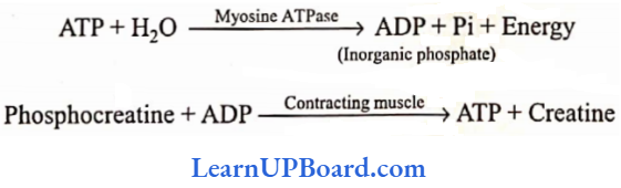
- At the end of muscle contraction, the conversion of ADP into ATP takes place.
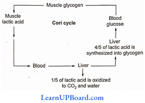
- The muscle is rich in glycogen which is broken down into lactic acid through a series of reactions (glycolysis) and liberates energy.
- Some of this energy is used for the reformation of phosphocreatine and also for the conversion of four-fifths of lactic acid back into glycogen.
- One-fifth of lactic acid is oxidized to water and carbon dioxide.
- These reactions taking place in the muscle and liver were proposed by Cori and Cori. Hence, known as the Cori cycle.
Rigor mortis: Extreme rigidity of the body after death is called rigor mortis. It is due to the complete depletion of ATP and phosphocreatine.
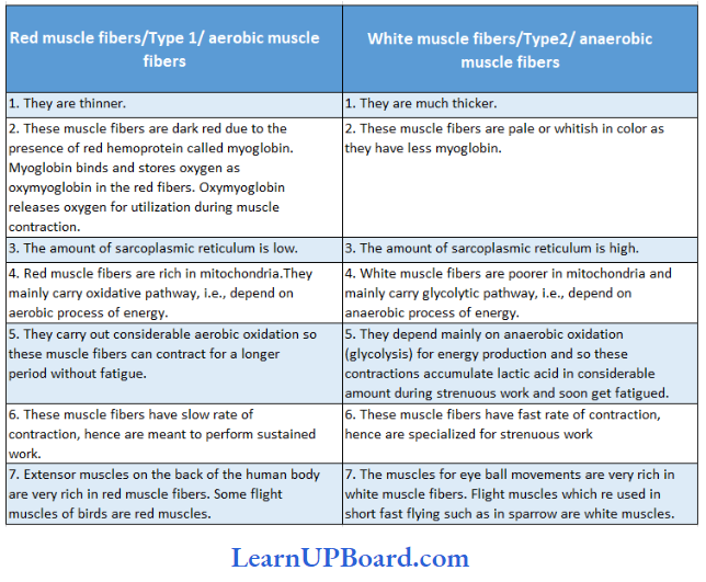
Red and white muscle fibers: Birds and mammals have two kinds of striated muscle fibers in their skeletal muscles: red or slow muscle fibers and white or last muscle fibers. Explains the difference between red and white muscle fibers.
Differences between red and white muscle fibers White muscle fibers:
Contraction In Smooth Muscles: In comparison with contraction in a skeletal muscle fiber, contraction in a smooth muscle liber starts more slowly and lasts much longer. Troponin is absent in smooth muscles, so they have a regulator protein called calmodulin that binds to Ca2+ in the tool.
- Using ATP. myosin head part can bind to actin and contraction can occur.
- Most smooth muscle fibers contract or relax in response to action potentials from the autonomic nervous system.
Contraction In Cardiac Muscles: Cardiac muscle fibers have the same arrangement of actin and myosin and the same bands, zones, and z-discs as skeletal muscle fibers,
- Gap junctions allow muscle action potential to spread from one muscle fiber to another. As a consequence, when a single muscle fiber is stimulated, all other fibers in the network become stimulated as well. Thus, each network contracts as a functional unit.
- Cardiac muscle tissue has a long refractory period and can use lactic acid produced by skeletal muscle fibers to make ATP.
“types of locomotion “
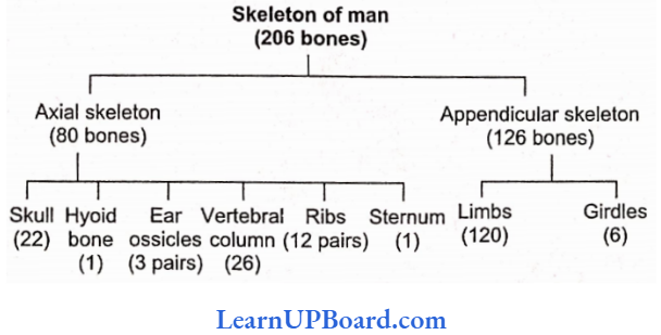
NEET Biology NotesLocomotion And Movement Skeletal System In Human
The skeletal system is divided into two main parts: axial skeleton and appendicular skeleton.
Axial skeleton: It lies along the principal axis of the body. It includes a skull, vertebral column, sternum, and ribs.
Appendicular skeleton: It is made up of girdles and limb bones. The distribution of bones in the whole human body and the distribution of human skull bones.
Distribution Of Bones In Human
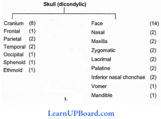
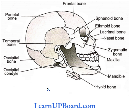
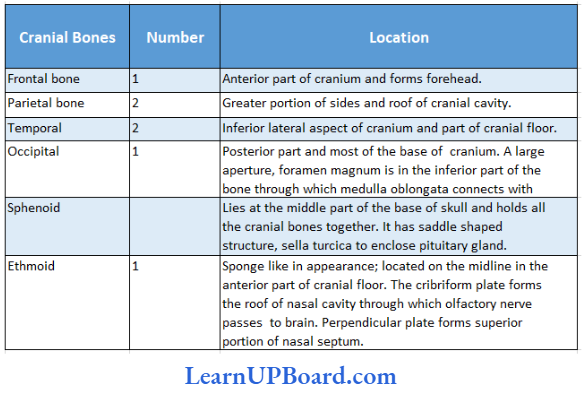
Location And Number Of Cranial Bones In Humans:
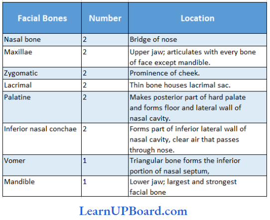
Location And Number Of Facial Bones In Human:
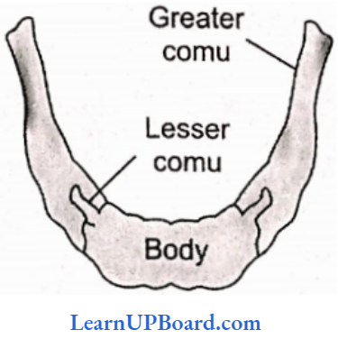
Hyoid: It serves as a point of attachment for some of the cells of the tongue and floor of the mouth but does not articulate with any other bone.
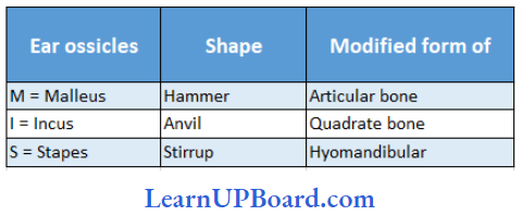
Ear ossicle: Ear ossicles are three in number and arc discussed.
Types Of Ear Ossicles And Their Shape:
NEET Biology NotesLocomotion And Movement Vertebrae
The vertebral column protects the spinal cord, supports the head, and serves as the point of attachment for the ribs and musculature of the back.
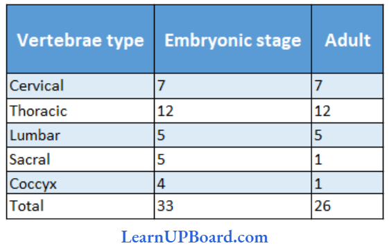
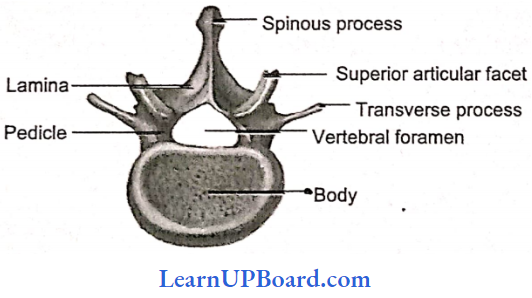
Types Of Vertebrae And Their Count At The Embryonic And Adult Stages:
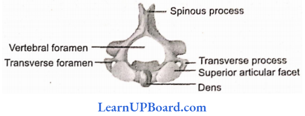
Cervical Vertebrae: 1, 2, and 7 cervical vertebrae are atypical and 3-6 are typical.
- The transverse processes are perforated by foramina (foramen transversarium) for the passage of the vertebral arteries except in the seventh vertebra.
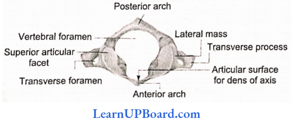
- The first cervical vertebra or atlas supports the head and consists of a complete ring of bone.
- On its upper surface, it presents kidney-shaped facets for articulation with the condyles of the occipital bone, forming a condyloid joint, the atlanto-occipital joint, at which the nodding movements of the head take place. The atlas articulates with the second cervical vertebra.
The second cervical vertebra or axis is the pivot which the atlas turns in the rotary movements of the head. From the body of the axis, a process of bone rises called an odontoid peg, which articulates with the back of the anterior arch of the atlas and is held in position by the transverse ligament of the atlas. The lateral masses of the atlas articulate with corresponding facets on the axis placed on each side of the odontoid peg. The atlas moves around the odontoid peg of the axis, forming a pivot joint at which the head rotates.
The seventh cervical vertebra is the first vertebra with an undivided spinous process. This process has a tubercle at its tip. It forms a distinct projection in the neck and can be seen at the lower part of the back of the neck. Because of this characteristic, the bone is called vertebra prominent.
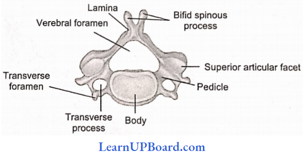
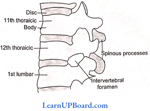
Thoracic Vertebrae: Thoracic 2-8 are typical. 1, 9-12 are atypical. These are larger than the cervical and they increase in size as they extend downwards.
- The body is heart-shaped, with facets on each side for attachment of the ribs.
- The neural arch is relatively small; the spinous process is long and is directed downwards.
- The transverse processes that help to support the ribs are thick, and strong, and carry articular facets for the ribs.
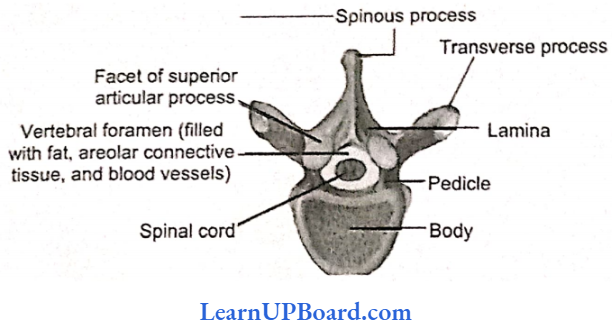
Lumbar Vertebrae: These are the largest vertebrae as compared with the bodies of the other vertebrae and are kidney-shaped. The spinous process is broad and hatchet-shaped.
- The transverse processes are long and slender.
- The fifth lumbar vertebra articulates with the sacrum at the lumbosacral joint.
“locomotion and movement neet notes “
Sacrum: It is a triangular bone situated at the lower part of the vertebral column, wedged in between the two innominate bones and forming the back of the pelvic cavity.
- The base of the sacrum lies above and articulates with the fifth lumbar vertebra, forming a typical intervertebral joint.
- The junction between the fifth lumbar vertebra and the sacrum forms the sacro-vertebrcA angle.
- At the extremities of transverse ridges, on each side, sacral foramina is present for the passage of nerves. The apex of the sacrum articulates with the coccyx. At the sides, the sacrum articulates with the innominate bones, forming the right and left sacroiliac joint.
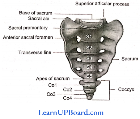
Coccyx: It is composed of four or five rudimentary vertebrae, fused to form one bone. It articulates above with the sacrum.
Joints of the Vertebral Column: The intervertebral discs are thick pads of fibro-cartilage between the bodies of tire movable vertebrae.
- It is strengthened by ligaments running in front and behind the vertebral bodies throughout the entire length of the column.
- Masses of muscle on each side materially aid in the stability of the spine.
- Movement: The joints formed between the discs and the vertebrae are only slightly movable joints of the symphysis variety. But their number count gives considerable flexibility to the column as a whole.
- The movements possible are flexion, forward bending, extension, backward bending, lateral bending to each side, and rotation to the right and left. The joint is called a cartilaginous joint or amphiarthroses.
Curves of the Vertebral Column
Looking from the side, the vertebral column presents four anthro-posterior curves: a cervical curve in the neck which is convex forwards; a thoracic curve, convex backward; a lumbar curve, convex forwards; and pelvic curve, convex backward.
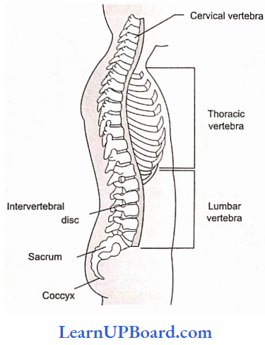
The two antero-convex curves are secondary. The cervical curve is developed when an infant raises his head to look about and investigate his surroundings, and the lumbar curve forms when he crawls and leams to stand and walk and keep himself erect.
NEET Biology NotesLocomotion And Movement Ribs
There are 12 pairs of ribs. Each rib is a thin flat bone connected dorsally to the vertebral column and ventrally to the sternum.
- It has two articulation surfaces on its dorsal end and is called bicephalic.
- The first seven pairs of ribs are called true ribs.
- Dorsally, they are attached to the thoracic vertebrae and ventrally connected to the sternum with the help of hyaline cartilage called vertebrosternal (true) ribs.
- The eighth, ninth, and tenth pairs of ribs do not articulate directly with the sternum but join the seventh rib with the help of hyaline cartilage.
- These are called vertebrochondral (false) ribs. The last two pairs (11th and 12th) of ribs are not connected ventrally and are, therefore, called floating (vertebral) ribs.
- Thoracic vertebrae, ribs, and sternum together form the rib cage.
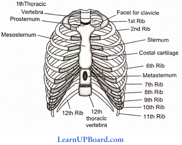
Sternum: It is a flat bone on the ventral midline of thorax.
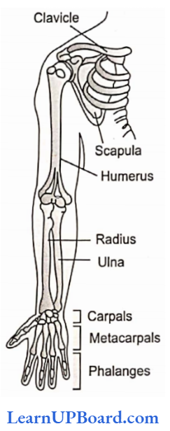
NEET Biology NotesLocomotion And Movement Skeleton Of The Upper Limb
The skeleton of the upper limb is attached to the skeleton of the trunk by means of the shoulder girdle, which consists of a clavicle and scapula.
Pectoral Girdle/Shoulder Girdle
It consists of two bones: A clavicle and a scapula.
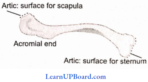
Clavicle
- The clavicle or collar bone is a long curved horizontal bone forming the anterior part of the shoulder girdle.
- Its sternal extremity articulates with the sternum and acromial extremity and articulates with the acromion process of the scapula.
- The clavicle is often broken by direct or indirect violence such as falling on the hand or shoulder. The bone is usually fractured in the middle or lateral third.
Scapula: The scapula forms the posterior part of the shoulder girdle and lies at the back of the thorax superficially to the ribs.
It is a triangular Hat bone. Its anterior or costal surface is called the subscapular fossa and lies nearest to the ribs.
The posterior or dorsal surface is divided by a prominent ridge of bone, called the spine of the scapula. which passes across it to end in the acromion process, which overhangs the shoulder joint.
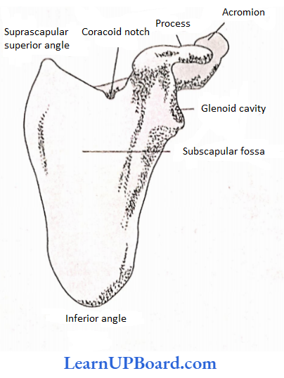
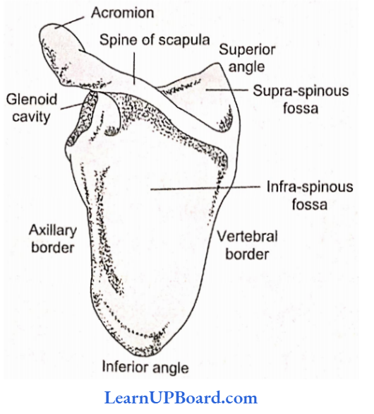
- The scapula shows a glenoid cavity, which is a shallow directed outwards to receive the head of the humerus in the formation of the shoulder joint, humor-scapular joint.
- The joint between the head of the humerus and the glenoid cavity is also called a ball-and-socket joint.
- The coracoid process of the scapula arises internal to the glenoid cavity and projects forward. It gives attachment to the short head of the biceps and pectoralis minor.
Bones Forming Upper Limb: The bones that form the skeleton of the arm, forearm, and hand, making altogether 30 bones, arc humerus, ulna, and radius (1 +1), carpal bones (8), metacarpals (5), and phalanges (14).
Humerus: It is the longest bone of the upper limb. It presents a shaft and two extremities.
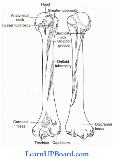
- The upper extremity of the humerus consists of one-third of a sphere—the head, which articulates with the glenoid cavity of the scapula in the formation of the shoulder joint.
- Immediately below the head is a slightly constricted part called the anatomical neck.
- Below the anatomical neck is a rough prominence, the greater tuberosity, ami at the front is a smaller prominence, lire lesser tuberosity,
- The bone becomes narrower below the tuberosities, and at this point, it is called the surgical neck, because of the liability of fracture at that part.
- A rough tubercle on the lateral aspect of the shaft, just above the middle, is called the deltoid tuberosity, ft receives the insertion of the deltoid muscle. So, the characteristic feature of the humerus is a deltoid ridge.
- The lower extremity is broad and flat. At its lowest part, the articulating surfaces for the bones of the fore- ami lie.
- The trochlea on the inner side is a pulley-shaped surface for articulation with the ulna and the capitulum on the outer side for the radius,
- Above the articulating surface of the ulna is a depression in front called the coronoid fossa of the humerus, into which the coronoid process of the ulna is received when the elbow’ is flexed or bent.
- A large cavity, the olecranon fossa, lies in a similar position at the back of the bone, which receives the olecranon process of the ulna when the elbow is extended or straight.
Ulna: The ulna is a long bone having a shaft and two extremities.
- It is the medial bone of the forearm and is longer than the radius.
- The head of the ulna is at the lower end.
- The upper extremity of the ulna is strong, and thick, and enters into the formation of the elbow joint.
- The trochlear notch of the ulna is formed by two processes; it articulates with the trochlear surface of the humerus in the tine formation of the elbow joint, i.e., the hinge joint.
- The radial notch is on the lateral aspect of the upper extremity of the bone, near the coronoid process.
- The side of the head of the radius articulates with the radial notch as the radius rotates around the ulna, thus forming die superior radio-ulnar articulation.
- The upper ends of forearm bones articulate with each other forming a pivot joint.
- The flexors come from the anterior and the extensors from the posterior surface.
- The muscles pronating and supinating the forearm are also attached to the shaft.
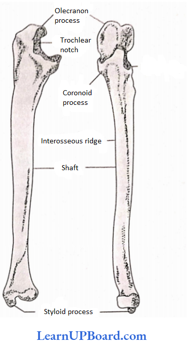
- The lower extremity is small.
- Two eminences arise from it.
- A small rounded eminence, head of the ulna, articulates with the medial side of the lower extremity of the radius in the formation of the inferior radio-ulnar joint.
- A pointed process, a styloid process, projects downward from the back of the lower extremity. Ulna is toward the little finger.
Radius: The radius is the lateral bone of the forearm.
- It is a long bone with a shaft and two extremities.
- It is shorter than the ulna. The radius is toward the thumb.
- Colies fracture is the breaking of the lower end of the radius, by falling on the outstretched hand. This results in the characteristic deformity of the wrist and hand.
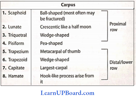
Carpal Bones: The carpus is composed of eight bones arranged in two rows, four bones in each row, as mentioned.
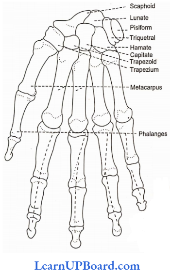
Metacarpals: There are five metacarpal bones. Each bone has a shaft and two extremities.
- The extremity articulating with the carpal bones is called the carpal extremity, and the joint so formed is the carpometacarpal joint which is the gliding joint.
- The distal extremity articulates with the phalanges and is called the head.
Phalanges: These are also long bones, having a shaft and two extremities. The shaft tapers toward the distal end.
There are 14 phalanges, three in each finger and two in the thumb. The joint between metacarpals and phalanges is an ellipsoid or condyloid joint.
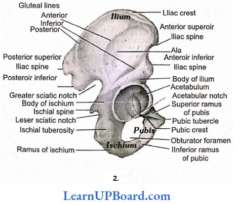
NEET Biology NotesLocomotion And Movement Pelvic Girdle Or Body Pelvis
The pelvic girdle is the means of connection between the trunk and lower extremities.
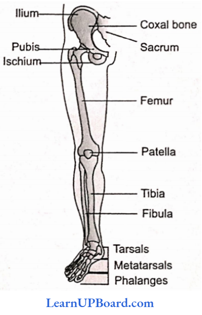
- It is formed by a part of the axial skeleton—the sacrum and coccyx being wedged in between the two innominate bones.
- Ischium is the thickest and strongest portion of the bone.
- The tuberosity of ischium lies at its lowest point, and on this, the trunk rests when sitting.
- The tuberosity is marked by two facets that give attachment to the hamstring muscles.
- A pointed eminence, the spine of ischium, arises from the back of the bone and marks the lowest part of the sciatic notch.
- The body of the ischium forms the posterior boundary of the obturator foramen; from this, the ramus passes forward, to join the descending ramus of the pubis.
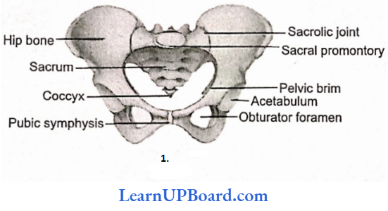
- While sitting, the entire weight of the body falls on the ischium.
- The obturator foramen is a large oval foramen lying below the acetabulum and bounded, as described, by the pubis and ischium.
- It is filled in with membrane, and at its upper part, the obturator vessels and nerves pass from the pelvis into the thigh.
- The acetabulum is a deep, cup-shaped cavity formed by the union of the three bones: the pubis forms the front part. ilium the upper part, and ischium the back part.
- The acetabulum articulates with the femur in the formation of the hip joint.
Bones Forming Lower Limb: It is altogether made up of 30 bones, namely, the femur, patella, tibia, fibula, tarsals (7), metatarsals (5), and phalanges (14).
Femur: The femur is the longest bone in the body.
- It articulates with the acetabulum in the formation of the hip joint, and from here the bone inclines medially to the knee, where it articulates with the tibia.
- It is a long bone with a shaft and two extremities. The hip joint is a ball and socket joint.
- The great trochanter is a prominent process of bone that gives attachment to several muscles, including the gluteal muscles and is a characteristic feature of the femur.
- The lesser trochanter is distinctly raised.
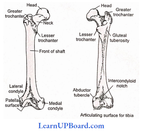
- The intercondylar notch separates the condyles behind. The surfaces of this notch give attachment to the cruciate ligaments of the knee joint.
- The condyles are separated in front by the patellar surface which extends over the anterior aspect of both condyles; on this surface, the patella rests.
- The tibial surface of the femoral condyles lies below and rests on the upper articulating surface of the condyles of the tibia.
- The femur articulates with three bones, the innominate bone, tibia, and patella, but it does not articulate with fibula.
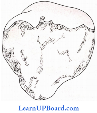
Patella: Patella is a sesamoid bone developed in the tendon of the quadriceps extensor muscles. The apex of the patella points downwards. The anterior surface of the bone is rough.
The posterior surface is smooth and articulates with the patellar surface of the lower extremity of the femur. This articulating surface is divided by a line into two facets.
Tibia: The lower extremity of the tibia enters into the formation of the ankle joint.
- It is slightly expanded and is prolonged downward on the medial side as medial malleolus.
- The lower extremity articulates with the talus, the margins of the bone giving attachment to the ligaments of the joint.
- The front of the tibia is smooth, and tendons passing to the foot glide over it.
- The lateral surface of the lower extremity articulates with the fibula at the inferior tibiofibular joint. The tibia articulates with three bones: femur, fibula, and talus.
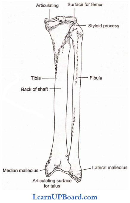
Fibula: The fibula is the lateral bone of the leg. It is a long bone with a shaft and two extremities.
- The upper extremity forms the head, and articulates with the back of the outer condyle of the tibia, but does not enter into the formation of the knee joint.
- The shaft is slender and deeply embedded in the leg muscles, to which it gives numerous attachments,
- The lower extremity is prolonged downward as lateral malleolus.
- A rough depression lies behind the lateral malleolus called the malleolar fossa, which provides a surface for the attachment of some of the powerful ligaments of the ankle joint.
- The lateral malleolus extends lower than the medial malleolus of the tibia.
- Its lateral surface is subcutaneous and its medial surface articulates with the lateral surface of the talus in the formation of the ankle joint.
NEET Biology NotesLocomotion And Movement Bones Of The Foot
Tarsal bones: There are seven bones known collectively as tarsus. They are short bones made up of cancellous bone tissue, with a covering of compact tissue. These bones support the weight of the body in standing.
Types Of Bones Forming The Tarsus:
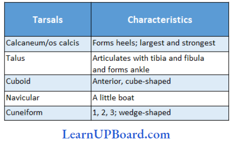
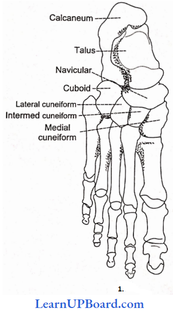
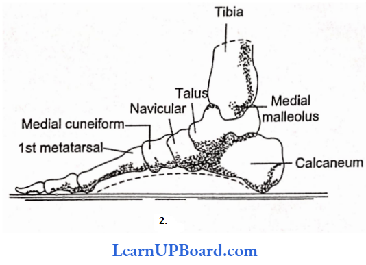
The calcaneum helps to support the talus and it also gives attachment to the spring ligaments, which is important in maintaining the medial arch of the foot. The ankle joint is also a hinge joint.
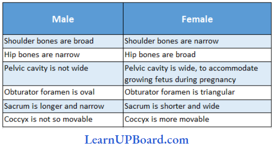
General Differences In The Skeleton Of Male And Female:
NEET Biology NotesLocomotion And Movement Articulation Of Bones And Joints
A bone joint or articulation may be defined as the junction of two bones. The study of such joints is known as arthrology. There are three principal types of bone joints: fibrous joints or immovable or fixed joints/synarthrosis, cartilaginous joints/amphiarthrosis, and synovial joints/diarthrosis.
- Fibrous joints or immovable or fixed joints/synarthrosis
- These joints are immovable or fixed.
- They do not show any movement due to the presence of strong and tough white collagenous fibers, and there is no joint cavity.
- These joints include:
- Sutures: Found between skull bones; articulating bones are held together by white fibrous tissue.
- Gomphoses: Teeth in mandibles and in maxillary bones.
- Schindyleses: One bone fits into the slit of another, for example, ethmoid bone in vomer.
- Cartilaginous joints/amphiarthrosis
- They are slightly movable joints.
- Discs of white fibrocartilage; are strong but more elastic and compressible than the white fibrous tissue. Hold the bones together at the joints between the bodies of vertebrae, at the symphysis pubis, and between the sternum and ribs.
- The bones make some movements at such joints through compression of the cartilage discs.
- Synovial joints: Synovial joints are of different types depending upon the nature of articulation and degree of movement:
- Ball and socket joints (enarthroses): The “head” of one bone fits in the “socket” of another bone and allows free movement in all planes, for example, shoulder joint and hip joint.
- Hinge joints (ginglymi): Perfect joints that allow movements only in a single plane, for example. elbow joint, knee joint, and ankle joint.
- Pivotal joints (rotary joints or rotary): One of the two bones is fixed in its place and bears a peglike process over which rotates the other bone, for example, atlas along with the skull rotating over the odontoid process of axis vertebra in mammals.
- Saddle joints: It is similar to ball-and-socket joints, but are poorly developed and movements are comparatively less free, for example, the joint between the metacarpal of the thumb with the carpals below.
- Gliding joints: The joints that permit sliding of the articulating bones on each other, for example, a joint between the zygapophyses of successive vertebrae, and between the sternum and clavicle.
- Angular joints (or ellipsoid or condyloid): These joints allow movements in two directions, i.e., side to side and back and forth, for example, metacarpophalangeal joints.
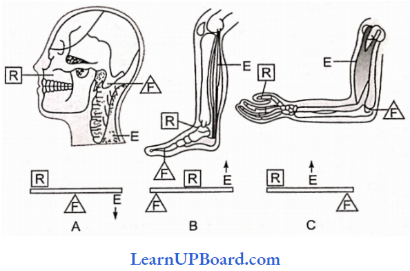
NEET Biology NotesLocomotion And Movement Lever System
In producing movement, bones act as levers, and joints function as fulcums. A lever may be defined as a rigid rod that moves around a fixed point called a fulcrum (F).
- The lever is acted at two different points by two different forces, the effort (E) which causes movement, and the resistance or load (R) which opposes movement.
- The effort is the force exerted by muscular contraction, whereas the resistance is typically the weight of the body part that is moved.
- Motion occurs when effort applied to the bone at insertion exceeds resistance.
- Functioning of all three types of levers can be observed in the human skeleton:
First-class lever: The joint between the first vertebra (atlas) and the occipital bone of the skull exhibits the example of a first-class lever in which the joint is the fulcrum, contraction of a back muscle is the effort, and facial part of the skull on raised head acts as the resistance.
Second-class lever: A human body resting on the tip of the toes is an example of a second-class lever, as the toe forms the fulcrum, and contracting calf muscle provides effort distally. The body functions as resistance exerting in between the fulcrum and effort.
Third-class lever: The flexing movements of the elbow of the forearm are based on the principle of the third-class lever. Here, the elbow joint acts as a fulcrum and the distal part of the hand provides resistance. The contracting biceps muscles attached near the elbow joint exert the effort in between the fulcrum and resistance.
NEET Biology NotesLocomotion And Movement Fracture
Fracture is the breakage of bone, either complete or incomplete. The following are the types of fractures:
- Simple or closed fracture: A fracture breaking the bones into two frilly separate parts with little damage to surrounding tissues and no break in the overlying skin.
- Greenstick fracture: A break of the bone in the form of only a crack, with broken parts still holding together.
- Comminuted fracture: In this fracture, the bone is broken into more than two fragments with some of the fragments losing any connection with blood circulation.
- Compound open fracture: The broken ends of the fractured bone protrude through the skin.
NEET Biology NotesLocomotion And Movement Various Types Of Bones
Types of Bones Based on the Basis of Their Formation
Cartilaginous or replacing bones or endochondral bones: These bones develop from the pre-existing cartilage, for example, the humerus, and femur.
- Investing or dermal or membrane bone: These bones develop in the dermis of skin as thin plates and sink to get attached over original cartilages, for example., frontal, nasals, vomers, and parietals of the skull.
- Sesamoid bones: These bones are formed in tendons at the joint clavicle, for example., patella (knee cap), pisciform.
- Viscerai bones: These bones are present in organs and dissociated from the rest of the skeleton. In the heart of some ungulates (‘ruminates), bones develop in the connective tissue of the cardiac skeleton as cordis.
Types of Bones Based on the Basis of Their Shape and Size
- Long bones: Humerus, radius, ulna, femur, tibia, and fibula
- Short bones: Carpais and tarsals
- Flat bones: Skull bones, sternum, and ribs
- Irregular bones: Ear ossicles (malleus, incus, stapes) and vertebrae
- Sesamoid bones: Patella (knee cap), fabellae, and pisciform
NEET Biology NotesLocomotion And Movement Disorders Of Bones, Joints, And Muscles
- Rheumatoid Arthritis:
- It is diagnosed by the presence of rheumatoid factor (a type of immunoglobulin IgM).
- It is the primary symptom of inflammation of the synovial membrane.
- If it is left untreated, then the membrane thickens, and synovial fluid increases, exerting pressure that causes pain.
- The membrane then starts secreting abnormal granules, called pannus, which after accumulating on the surface of the cartilage causes its erosion. As a result, the fibrous tissues are attached to the bones and become ossified, making the joints immovable. Heat treatment and physiotherapy for pain and inflammation and, in extreme cases, replacement of the damaged joints are recommended.
- Osteoarthritis
- It is a degenerative joint disease characterized by the degeneration of the articular cartilage and the proliferation of new bones.
- Usually, affected joints are of spine, knees, and hands.
- Gouty arthritis or gout
- It is caused either by to excessive formation of uric acid or the inability to excrete it.
- It gets deposited in joints as monosodium salt.
- Osteomalacia and rickets
- Osteomalacia is called rickets when it occurs in childhood.
- In this disorder, the bones contain insufficient amounts of calcium and phosphorus.
- Osteoporosis
- Osteoporosis is a disease in which the bone loses
- minerals and fibers from its matrix.
- Individuals taking hydrocortisone for arthritis, allergies, or other disorders are especially prone to bone loss.
- Bursitis
- The bursae of joints often become inflamed, a condition known as bursitis.
- The inflammation can be caused by a physical injury or by constant pressure to the same joint over a long period of time.
- Dislocation
- A dislocation is a displacement of the articular surfaces of a joint; it usually involves damage to the ligaments surrounding the joint.
- Most dislocations result from falls, blows, or extreme exertion and are most often seen in the joints of the thumb, fingers, knee, or shoulder.
- Symptoms of dislocation include swelling, pain, and loss of motion.
- Sprain and strains
- A sprain is the twisting of a joint without dislocating it. Such an injury causes damage to ligaments and also often damages tendons, muscles, blood vessels, and nerves.
- Severe sprains are quite painful and require immobilization during the healing process.
- In contrast to a sprain, a strain is a less severe stretching or twisting of a joint.
- Muscles and tendons may be stretched and become somewhat painful, but only minor damage is done to the joint tissues.
- Myasthenia gravis: Autoimmune disorder affecting neuromuscular junction leading to fatigue, weakening, and paralysis of skeletal muscle.
- Muscular dystrophy: Progressive degeneration of skeletal muscle mostly due to genetic disorder.
- Tetany: Rapid spasm (wild contraction) in muscles due to low Ca++ in body fluid.
NEET Biology NotesLocomotion And Movement Locomotion And Movement Points To Remember
- Kinesiology: Study of body movements.
- The longest bone in the human body is the femur.
- The longest bone in frogs is the tibia-fibula.
- Largest foramen: Foramen magnum.
- Motor unit: A single nerve fiber with its supply to muscle fiber (as many as it covers).
- General divisions of endoskeleton in a land vertebrate:
img
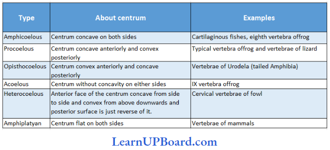
Types of vertebrae:
NEET Biology NotesLocomotion And Movement Assertion-Reasoning Questions And Answers
In the following questions, an Assertion (A) is followed by a corresponding Reason (R). Mark the correct answer.
- If both Assertion and Reason are true and the Reason is the correct explanation of the Assertion.
- If both Assertion and Reason are true, but the Reason is not the correct explanation of the
Assertion. - If Assertion is true, but Reason is false.
- If both Assertion and Reason are false.
Question 1.
Assertion: Maximum movement is possible at the am-phi arthrosis joint.
Reason: Such joints are also called synovial joints and have almost frictionless movement due to
synovial fluid.
Answer: 4. If both Assertion and Reason are false.
Question 2.
Assertion: Ca2+ plays an important role in muscle contraction.
Reason: Ca2+ combines with the troponin chain, displacing tropomyosin and allowing the myosin head part to combine with actin to form an actomyosin complex.
Answer: 1. If both Assertion and Reason are true and the Reason is the correct explanation of the Assertion.
Question 3.
Assertion: On repeated application of stimuli, involuntary striped muscles undergo fatigue.
Reason: This is due to the non-availability of ATP molecules.
Answer: 4. If both Assertion and Reason are false.
Question 4.
Assertion: All muscles follow the “all or none” principle.
Reason: All muscles contract either fully or do not contract at all depending upon the threshold stimulus availability.
Answer: 4. If both Assertion and Reason are false.
Question 5.
Assertion: The Tibia is stronger and inner whereas the fibula is the slender outer bone of the lower leg or shank.
Reason: Tibia has a sharp crest in the shaft and a projection on the inner side of the ankle called the lateral malleolus.
Answer: 3. If Assertion is true, but Reason is false.
Question 6.
Assertion: Skeleton helps in blood cell formation.
Reason: Blood flows through the skeleton.
Answer: 3. If Assertion is true, but Reason is false.
Question 7.
Assertion: The skeleton serves as a storage depot.
Reason: Skeleton stores carbohydrates and protein.
Answer: 3. If Assertion is true, but Reason is false.
Question 8.
Assertion: Ball-and-socket joints are the most mobile joints.
Reason: Synovial fluid is present in ball-and-socket joints.
Answer: 2. If both Assertion and Reason are true, but the Reason is not the correct explanation of the Assertion.
Question 9.
Assertion: Arthritis or inflammation of a joint makes the joint painful.
Reason: Some toxic substances are deposited at the joint.
Answer: 3. If Assertion is true, but Reason is false.
Question 10.
Assertion: The contraction and relaxation of muscle fiber arc controlled by nerve impulses.
Reason: The threshold stimulus is the minimum stimulus required for the beginning of contraction.
Answer: 2. If both Assertion and Reason are true, but the Reason is not the correct explanation of the Assertion.