
Neural Control And Coordination Introduction
Different activities of an animal’s body are controlled and coordinated through two systems—the nervous system (neural system) and the endocrine system.
Nervous System
The nervous system of all animals is composed of highly specialized cells called neurons, which can detect, receive, and transmit different kinds of stimuli.
- The neural organization is very simple in lower invertebrates. For example, Hydra is composed of a network of neurons.
- The nervous system is better organized in insects, where a brain is present along with the number of ganglia and neural tissues.
- The vertebrates have a more developed neural system.
- The human nervous system is divided into two parts: The central nervous system (CNS) and the peripheral nervous system (PNS).
- The CNS includes the brain and spinal cord and is the site of information processing and control.
- The PNS comprises all the body nerves associated with the CNS (brain and spinal cord).
- The nerve fibers of PNS are of two types: afferent- fibers and efferent fibers. Afferent fibers transmit impulses from tissues/organs to the CNS and efferent fibers transmit regulatory impulses from the CNS to concerned peripheral tissues/organs.
- The PNS is divided into two divisions called somatic and autonomic neural systems.
- The somatic neural system relays impulses from the CNS to skeletal muscles while the autonomic neural system transmits impulses from the CNS to involuntary organs and smooth muscles of the body.
- The autonomic neural system is further classified into sympathetic and parasympathetic neural systems.
” neural control”
Types Of Neuron
Nerve cells or neurons are the functional units of the nervous system. These include multipolar nerve cells, with many short dendrites and one long axon (for example, pyramidal cells in the cerebral cortex), bipolar nerve cells, where the long axon extends on either side of the cell body (for example, bipolar neurons in the retina of the eye), and pseudounipolar nerve cells, with cell body on a side-branch of the main axon (for example, cells of dorsal root ganglion).
- Surrounding neurons are special companion cells, known as glia (Gk: glue).
- The glial cells perform many housekeeping functions; astrocytes provide nutritional support to neurons; microglia (phagocytic or scavenger cells) consume waste products.
- Oligodendrocytes insulate by forming myelin sheath in CNG, separating each neuron from others.
- In the PNS, Schwann cells or neurolemmocytes, a type of glial cell, wrap around the axons of neurons, thereby covering the axon with concentric layers of insulating plasma membrane, i.e., the myelin sheath.
Read and Learn More NEET Biology Notes
Nerve Impulse And Its Transmission
Nerve cells have polarized membranes, i.e., they have electrical potential difference or membrane potential. This is because of a variety of ion channels (pores formed by proteins) specific to a particular type of ion.
- Some ion channels remain open while most of them open under one condition and close under another condition.
- Because of such regulated or voltage-gated channels, membranes become excitable as these channels respond to different types of stimuli, for example, light, touch, sound, etc.
- When a neuron is not sending any signal, it is said to be at rest and its membrane has resting membrane potential.
Resting Membrane Potential: In a resting nerve fiber, the cytoplasm just beneath its membrane is electronegative relative to the layer of extracellular fluid (ECF) just outside the membrane.
If the two sides of the membrane are connected by a galvanometer (double beam cathode ray oscilloscope), the inner side is seen to possess a negative potential of about 70 mV relative to the outer side. This is called the resting membrane potential. This results from two factors:
Epithelium
- The resting membrane has a poor permeability for Na% although it has a higher permeability for K+. Therefore, K+ can cross more easily while Cl– and Na+ have more difficulty in crossing.
- A negatively charged protein molecule inside the neuron cannot cross the plasma membrane because of its semipermeability.
- The differential flow of the positively charged ions and the fact that the negatively charged organic ions within the nerve fiber cannot pass out cause an increasing positive charge on the outer side of the membrane and a negative charge on the inner side of the membrane.
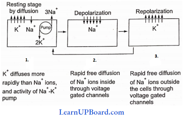
- This makes the membrane of the resting nerve fiber polarized (i.e., its outer side is positively charged with respect to its inner side). Such electrochemical gradients are maintained by the active transport of ions involving the Na+-K+ ion transmembrane pump. It pumps out 3Na+ for every 2K+ ions passed inwardly.
- K+ concentration is 30 times more inside a neuron than outside and Na+ concentration is 10 times more in interstitial fluid as compared to the inner side of neuron.
.
class 11 neural control and coordination
Conduction of Nerve Impulse: It involves initiation of the impulse followed by conduction along the axon to be transferred to the target muscle/tissue.
Initiation of Impulse: When stimulated, voltage-gated Na+ channels open which causes a rapid, very localized, temporary inflow of Na+ into the cell, which further causes the development of the net positive charge on the inner side of the membrane in that area.
- This is called depolarization. It occurs at a particular region of the neuron called the trigger zone. Voltage-gated ion channels are clustered in the area of the trigger zone.
- The stimulus of the threshold value causes the stoppage of the Na+-K+ ATPase pump.
- Continued passage of Na+ ions into the neuron creates a reverse potential of +20 mV to +30 mV, rarely to +60 mV.
- The total change occurs in a spike-like fashion which is also called spike potential.
- Na+ ion channels open for about 0.5 ms. This creates a potential that sets in a wave of depolarization through the nerve fiber.
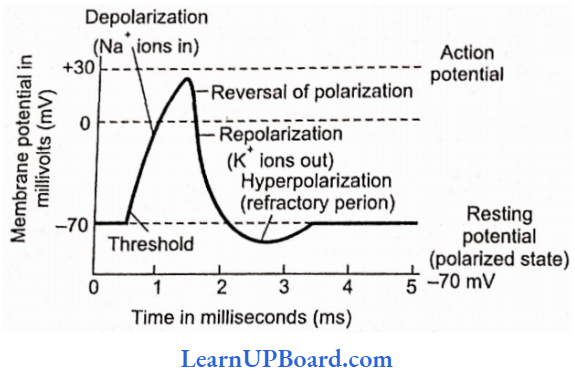
- The membrane potential that sets in a wave of depolarization is called action potential.
- For most excitable cells, the threshold is about -55 mV to -60 mV.
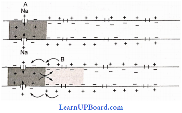
Conduction Of Impulse: In the area of depolarization, the potential difference across the membrane is small while its nearby region has a large difference in membrane potential. This produces a small local current in the area.
- The local current becomes a stimulus and causes the gated Na+ channels of the next region to open and depolarize the area to produce a fresh action potential.
- The process continues till the impulse reaches the end of the neuron.
Repolarization: As Na+ channels close, after 0.5 ms, the membrane becomes extra permeable to K+ ions due to the opening of the K+ ion gates.
- With the pumping out of K+ ions, the neuron interior becomes negative and the potential falls back to the resting potential.
- The phenomenon of change of membrane potential from an excited state to a resting state is called repolarization. However, K+ ion channels remain open for a bit longer period so that the membrane potential becomes more negative than -70 mV. This is called hyperpolarization.
- It takes about 1-5 ms for repolarization.
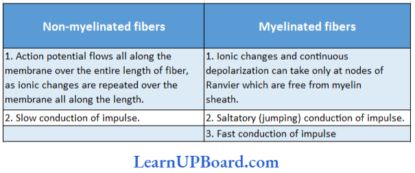
Difference between non-myelinated and myelinated fibers:
Synaptic Transmission
Synapses are the neuronal junctions through which information from one neuron can pass to the other. There are mainly two types of synapses depending upon the nature of the transfer of information across the synapse: electrical and chemical.
Electrical Synapses: At an electrical synapse, ionic current spreads directly from one cell to another through gap junctions.
- Each gap junction contains a hundred or so tubular protein structures called connexons that form a tunnel to connect the cytosol of the two cells. This provides a path for ionic current flow.
- Gap junctions are common in visceral (single-unit) smooth muscle, cardiac muscle, and a developing embryo.
- They also occur in the CNS.
Epithelium
Electrical synapses have three obvious advantages:
- They allow faster communication than do chemical synapses since impulses conduct across gap junctions.
- They can synchronize the activity of a group of neurons or muscle fibers. The value of synchronized action potentials in the heart or in visceral smooth muscles is to achieve coordinated contraction of these fibers.
- They may allow two-way transmission of impulses in contrast to chemical synapses, which function as one-way points of communication.
Chemical Synapses: Chemical synapses have a 10-20 nm gap which is too great a distance for such direct electrical coupling.
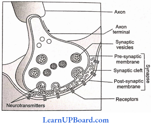
- Chemical synapses are the most common type of synapses. These consist of a bulbous expansion of a nerve terminal called a synaptic knob lying in close proximity to the membrane of a dendrite or other part of a neuron.
- The cytoplasm of the synaptic knob contains numerous tiny, round sacs, called synaptic vesicles.
- Each vesicle has a diameter of approximately 50 nm and contains as many as 10,000 molecules of a neurotransmitter substance responsible for the transmission of nerve impulses across the synapse.
- The membrane of the synaptic knob on the axon side, thickened as a result of cytoplasmic condensation, is called the presynaptic membrane.
- The mechanism of transmission of nerve impulses through chemical synapse is as follows:
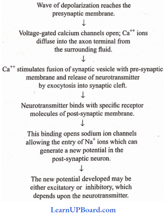
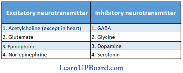
Examples Of Excitatory And Inhibitory Neurotransmitters:
Points To Remember
- Acetylcholinesterase is present in the muscle cell or post-synaptic neuron. It breaks down acetylcholine into acetate and choline and terminates the action of the transmitter.
- Nor-epinephrine secreted by the sympathetic neural system and also by some neurons of the central neural system is inactivated by the enzyme monamine oxidase.
” neural control and coordination class 11 notes pdf download”
Central Nervous System Of Humans
The structures of the CNS arise from their embryological components.
- Prosencephalon: Becomes thalamus and hypothalamus (diencephalon); cerebral cortex, corpus striatum, hippocampus, and amygdala (telencephalon).
- Mesencephalon: Becomes midbrain.
- Rhombencephalon: Develops into the medulla (myelencephalon) and pons and cerebellum (metencephalon).
Brain
Meninges: The brain is surrounded by three protective coats of connective tissue besides the bony cranium. These are known as meninges (singular, meninx).
- Pia mater: It is the inner meninx. It is very thin, highly vascular, and closely invests the brain. It is covered by simple squamous epithelium.
- Arachnoid mater or membrane: It is the middle meninx. It is also thin but is non-vascular. It is covered with simple squamous epithelium on both (internal and external) surfaces. There is a narrow space between the pia mater and the arachnoid membrane. It is called the subarachnoid space. It contains cerebrospinal fluid and is crossed by a number of connective tissue strands.
- Dura mater: It is the outer meninx. It is thick, and tough, and lines the cranial cavity. Its internal surface is covered with simple squamous epithelium. A very narrow space also exists between the dura mater and the arachnoid membrane. It is called the subdural space. It contains a little fluid which is not the cerebrospinal fluid.
An adult human brain contains more than 100 billion neurons and almost 10 times neuroglia cells. The brain is divided into three main sections: (I) forebrain, midbrain, and hindbrain.
Epithelium
Different Regions of the Brain
Forebrain: It consists of two main parts, cerebrum and diencephalon.
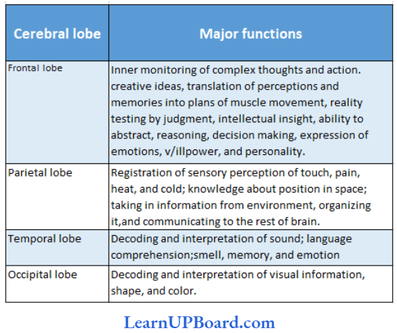
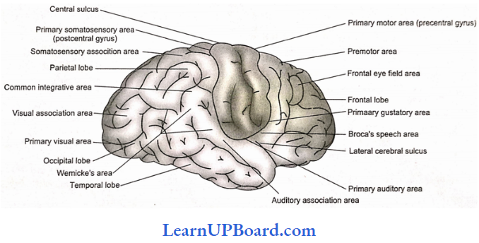
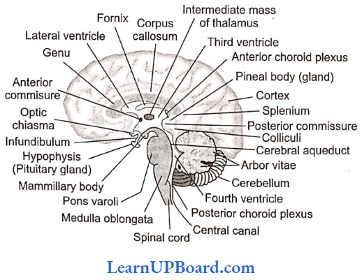
Cerebrum: By far the largest and most highly developed part of the brain is the cerebrum. It is divided into two hemispheres by a prominent longitudinal fissure. The two hemispheres are connected by a bundle of transverse fibers called the corpus callosum. The anterior part of the corpus callosum is curved and is called genu, while the posterior part is called splenium. Each cerebral hemisphere is divided into four lobes. These are frontal at the front, parietal toward the top of the head, temporal on the side, and occipital at the rear.
Cerebral Lobes And Their Major Functions:
Cerebral cortex: The outer surface of the cerebrum, called the cortex, is a layer only 2-4 mm thick.
- Because the six layers are packed with 10 billion pyramidal, spindle, and stellate neurons with a greyish-brown appearance, it is referred to as grey matter.
- The cerebral cortex contains roughly 10% of all neurons of the brain.
- Much of the neural activities occur here, for example, from the touch of a feature to the movement of an arm.
- Unlike the mouse brain, the human brain is greatly convoluted.
- These convolutions or folds consist of sulci (sing, sulcus: small groove), fissures (large grooves), and gyri (sing, gyrus: bulge between adjacent sulci or fissures).
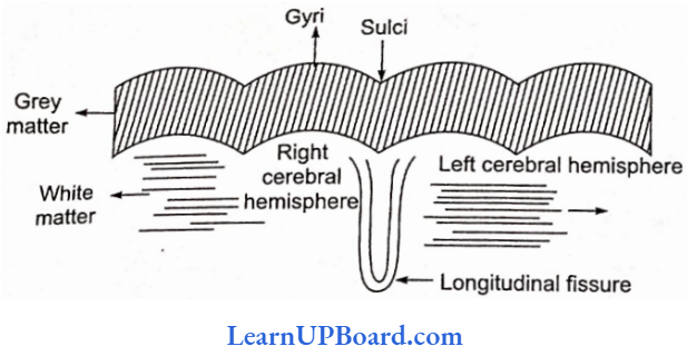
- These greatly enlarge the surface area of the cortex.
- In fact, two-thirds of the surface of the cortex is hidden in sulci and fissures. Thus, their presence triples the area of the cerebral cortex.
- Beneath this run millions of axons comprising nerve fiber tracts, connecting the neurons of the cerebral cortex with those located elsewhere in the brain.
- The large concentration of myelin gives this tissue an opaque white appearance. Hence, they are referred to by the term white matter.
- By examining the effect of injuries or lesions and the effect of electrical stimulation on the behavior, it has been possible to map roughly the location of its various associative activities on the cerebral cortex.
- Each area is referred to as a specialized cortex.
- There are three general kinds of cortex: sensory, motor, and associative.
Diencephalon: The diencephalon contains epithalamus, thalamus, and hypothalamus.
- The epithalamus is a thin, non-nervous roof of the diencephalon.
- Its anterior region is folded and fused with pia mater to form the anterior choroid plexus. This is responsible for the formation of cerebrospinal fluid (CSF).
- Above it is present pineal stalk bearing the pineal body at the top of it.
- The pineal body is an endocrine gland and is also taken as a vestige of the third eye.
- The thalamus directs sensory impulses from the lower parts of the brain and spinal cord to appropriate parts of the cerebrum.
- Limited sensory awareness of pain, temperature, touch, and pressure is provided by the thalamus.
Hypothalamus: As the name implies, the hypothalamus nestles at the base of the thalamus, and so of the brain.
- Although relatively small, just 4 g, about 1/300 of the total brain mass is highly vascularized.
- It integrates and controls visceral activities.
- The hypothalamus, through its connection with the brain stem, maintains homeostasis and the body’s internal equilibrium, specializing in involuntary behavior control.
- The nuclei in it signal the body to eat, drink, get angry, keep cool, etc.
- The hypothalamus organizes behavior related to the survival of species: fighting, feeding, fleeing, and mating.
- It keeps the body temperature at roughly 37°C by means of a complex thermostat system.
- It also influences respiration and heartbeat and sends, out signals to correct them when they are wrong.
- Through connections with the pituitary gland, it controls growth and sexual behavior.
neural control and coordination notes pdf download
Basal ganglia: The inside of the human brain is not so densely packed, but there are all kinds of different collections of neurons, called nuclei, each with its specific functions.
- These control different body activities automatically.
- Basal ganglia are a collection of subcortical nuclei in the forebrain, at the base of the cortex.
- The largest nucleus in it is the corpus striatum.
- It regulates the planning and execution of stereotyped movements.
- Other basal ganglia perform at a subconscious level learned patterns of movements such as slow and fast pedaling, slow and fast writing/typing, etc.
- Destruction of the dopamine-secreting par compacta part of the basal nucleus called substantia nigra leads to paralysis agitans, Parkinson’s disease. Huntington’s chorea is due to the degeneration of GABA-secreting neurons of the corpus striatum and acetylcholine-secreting neurons of other parts.
Limbic system: Flared like a wishbone, a ring or fork through extensive neural links with the cerebrum and the brain stem below constitute what is called limbic system (meaning lip-like).
- The limbic system sends out signals to the rest of the brain and the body which have a great effect on your behavior.
- It includes the hypothalamus, amygdala, hippocampus, septum, anterior nucleus of the thalamus, and a portion of basal ganglia.
Amygdala: above the hypothalamus, attached to the inner lips of both forks, is like an almond-shaped amygdala. This bulge of neurons is like a defense castle controlling moods, especially anger and rage. Various regions of the amygdala play important roles in emotional behavior such as aggression and remembering fear.
This remarkable organ deals with a strange mix of signals about smells and memories.
The hippocampus functions as a kind of index for the recall of an event with its associated memory.
It converts information from short-term to long-term memory, essential in learning.
The septum linked to the hypothalamus contains yet another emotional center for sexual arousal.
Epithelium
Midhniln: It has two structures: corpora quadrigemina and crura cerebri.
Corpora Quadrigemina: It contains four lobes, therefore, corpora quadrigemina.
- Its principal structures are superior and inferior colliculi.
- The superior pair of colliculi receives sensory impulses from the eyes and muscles of the head and controls visual reflexes. For example, they control and coordinate the movement of the head and eyes, to fix and focus on an object.
- The inferior pair of colliculi receives sensory impulses from the ears and muscles of the head and controls auditory reflexes such as the movement of the head to locate and detect the source of a sound.
Crura cerebri (cerebral peduncle): These are two heavy fibrous tracts on the inferior side of the midbrain and connect the hindbrain with the forebrain.
- Crura cerebri is involved in controlling muscle tone and modifying some motor activities.
- These relay sensory as well as motor impulses between forebrain and hindbrain.
Hindbrain: It consists of the cerebellum, pons, and medulla.
- Cerebellum
- To the rear of the brain and placed under the cerebrum, the cerebellum is the second largest part of the brain, which means simply “little cerebrum.
- Wedged between cerebral hemispheres and the brainstem, the cerebellum is made up of two cerebellar hemispheres.
- Like the cerebrum, the cerebellum has its grey matter on the outside, comprising three layers of cells and fibers.
- The middle layer contains characteristically large flask-shaped Ptirkinje cells.
- Tree-like structure with a myriad of dendrites, Purkinje cells rank among the most complex of all neurons.
- The white and grey matter form arbor vitae.
- The central portion of the cerebellum has a worm-like appearance as it is narrow and furrowed. It is called vermis.
- Three paired bundles of myelinated nerve fibers, called cerebellar peduncles, form communication pathways between the cerebellum and other parts of the CNS.
- The superior cerebellar peduncles connect the cerebellum to the midbrain, middle cerebellar peduncles communicate with pons, and inferior cerebellar peduncles consist of pathways between the cerebellum and medulla oblongata as well as the spinal cord.
- The cerebellum does not initiate movement but modulates or reorganizes motor commands.
- The cerebellum’s unconscious directions and cerebrum’s conscious instructions determine how and when to move body parts.
- The cerebellum is vital to the control of rapid muscular activities such as running, typing, and even talking.
- All the activities of the cerebellum are involuntary but may involve learning in their early stages.
- Pons: Pons (Latin: bridge) forms the floor of the brain stem.
- It serves as a neuronal link between the cerebral cortex and cerebellum.
- It has a pneumatic center and a switch-off center for inspiration.
- Medulla oblongata: Literally meaning oblong mar- mu-, medulla oblongata is the posteriormost part that connects the spinal cord and various parts of the brain.
- It lies with its breathing center, cardiovascular center, and vomiting center.
- The Vagus nerve arises from the medulla.
- Its roof is thin and non-nervous and constitutes posterior choroid plexus.
- Below the plexus, the roof has three openings, a pair of lateral apertures called foramina Luschka, and a single median foramina Magendie.
- These apertures connect the external and internal components of the CSF of the brain.
- Most of the sensory as well as motor nerve tracts cross over to the other side of the medulla. Therefore, the right half of the cerebrum controls the left half of the body and vice versa.
The reticular formation that connects to the thalamus and major nerves in the spinal cord is the gatekeeper to consciousness.
Brain stem: It is the area of the brain between the thalamus and spinal cord and includes the medulla, pons, and midbrain. Diencephalon may or may not be included.
Ventricles of the Brain and Cerebrospinal Fluid:
- The ventricles consist of four hollow, fluid-filled spaces inside the brain.
- A lateral ventricle lies inside each hemisphere of the cerebrum.
- Each lateral ventricle is connected to the third ventricle by an interventricular foramen (foramen of Monro).
- The third ventricle consists of a narrow channel between the hemispheres through the area of the thalamus.
- It is connected by a cerebral aqueduct or aqueduct of Sylvius or iter in the midbrain portion of the brain stem to the fourth ventricle in the pons and medulla.
- The fourth ventricle continues with the central canal of the spinal cord.
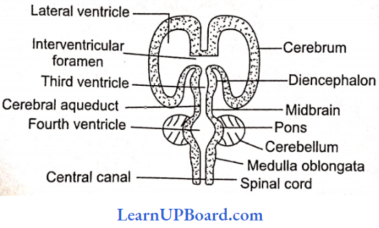
- Three openings in the roof of the fourth ventricle, a pair of lateral apertures (foramina or Luschka), and a median aperture (foramen of Magendie) allow CSF to move upward to the subarachnoid space that surrounds the brain and spinal cord.
- The CSF is secreted by the anterior choroid plexus and posterior choroid plexus and is found inside the ventricles of the brain, the central canal of the spinal cord.
- The CSF acts as a shock absorber for the brain and spinal cord and may also contribute to nourishing brain tissue. It contains protein, glucose, chloride, and water.
Spinal Cord: It is an elongated cylindrical structure that lies in the neural canal of the vertebral column and is continued with the medulla oblongata through the foramen magnum to the skull.
- It measures about 45 cm in length.
- It extends down up to the first lumbar vertebra where it tapers to a point called conus modular/conus termi-nalis. However, the meninges of the spinal cord continue as the film terminates starting from the conus and running up to the coccygeal region.
- The spinal cord shows two enlargements: brachial swelling (from 4th cervical to 1st thoracic vertebrae) and lumbar swelling (from 9th thoracic to 12th thoracic vertebrae).
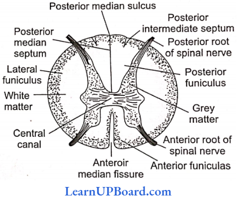
- The spinal cord possesses an anterior and a posterior median fissure running along its length.
- The grey matter of the spinal cord is internal and present around the central canal.
- It is produced into posterior and anterior pairs of grey columns/roots.
- Each dorsal root has a ganglion called dorsal root ganglion.
- The dorsal root is sensory and the ventral root is motor in nature. Both get combined before coming out of the vertebral column through intervertebral foramina.
- The white muter is outer and is divided into four funiculi one dorsal, one ventral, and two lateral
- The spinal cord conducts impulses to and from the brain.
- The dorsal funiculus has an ascending nerve tract for conducting sensor impulses toward the brain.
- Latetal and ventral funiculi conduct motor impulse Hour brain to spinal coni.
- It controls most of the reflex activities.
Peripheral Nervous System Of Humans
The PNS is constituted by the nerves that arise from the brain and spinal cord. They are, respectively, called cranial (cerebral! and spinal nerves, PNS is subdivided into the somatic neural system and the autonomic nervous system.
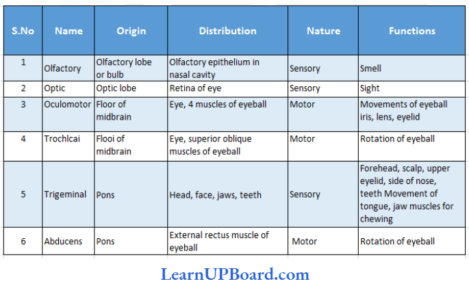
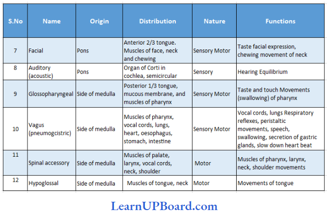
Cranial nerves:
- There are 12 pairs of cranial nerves in man.
- They leave the cranium through foramina and mainly innervate the head region.
- Their origin, supply, and nature are given in
Origin, Supply, And Nature Of Cranial Name Nerves:
Spinal Nerves:
- In man, there are 31 pairs of spinal nerves. They are classified into live groups.
- They include eight pairs of cervical nerves, 12 pairs of thoracic nerves, five pairs of lumbar nerves, five pairs of sacral nerves, and one pair of coccygeal.
- They leave the vertebral column through intervertebral foramina, and all of them are mixed. It is divided in the way into three to four branches.
- Posterior or dorsal branch: It supplies muscles and skin of the back.
- Anterior or ventral branch: It supplies organs in the front and sides of the body. It forms the main spinal nerve.
- Meningeal or recurrent branch: It supplies meninges, ligaments, blood vessels, and other parts of the vertebral column.
- Visceral or ramus communicans: It occurs from the first thoracic to third lumbar nerves. It joins the sympathetic chain of its side.
- The main spinal nerves are anterior or ventral branches of spinal nerves joined at places to form plexuses. These plexuses are five in number.
- Cervical plexus: It occurs in the neck region. The cervical plexus is formed by the first four cervical spinal nerves and the phrenic nerve. The plexus innervates the neck and diaphragm.
- Brachial plexus: It is formed by the fifth, sixth, seventh, and eighth cervical spinal nerves and the first thoracic spinal nerve. The plexus innervates the chest and arms.
- Lumbar plexus: It is formed by the first four lumbar spinal nerves. It supplies nerves to the legs.
- Sacral plexus: The plexus is formed by a branch of each of the fourth and fifth lumbar nerves and the first four sacral nerves. It innervates the hip joint and a part of the pelvis.
- Coccygeal plexus: It is formed by the fourth and fifth sacral spinal nerves and the coccygeal nerve for innervating skin and parts of the pelvis.
Peripheral Nervous System Of Humans Points To Remember
Spinal accessory (XI) and hypoglossal (XII) are lacking in amniotes (cyclostomes, fishes, and amphibians)
Wrist drop: Due to injury in the brachial plexus.
Foot drop: Due to injury in the sciatic nerve.
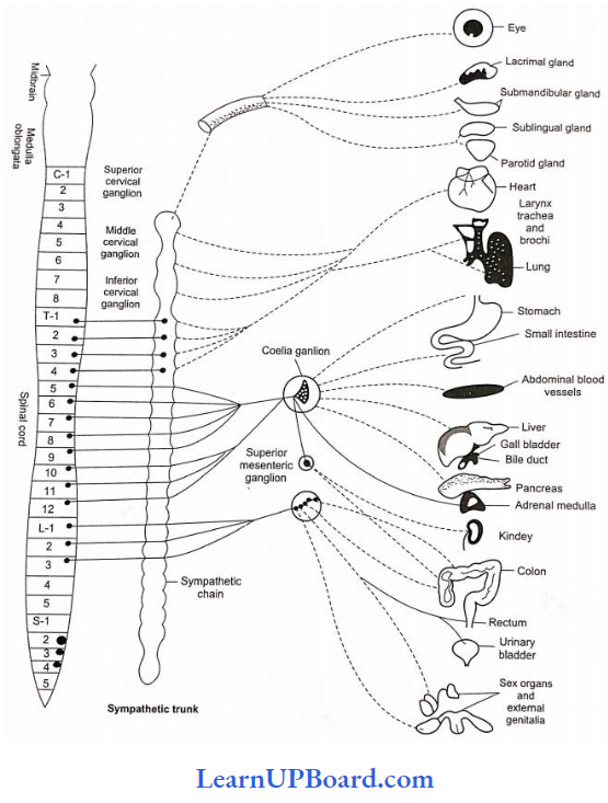
Autonomic (or Visceral) Nervous System
The autonomic nervous system consists of two antagonistic components—sympathetic and parasympathetic.
- Sympathetic nervous system
- The sympathetic nervous system is represented by a chain of 21 sympathetic ganglia on either side of the spinal cord.
- It receives preganglionic sympathetic fibers from the spinal cord, which make their exit along with thoracic and lumbar nerves and constitute thoracolumbar outflow.
- These fibers synapse with the neurons present in the sympathetic ganglia.
- From the sympathetic ganglia arise postganglionic fibers which terminate on the viscera.
- Each sympathetic ganglion is connected to the spinal cord by white communicants and the spinal nerve by gray communicants.
- Some preganglionic fibers pass through the sympathetic chain without synapsing and then join to form splanchnic nerves emanating in some collateral ganglia which include a celiac ganglion, an anterior mesenteric ganglion, and a posterior mesenteric ganglion.
- The postganglionic fibers arising from collateral ganglia supply the digestive system and urinogenital system.
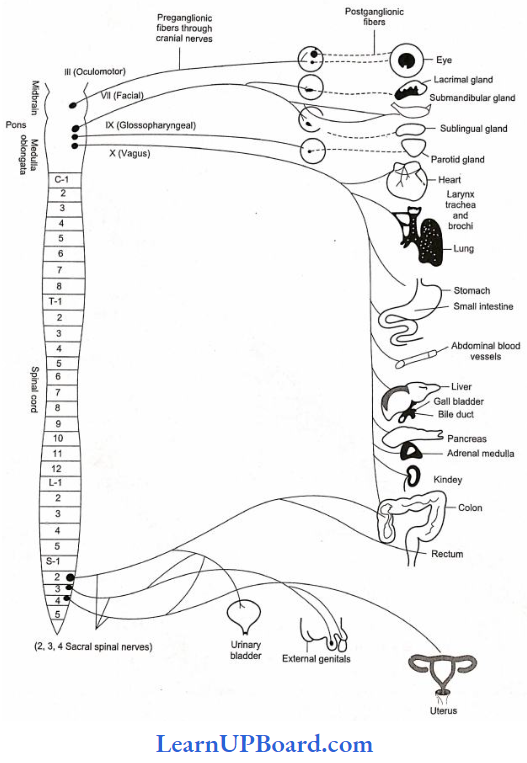
- Parasympathetic nervous system
- It consists of preganglionic parasympathetic fibers, parasympathetic ganglia, and postganglionic parasympathetic fibers.
- Preganglionic parasympathetic fibers make their exit along with the 3, 4, 9, and 10 cranial nerves, and 2, 3, and 4 sacral nerves.
- They together form craniosacral outflow.
- The parasympathetic ganglia do not form any chains and instead lie on or near the viscera.
- The postganglionic parasympathetic fibers arise from these ganglia and supply the viscera.
- The neurotransmitter within the ganglion is acetylcholine for both sympathetic and parasympathetic nerves. However, the neurotransmitter between the terminal autonomic neuron axon and the target organ is different in the two antagonistic autonomic nervous systems.
- In the parasympathetic system, the neurotransmitter at the terminal synapse is acetylcholine, just as it is in the ganglion. In the sympathetic system, the neurotransmitter at the terminal synapse is either adrenaline or noradrenaline, both of which have an effect opposite to that of acetylcholine.
- There is one exception: the sympathetic postganglionic neuron that terminates on sweat glands uses acetylcholine. Thus, depending on which of the two paths is selected by the CNS, an arriving signal will either stimulate or inhibit the organ. Thus, an organ receiving nerves from both visceral nervous systems is subjected to the effects of two opposing neurotransmitters.
- If the sympathetic nerve ending excites a particular organ, the parasympathetic usually inhibits it.
- With few exceptions, organs of the body are innervated by “dual innervations,” and each has a different effect.
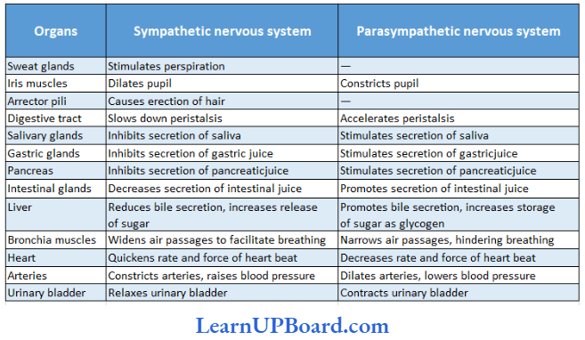
- The function of the autonomic nervous system is to control and coordinate the activities of visceral organs. The two components work against one another.
- The roles of sympathetic and parasympathetic nervous systems are mentioned
Antagonistic Role Of Sympathetic And Parasympathetic Nervous Systems:
Nervous System Points To Remember
- A gasserian ganglion is associated with the trigeminal nerve.
- Geniculate ganglion is the swelling of the facial nerve.
- Parasympathetic fibers do not travel in the dorsal and ventral rami of the spinal nerve.
- As a result, the effector in the skin, sweat gland, arrector pili muscle, and cutaneous blood vessels receive no parasympathetic innervation.
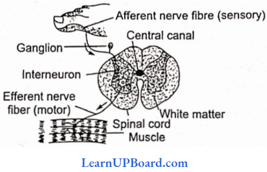
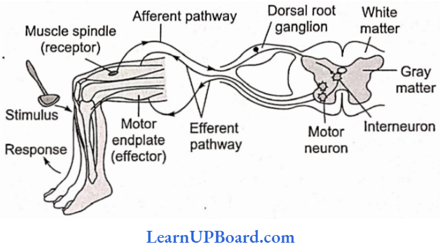
Reflex Action
Reflex action is the simplest kind of activity which can be defined as an integrated activity occurring involuntarily in response to a stimulus applied to a receptor. The reflex arc is composed of the following: a receptor organ, an afferent neuron, synapse involving some cells in the CNS.
Reflexes can be classified as unconditioned and conditioned reflexes. Unconditioned reflexes are inborn, for example, knee jerk, salivation on tasting the food, peristalsis, and closing of eyes on being approached by an object. Conditioned reflexes are acquired, i.e., developed after birth through conditioning or learning. For example, playing a musical instrument, knitting without looking, writing as well as reading.
Reflexes can also be classified according to the number of synapses in the reflex path. These are as follows:
- Monosynaptic reflexes: When there is only one synapse in the reflex path. For example, knee jerk.
- Polysnaptic reflexes: When there is more than one synapse in the reflex path.
Reflex Action Points To Remember
Salivation on seeing hearing or smelling delicious food is an example of cerebral reflex action, whereas withdrawal of legs when a drop of HCl is dropped over the sciatic nerve of a decapitated frog is an example of spinal reflex action.
- Characteristics Of Reflexes: Although the reflexes are involuntary functions, they have certain features that make them highly complicated. Some important characteristics are as follows:
- Predictability: Once the response of an organ to a specific stimulus is observed, one can predict that the same stimulus will always elicit the same response.
- Purposefulness: Generally all reflex actions are useful to the organism and are performed with a definite purpose.
- Localization: In performing a reflex action, a specific effector is involved in response to the stimulus applied to a specific receptor.
- Delay: Reflex time is the interval between the application of the stimulus to a receptor and the beginning of a response by an effector organ. A synaptic delay occurs due to latent period and reflex time at the synapse. This depends upon the number of synapses in the nerve pathway.
- Unlearned: To activate spinal effector mechanisms, no experience is needed to bring them into operation.
- Adjustive and protective: Reflexes serve adjustive and protective purposes and become an important part of animal behavior.
- Fatigue: Reflex responses arc readily fatigued after prolonged and continuous work. As a consequence, the latent period of contraction becomes longer and the rise of tension smaller and more gradual.
Sense Organs
Stimuli are received by certain structures in the body. These are called receptors or sense organs. A receptor may be extremely simple such as those of touch, taste, and smell, or they may be highly complex in their structure as well as working, for example, the sense organs of sight and hearing. Sense organs can be classified based on the following criteria:
- According to their position
- Exteroceptors: The external sense organs that receive the stimuli from the outer environment.
- Proprioceptors: Simple receptors present in joints, skeletal muscles, tendons, etc. They are not in direct contact with the environment but are affected by the changes in the environment.
- Visceroceptors or internal receptors: The receptors present within the viscera. They receive stimuli originating within the body itself. They are simple and mostly represented by free nerve endings. Perception is conscious awareness and interpretation of sensation.
- According to the form of stimulus they receive: The sense organs are classified on this basis.
Different Types Of Receptors And The Form Of Stimulus They Receive:
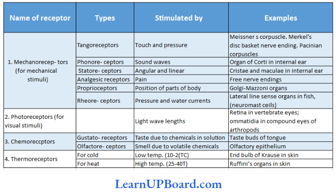
Tangoreceptors: These are the sense organs for touch, pressure, and pain. heat, or cold. They are located in the skin and include:
Meissner’s corpuscles: They are present immediately below the epidermis and receive the stimulus of touch/gentle pressure.
Pacinian corpuscles: Situated deep in the dermis of skin, joints, tendons, and muscles. Each corpuscle has a nerve ending surrounded by connective tissue. They respond to pressure changes.
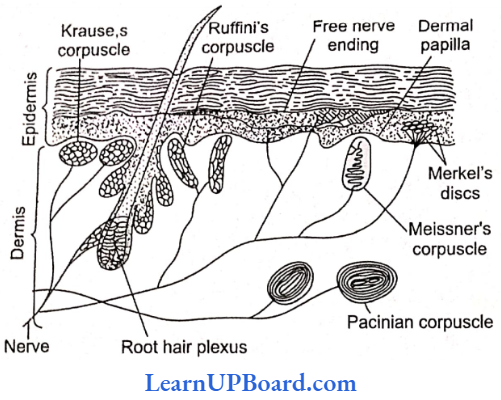
Merkel’s disc: Occurs in the epidermis and is responsible for touch.
Skin is often called hypothermic because it has more cold receptors. The regulation of temperature in the human body is mediated by the hypothalamus which has a “set point” (96.4°F or 37°C) around which the core temperature oscillates.
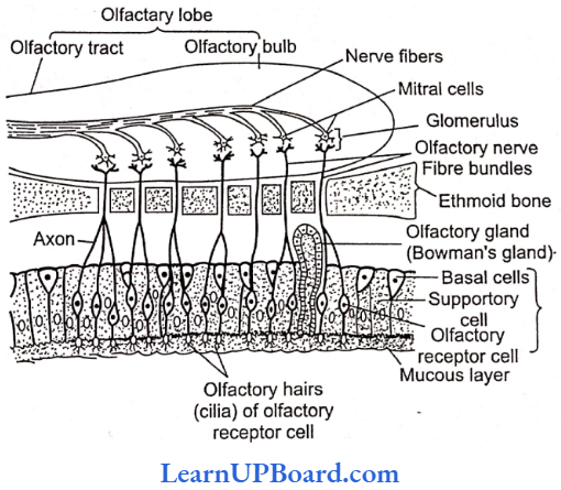
Smell Receptors (Olfactoreceptors): The receptors of smell occur in a small patch of olfactory epithelium (pseudostratified epithelium) located in the roof of the nasal cavity. Smell receptors are of the following types:
- Olfactory receptor cells: They act as sensory receptors as well as conducting neurons. The olfactory receptor cells are “unusual” bipolar neurons. Each cell is spindle-shaped and has a thin apical dendrite that terminates in a knob that bears non-motile cilia called olfactory hairs. Olfactory receptor cells are unique in that they are the only neurons that undergo turnover throughout adult life.
- Supporting cells: These are columnar cells that lie between the olfactory receptor cells to support them. They have brownish-yellow pigment (similar to lipofuscin) which gives the olfactory epithelium its yellowish color.
- Basal cells: These are small cells that do not reach the surface. They give rise to new olfactory receptor cells to replace the worn-out ones. This is an exception to the fact that neurons are not formed in the postnatal (after birth) life. The olfactory receptor cells survive only for about two months.
- Olfactory glands (Bowman’s glands): Many olfactory glands occur below the olfactory epithelium that secrete mucus to spread over the epithelium to keep it moist. The mucus also protects the cells from dust and bacteria.
- Working on smell receptors: The dissolved chemicals stimulate the olfactory receptors by binding to protein receptors in the olfactory hairs (cilia) membranes and opening specific Na+ and K channels. This leads ultimately to an action potential that is conducted to the first relay station in the olfactory bulb.
The fibers of the olfactory nerves synapse with mitral cells (second-order neurons) in complex structures called glomeruli (balls of yam). When mitral cells are activated, impulses travel from the olfactory bulbs via olfactory tracts to main destinations (for example, the temporal lobe of the cerebrum).
Sense Organs Points To Remember
- Women often have a keener sense of smell than men, especially at the time of ovulation.
- Smoking damages the olfactory receptors.
- With aging, the sense of smell deteriorates.
- Hyposmia (hypo: less, osmi: smell) is a reduced ability to smell.
- In addition to smell receptors, the nose, mouth, and tongue contain a network of nerves that form the trigeminal nerve (fifth cranial nerve) also known as the dentist’s nerve reacts to messages of path,
- The brain combines the trigeminal signals with those of smell to identify some odors when exposed to irritants such as ammonia or vinegar.
- The trigeminal can protect by warning about harmful chemicals in the air. Bowman’s glands inside the nose release fluids to get rid of the irritating substances.
Taste Receptors (Gustatoreceptors)
Location: The receptors for taste are found in the taste buds, mostly located on the tongue, but are also found on the palate, pharynx, epiglottis, and even in the proximal part of the esophagus. The number of taste buds declines with age.
Structures: Each taste bud is an oval body consisting of three kinds of cells.
Gustatory receptor cells: They bear at the free end microvilli projecting into the taste pore. The microvilli have special protein receptor sites for taste-producing molecules and come in contact with the food being eaten. Nerve fibers of the cranial nerves 7 (facial),9 (glossopharyngeal), or 10 (vagus) end around the gustatory receptor cells, forming synapses with them. The gustatory receptor cells (taste cells) survive only about 10 days and are then replaced by new cells.
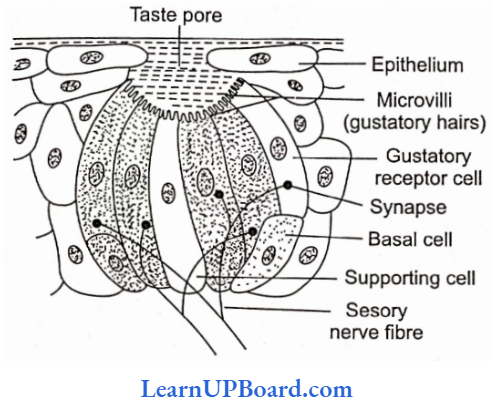
Supporting cells: These cells lie between the gustatory receptor cells in the taste bud. They bear microvilli but lack nerve endings.
Basal Cells: These cells are found at the periphery of the taste bud. They produce supporting cells, which then develop into gustatory receptor cells.
Working: Specific chemicals in solution pass into the taste bud through the taste pore to come in contact with the protein receptor sites on the microvilli of the gustatory receptor cells. The latter set up nerve impulses in the sensory nerve fibers. The nerve fibers transmit the impulses to the taste center in the brain (for example, the parietal lobe of the cerebrum) where the sensation of taste arises.
The facial nerve (8) serves the anterior two-thirds of the tongue, the glossopharyngeal nerve (9) serves the posterior one-third of the tongue and the vagus nerve (10) serves the pharynx and epiglottis but not the tongue.
Organs of Sight (Eye)
Location: The organs of sight in man are a pair of eyes located in the eye orbits of the skull.
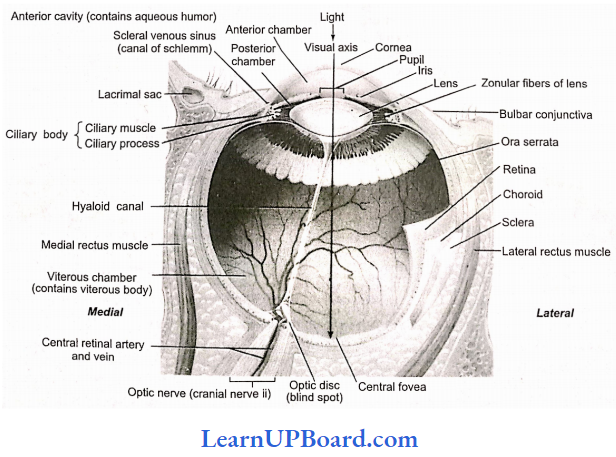
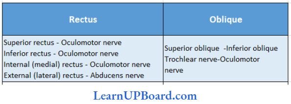
Structure: The exposed part of the eye is protected by an upper and a lower eyelid which are provided with eyelashes. Each eye is represented in the form of a spherical eyeball which is moved in the eye orbit with the help of six eye muscles, namely, superior oblique, inferior oblique, superior rectus, inferior rectus, external rectus, and internal rectus. An eyeball measures about 2.5 cm in diameter and is hollow. Its wall is formed of three layers or coats. The outermost is called the fibrous coat, the middle one is a vascular coat, and the inner one is a retina.
- Fibrous coat: The outer coat of the eyeball is thick and tough. It provides form and shape to the eyeball. The fibrous coat consists of two parts, sclera and cornea.
- The sclera constitutes about five-sixth of the outer coat. It is white (made up of tough but clastic sheath of fibrous connective tissue containing collagen fibers) and opaque, popularly called white of the eye. Most the of sclera is concealed in the orbit.
- The cornea is the anterior transparent part of the sclera and constitutes about one-sixth of the fibrous coat, ft is non-vascular and convex anteriorly. The cornea is covered by a thin and transparent membrane called conjunctiva composed of stratified epithelium and continued over the inner surface of lids.
- Vascular coat: The middle coat of the eyeball is differentiated into three regions, namely, choroid, ciliary body, and iris.
- The choroid is a delicate, highly vascular, and pigmented part that lies in contact with the sclera. It provides a dark color to the interior of the eyeball, it is black in color. It prevents internally reflected light within the eye. The blood vessels of the choroid nourish the retina.
- The ciliary body is the part of the vascular coat immediately behind the peripheral margin of the iris. The ciliary body is thicker and less vascular than the choroid. Its inner surface is folded to form ciliary processes. Present within the ciliary body are ciliary muscles.
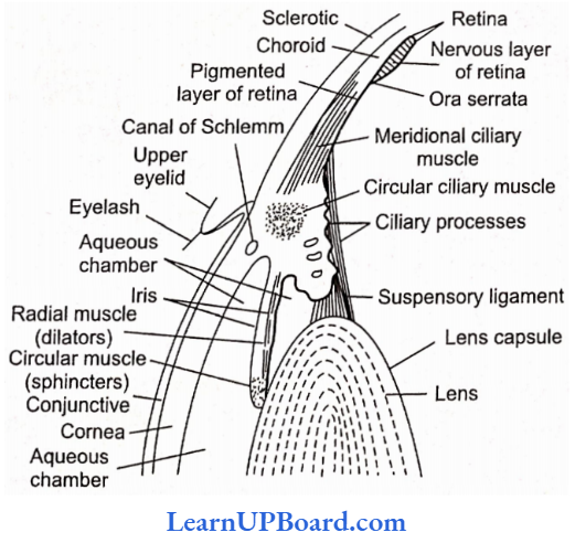
Iris is the anterior part vascular coat that lies behind the cornea. It is centrally perforated by the pupil, the size of which is regulated by iridial muscles arranged radially and circularly. The iris, being pigmented, provides color to the eye.
Mirror-like tapetum layer of carnivores such as cats, etc.. increases sensitivity by reflecting unabsorbed light through the photoreceptor layer to shine in the dark.
Retina (nervous tunic):
- The third and inner coat of the eyeball, the retina (nervous tunic), lines the posterior three-quarters of the eyeball and is the beginning of the visual pathway.
- The optic disc is the site where the optic nerve exits the eyeball.
- Bundled together with the optic nerve are the central retinal artery, a branch of the ophthalmic artery, and the central retinal vein.
- Branches of the central retinal artery fan out to nourish the anterior surface of the retina.
- The central retinal vein drains blood from the retina through the optic disc.
- The retina consists of a pigment epithelium (nonvisual portion) and a neural portion (visual portion).
- The pigment epithelium is a sheet of melanin-containing epithelial cells that lays between the choroid and the neural portion of the retina some histologists classify it as part of the choroid rather than the retina.
- Melanin in the choroid and pigment epithelium absorbs stray light rays which prevent reflection and scattering of light within the eyeball. This enables the image cast on the retina by the cornea and the lens to remain sharp and clear.
- The pigmented layer is continuous over the choroid, ciliary body, and iris, while the nervous layer terminates just before the ciliary body. This point is called orra serrata.
- Albinos lack melanin pigment in all parts of the body, including the eye.
- The neural portion of the retina is a multilayered outgrowth of the brain.
- It processes visual data extensively before transmitting nerve impulses to the thalamus, which then relays nerve impulses to the primary visual cortex.
- Three distinct layers of retinal neurons are separated by two zones where synaptic contacts arc made—inner and outer synaptic layers.
- The three layers of retinal neurons, in the order in which they process visual input, are the photoreceptor layer, bipolar cell layer, and ganglion cell layer.
- Note that light passes through the ganglion and bipolar eel! layers before reaching the photoreceptor layer.
- Two other types of cells present in the retina are called horizontal cells and endocrine cells. These cells form laterally directed pathways that modify the signals being transmitted along the pathway from photoreceptors to bipolar cells to ganglion cells.
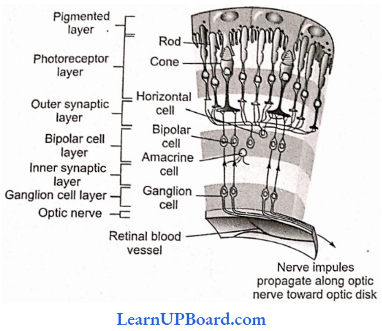
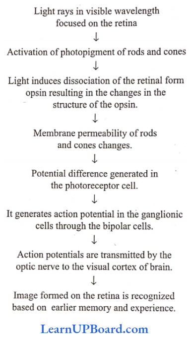
- Photoreceptors are specialized to transduce light rays into receptor potentials.
- The two types of photoreceptors are rods and cones.
- Each retina has about 6 million cones and 120 million rods.
- Rods are the most important for seeing shades of grey in dim light.
- They also allow us to see shapes and movement.
- Cones provide color vision in bright light.
- The visual pigments for color vision are erythropsin (sensitive to red), chloropsin (sensitive to green), and cyanopsin (sensitive to blue).
- In moonlight, we cannot see colors because only the rods are functioning.
- Due to the low light level cones are not functioning.
- The macula lutea is in the exact center of the posterior portion of the retina, at the visual axis of the eye.
- The central fovea, a small depression in the center of the macula lutea, contains only cone photoreceptors.
- In addition, the layers of bipolar and ganglion cells, which scatter light to some extent do not cover the cones here; these layers are displaced to the periphery of the fovea.
- As a result, the central fovea is the area of highest visual acuity or resolution (sharpness of vision).
- Rods are absent from the fovea and macula and increase in number towards the periphery of the retina.
- From photoreceptors, information flows to bipolar cells through the outer synaptic, layer and then from bipolar cells through the inner synaptic layer to ganglion cells.
- The axons of ganglion cells extend posteriorly to the optic disc and exit the eyeball as optic nerves.
- The optic disc is also called the blind spot since it contains no rods or cones.
Accommodation:
- Accommodation (focusing) is the reflex mechanism by which light rays from objects at various locations in the near visual field are brought to focus on the retina.
- Altering the shape of the lens does this. In bright light, the circular muscle of the iris contracts, the radial muscle relaxes, the pupil becomes smaller, and less light enters the eye, preventing damage to the retina.
- In dim light, the opposite muscular contractions and relaxations occur.
- In the dark of night, your pupil may become up to 16 times bigger.
- The added advantage of reducing the pupil size is that it increases the depth of focus of the eye so that any displacement of the photosensors in the retina will not impair the focus.
- Light rays from distant objects (>6 m) are parallel when they strike the eye.
- Light rays from near objects (<6 m) are diverging when they reach the eye.
- In both cases, the light rays must be refracted or bent to focus on the retina and refraction must be greater for light from near objects.
- The normal eye can accommodate light from objects from about 25 cm to infinity.
- With the involuntary ciliary muscles at rest, the flatter lens has the correct optical properties to focus distant images on the retina, but not close images.
- The state of contraction of the ciliary muscles changes the tension of suspensory ligaments. This acts on the natural elasticity of the lens, which causes it to change its radius of curvature and, thus, the degree of refraction.
- As the radius of curvature of the lens decreases, it becomes thicker, and rounds up, and the amount of refraction increases.
- It is the tension of the suspensory ligaments applied to the lens that determines the shape of the lens.
- When the circular ciliary muscles are relaxed and the suspensory ligament becomes tout, the lens is pulled into a flattened shape suitable for focusing distant objects, decreasing the refraction.
- When the tension is decreased, the circular ciliary muscles are contracted and the suspensory ligaments slack. Consequently, the lens becomes a more spherical shape suitable for focusing objects.
- The image produced by the lens of the eye on the retina is inverted and reversed. However, objects are perceived the right way up because of the way in which the brain interprets the images.
- The region of the environment from which each eye collects light is called the visual field.
- Since both our eyes are frontally placed, there is an overlap between the visual fields of each eye. This is called binocular vision.
Image formation is a refractive process; maximum refraction takes place at the cornea.
Extra-ocular muscle of the eye: The eye is rotated in the orbit by six strap-shaped muscles inserted on the sclera. These are arranged in two groups—rectus and oblique.
Chambers of Eyeball
- The lens and suspensory ligament divide the interior of the eyeball into two chambers, the anterior small aqueous chamber containing a watery fluid, the aqueous humor, and the posterior larger vitreous chamber containing viscous fluid, the vitreous humor.
- Aqueous humor maintains intra-ocular pressure mainly whereas vitreous humor is responsible for the shaping of eyeballs.
Mechanism Of Vision
Protective Devices of Eye:
- Eyebrows: Two arched eminences of skin having numerous hairs project obliquely from the surface of the skin. The function of the eyebrows is to protect the anterior aspect of the eyeball from sweat, dust, and other foreign bodies.
- Eyelids (palpebrae) and eyelashes: The eyelids are two movable folds and have hairs on their free edges—the eyelashes. The third eyelid is vestigial and is called plica semilunaris (nictitating membrane). The inner surface of each eyelid and parts of the eyeball are covered with a mucous membrane, called the conjunctiva.
- Glands of Zeis: These are modified sebaceous glands that are associated with the follicles of eyelashes. They open into the follicles of eyelashes. Meibomian or tarsal glands arc also modified sebaceous glands (oil glands) which are present along the edges of eyelids. They produce an oily secretion that serves to lubricate the corneal surface and hold a thin layer of tears over the cornea.
- Glands of Moll: These are modified sweat glands at the edge of the eyelid.
- Conjunctiva: The palpebral conjunctiva is very vascular and has numerous papillae. Over the sclera the ocular conjunctiva is loosely connected to the eyeball; here it is thin, transparent, without papillae, and slightly vascular. Reaching the cornea, it continues as the corneal epithelium. The epithelium of the palpebral conjunctiva near the margin of the lids is a non-keratin-nized squamous stratified epithelium. The conjunctiva helps to protect the eyeball and keeps it moist. It is this membrane that becomes inflamed in conjunctivitis or “pink eye.”
- Lacrimal apparatus: The lacrimal apparatus of each eye consists of a lacrimal gland and its numerous ducts, the superior and inferior canaliculi, a lacrimal sac, and a nasolacrimal duct. The lacrimal gland secretes tears which are composed of water, salts, and a bactericidal protein called lysozyme. Lysozyme dc- stroys microorganisms present on the front of the eyeball.
- Adipose tissue (fat): A layer of adipose tissue surrounds the eyeball in the orbit. It serves as a soft, shockproof pad.
Disorders of Eye
- Myopia or nearsightedness: In this case, the eyeball is too posteriorly elongated so that the image of distant objects is formed in front of the yellow spot. The defect can be removed by using concave glasses.
- Hypermetropia or longsightedness: The person can see distant objects clearly, but not those which are closer. This is due to the Antero-posterior shortening of the eyeball. Hence, the image forms behind the yellow spot. The defect can be overcome by using a convex lens.
- Presbyopia: A common defect in old age people due to the loss of elasticity of the lens and reduced power of accommodation. The disorder can be corrected by convex lenses.
- Astigmatism: The disorder due to the rough curvature of the cornea or lens which can be corrected by the use of cylindrical glasses.
- Cataract: The sight is impaired due to the lens becoming opaque (said motia). The defect can be cured by surgical removal of the defective lens.
- Glaucoma: It occurs due to an increase in intra-ocular pressure as it may develop due to the blockage of the canal of Schlemm. It exerts pressure on the optic nerve causing its damage. It leads to permanent blindness (kala motia).
Organs of Hearing (Ear)
The organs (phonoreceptors) in man are a pair of ears situated on the head. Apart from their auditory function, the ears are also the organs of balance. Each ear has three portions: external ear, middle ear, and internal ear.
1. External ear
- It consists of a pinna and an external auditory canal. The latter is a curved passage that is lined by a profusion of hair and about 4,000 ceruminous glands.
- The glands secrete cerumen, a waxy material that entraps dust and also lubricates the tympanum.
- The tympanum or eardrum is a circular membrane present on the inner end of the external auditory canal and parts it from the tympanic cavity.
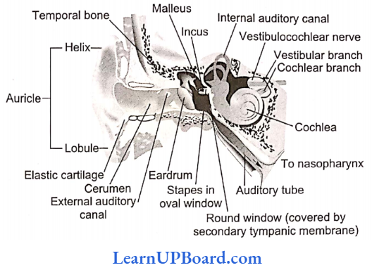
2. Middle ear
- The middle ear is represented by an air-filled tympanic cavity which communicates with the pharynx by a passage called the Eustachian canal.
- Present in the inner wall of the tympanic cavity are two openings, the upper fenestra ovalis and the lower fenestra rotunda, each covered by a membrane.
- The tympanic cavity contains three small bones, the ear ossicles, which from outside to inside include malleus, incus, and stapes.
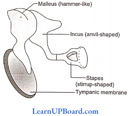
- The malleus is hammer-shaped; the incus is anvil-shaped, and the stapes are stirrup-shaped.
- The outer arm of the malleus is in contact with the inner surface of the tympanum, while the inner end of the stapes forms contact with the membrane on the fenestra ovalis.
- The middle ear is responsible for the amplification of the signal due to the leverage system of ossicle (10 times) by ear ossicles and 2.2 times by the smaller size of membrane covering fenestra ovalis. The oval window is the door to the internal ear.
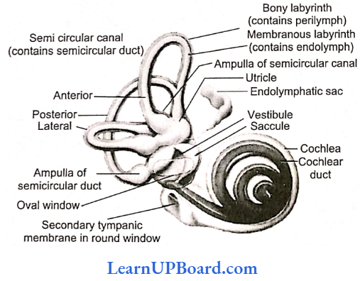
3. Internal car
- It is also called a membranous labyrinth and is surrounded by a bony labyrinth of almost similar shape.
- The space between the membranous labyrinth and the bony labyrinth is filled with a watery fluid, perilymph. The membranous labyrinth contains emdo- lymph.
- The internal ear is a delicate organ and is differentiated into vestibule, semicircular canals, and cochlear ducts.
- The vestibule is the central body and is formed of two chambers, the upper utriculus and the lower succulents.
- Semicircular canals are three-arched structures that emerge from the utriculus and open back into it. They include anterior and posterior vertical canals and a horizontal canal.
- The vertical canals join to form a common passage crus commune before they open into utriculus.
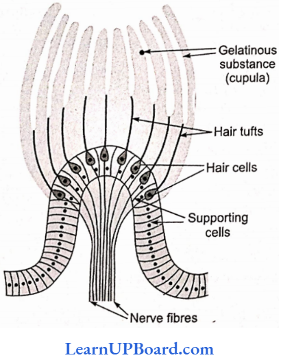
- Each semicircular canal is dilated at the base to form an ampulla which contains a sensory spot called a crista formed of receptor cells and supporting cells.
- The receptor cells bear sensory hair, which is embedded into a gelatinous cupitle above.
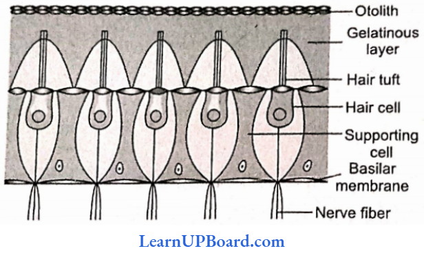
- The vestibule also contains two sensory spots called maculae, one in the sacculus and another in the utriculus.
- Maculae are similar to cristae, but there is no cupule.
- The sensory hair is embedded in an otolith membrane containing calcareous bodies called otoliths.
- Cristae and maculae are the receptors of balance.
- The auditory region of the internal ear is represented by a spirally coiled structure called the cochlea.
- lt consists of a cochlear duct arising from the sacculus, which is surrounded by a similarly shaped cochlear canal, a part of a bony labyrinth.
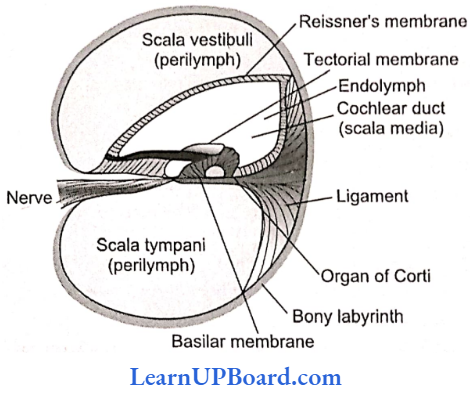
- The cochlear duets is fused with the cochlear canal on lateral sides, but is free laterally, therefore, in TS, the cochlea shows three chambers, upper scala vestibuli, middle scala media, and scala tympam.
- The scala media is partitioned from the live scala vestibuli by Reissner’s membrane and from the scala tympani by the basilar membrane.
- Scala vestibule and scala tympam contain perilymph while scala media is filled with endolymph.
- The upper and lower chambers communicate through lielicotrema, a narrow opening present at the distal end of the cochlea.
- The basilar membrane, sensory hair cells, and tectorial membrane make up live smallest unit of the ear, called the organ of Corti, first described by Italian microscopist, Alfonso Corti (1822 1888).
- Sensory hair cells inside the car resemble tracts left in the sand by truck tires.
- The cochlea contains 16,000-24,000 hair cells arranged in four rows.
- In three of the rows, the hairs form V-shaped patterns. In the fourth row, the hairs stand in a straight line.
- Each hair cell has up to 100 hairs.
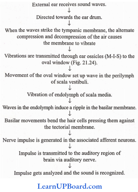
- When sound vibrations pass through the oval window, hairs create waves in the lymph fluid of the cochlea such as a sea wave in a tidal current.
- The waves cause the basilar membrane to ripple. This movement bends the hair cells, pressing against the tectorial membrane and setting off nerve impulses in their associated afferent neurons.
- More than 30,000 neurons and nerve fibers emerging from these convey electrical signals to the brain.
- just 2 cm away via the auditory (vestibulocochlear) nerve.
- The basal ends of hair cells synapse with the fibers of a cochlear branch.
- When the waves reach the round windows of the cochlea, they die away.
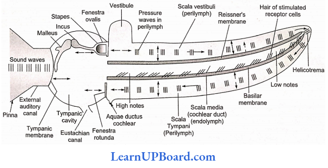
Mechanism Of Hearing
- The high-frequency resonance of the basilar membrane occurs near the base, where the sound waves enter the cochlea, while low-frequency resonance occurs near the apex mainly because of the stillness of the fibers of the basilar membrane.
- The three internal ossicles of car are malleus, incus, and stapes.
- In the case of non-mammals (amphibians, reptiles, birds), there is just one bone called columella auris.
Physiology Of Equilibrium
There are two kinds of equilibrium (balance):
- One is called static equilibrium, which refers to the maintenance of the body position (mainly the head) relative to the force of gravity.
- The second is dynamic equilibrium, which is the maintenance of body position (mainly the head) in response to sudden movements such as rotation, acceleration, and deceleration.
Collectively, the receptor organs for the equilibrium arc are called vestibular apparatus, which includes saccule, utricle, and semicircular ducts.
1. Static Equilibrium
- The walls of both utricle and saccule contain a small, thickened region called macula (plural maculae)
- Maculae are the receptors for static equilibrium and also contribute to some aspects of dynamic equilibrium.
- Static equilibrium provides sensory information on the position of the head and is essential for maintaining appropriate posture and balance.
- For dynamic equilibrium, they provide information about linear acceleration and deceleration. For example, the sensation you feel while in an elevator or a car that is speeding up or slowing down.
2. Dynamic equilibrium
- The vestibular apparatus contains three semicircular canals positioned at right angles to one another.
- The dilated portion of each duct, the ampulla, contains a small elevation called a crista.
- Each crista is composed of a group of hair cells, supporting cells covered by a mass of gelatinous material called cupula.
- Cristae in the three semicircular canals maintain dynamic equilibrium.
Diseases of the Ear
- Meniere’s disease: Due to increased amount of the fluid of internal ear, loss of hearing.
- Myringitis: inflammation of the tympanic membrane.
- Otitis media: Acute infection in the middle car.
- Vertigo: A type of dizziness where there is a feeling of motion when one is stationary.
- Cobyrinthine diseases: Improper functioning of the internal car.
- Most domestic mammals and sharks lack color vision.
- Tapetum lucidum: It is a part of the choroid adjacent to the retina in the eyes of a large number of elasmobranchs (cartilaginous fish). It possesses cells containing light-reflecting guanine crystals. It reflects light and causes the eyes to shine in the dark. It also reflects additional light on the retinal cells to enable the fish to see in water where light is poor.
- Accommodation: Fishes can see objects at different distances by changing the size of the eyeball.
- Pecten: It is a remarkable, highly vascular, and pigmented structure projecting into the vitreous chamber from the blind spot normally. It occurs in all birds except Kiwi (Apteryx). It is also found in some reptiles (e.g. Uromastix) but is absent in mammals. In Uromastix, it is like a cushion; however, in a pigeon, it is comb-like and folded like a fan. The actual function of pecten is unknown, but possibly it aids in the nutrition of the eyeball. In birds, it also helps in accommodation which is remarkably well developed in birds, by pressing the lens forward.
- Phaco-emulsification technique in cataract surgery is a “stitchless” technique. A foldable intraocular lens (IOL) is used.
- Most birds have only day vision as their retina has mainly cones.
- Owls have much better night vision as they contain a large number of rods and few cones in their retina.
- The taste of chilies is not a true sensation. It is mainly the sensation of burning pain produced by the stimulation of pain receptors of the tongue.
- Hordeolum: Inflammation of sebaceous glands of the eyelid.
- Owls and cats see only with the help of available light from the stars or moon at night.
- Frogs are short-sighted in air and long-sighted in water.
- Many insects such as honeybees possess gustatory receptors on their feet.
- Largest cranial nerve —trigeminal.
- Smallest/thinnest cranial nerve —pathetic/trochlear.
- Other names of various parts of the brain:
- Forebrain = Prosencephalon
- Midbrain = Mesencephalon
- Hindbrain* Rhombencephalon
- Olfactory lobes = Rhinencephalon
- Cerebrum = Telencephalon
- Diencephalon = Thalamencephalon
- Cerebellum and Pons = Metencephalon
- Medulla oblongata = Myelcncephalon
- Fourth ventricle = Metacoel
- Third ventricle = Diocoel
- Iter = Mesocoel and aqueduct ofsylvius.
- Lateral ventricle = Paracoel
- Spinal canal = Myelocoel
- The cavity of the olfactory lobe = Rhinocoel (absent in humans)
- The origin of CNS develops from a neural tube that is formed by the infolding of the ectoderm in the early embryo.
- Neopallium: The dorsal wall of the cerebrum/cerebral cortex of the brain.
- Monosynaptic/simple reflex involves a single sensory fiber and a single motor fiber, for example, knee jerk. No interneuron. Polysynaptic/compound reflex involves one (or more) sensory and more than one motor nerve fiber. A number of interneurons are present. Polysynaptic reflexes are more common. All our visceral reflexes are polysynaptic.
Neural Control And Coordination Assertion-Reasoning Questions
In the following questions, an Assertion (A) is followed by a corresponding Reason (R). Mark the correct answer.
- If both Assertion and Reason are true and the Reason is the correct explanation of the Assertion.
- If both Assertion and Reason are true, but the Reason is not the correct explanation of the Assertion.
- If Assertion is true, but Reason is false.
- If both Assertion and Reason are false.
Question 1.
Assertion: In the nervous system, the generation of an action potential depends upon the influx of sodium ions into the axoplasm.
Reason: The influx of sodium ions during nerve impulse generation is due to the efflux of potassium ions.
Answer: 3. If Assertion is true, but Reason is false.
Question 2.
Assertion: The presence of myelin sheath increases the rate of conduction of nerve impulses.
Reason: Ionic channels are absent in the area covered by the myelin sheath. Therefore, depolarization occurs only at the nodes of Ranvier, resulting in saltatory or jumping conduction.
Answer: 3. If Assertion is true, but Reason is false.
Question 3.
Assertion: Receptors in the tendon, and joints give information regarding the position and movements of different parts of the body.
Reason: These are termed as noci-receptors.
Answer: 3. If Assertion is true, but Reason is false.
Question 4.
Assertion: The sharpest vision is in fovea centralis.
Reason: The relationship of receptor to bipolar cells to ganglion cells is 1: 1: 1 within fovea centralis.
Answer: 2. If both Assertion and Reason are true, but the Reason is not the correct explanation of the Assertion.
Question 5.
Assertion: The postganglionic nerve fiber of the parasympathetic nervous system has acetylcholine while the sympathetic nervous system has adrenaline as the neurotransmitter.
Reason: The sympathetic nervous system inhibits intestinal peristalsis while the parasympathetic stimulates peristalsis.
Answer: 2. If both Assertion and Reason are true, but the Reason is not the correct explanation of the Assertion.
Question 6.
Assertion: The brain and spinal cord have a common covering.
Reason: Both the brain and spinal cord possess meninges.
Answer: 1. If both Assertion and Reason are true and the Reason is the correct explanation of the Assertion.
Question 7.
Assertion: Cerebrospinal fluid is present throughout the central nervous system.
Reason: CSF has no such function.
Answer: 3. If Assertion is true, but Reason is false.
Question 8.
Assertion: The brain stem contains centers for controlling activities.
Reason: The brain stem is very sensitive.
Answer: 2. If both Assertion and Reason are true, but the Reason is not the correct explanation of the Assertion.
Question 9.
Assertion: The spinal cord has a column of both grey and white matter.
Reason: Grey matter forms the central spinal canal.
Answer: 3. If Assertion is true, but Reason is false.
Question 10.
Assertion: The motor end plate is a neuromuscular junction.
Reason: The motor end plate is the junction between motor neurons and muscle fibers.
Answer: 1. If both Assertion and Reason are true and the Reason is the correct explanation of the Assertion.
Question 11.
Assertion: Corpus callosum is present in the space between the pia and arachnoid maters.
Reason: It serves to maintain a constant pressure inside the cranium.
Answer: 4. If both Assertion and Reason are false.
Question 12.
Assertion: With the evolution of multicellularity, it became imperative to develop a nervous system.
Reason: Special senses such as vision, and hearing are produced by sense organs associated with the nervous system.
Answer: 2. If both Assertion and Reason are true, but the Reason is not the correct explanation of the Assertion.
Question 13.
Assertion: The auditory ossicles help in hearing.
Reason: Auditory ossicles maintain the balance of air pressure between two sides of the eardrum.
Answer: 3. If Assertion is true, but Reason is false.
Question 14.
Assertion: The image focused on the fovea is seen most accurately.
Reason: The Fovea of the retina contains numerous photoreceptor rod cells.
Answer: 3. If Assertion is true, but Reason is false.
Question 15.
Assertion: A blind spot on the retina of the eye is devoid of the ability for vision.
Reason: The photoreceptor cone cells are absent at the blind spot.
Answer: 2. If both Assertion and Reason are true, but the Reason is not the correct explanation of the Assertion.