NEET Biology Notes – Plant Growth And Development

Plant Growth And Development

Growth

Growth is an irreversible increase in size, volume, and weight of a part or whole of an organism. An irreversible increase in size, volume, or weight is called apparent growth as it is the external manifestation of growth. Formation at cellular materials or protoplasm is the real growth. Growth is a measurable or quantitative phenomenon that can be measured in relation to time. The growth of living beings is internal or intrinsic.

Plant growth is diffused like that of animals only during the early embryonic stages. Later on, plants develop specific areas, called meristem, for growth. On account of meristems, plant growth is localized.

Characteristics Of Plant Growth

  1. Growth is localized.
  2. Growth continues throughout life.
  3. There is an increase in the number of parts.
  4. It is open-ended.
  5. The younger one or seedling can be quite different from an adult.
  6. The juvenile stage may have different traits.

Plant Growth Differentiation: Growth is invariably associated with differentiation. Differentiation is a permanent localized qualitative change in size, biochemistry, structure, and function of cells, tissues, or organs, for example, fiber, vessel, tracheid, sieve tube, mesophyll, leaf, etc. Some examples of differentiation are as follows

  1. Enlargement, lignocellulosic wall thickening, and emptying in case of tracheids
  2. Loss of end wall in case of vessel elements
  3. Loss of nucleus and perforation of end wall in sieve tube members
  4. Deposition of suberin and tannins in cells
  5. Differential wall thickening (in guard cells)
  6. Secretion of mucilage in root cap

Development is the sequence of changes that occur in the structure and functioning of an organism, organ, tissue, or cell involving its formation, growth differentiation, maturation, reproduction, senescence, and death.

Read and Learn More NEET Biology Notes

Characteristics Of Growth

  • Primary Growth: It is the formation of primary permanent tissues and organs. It is caused by the activity of apical and intercalary meristems.
  • Secondary Growth: It is an increase in girth. It occurs by two types of lateral meristems, vascular cambium and cork cambium.
  • Efficiency Index: It is the rate of growth. It is measured by calculating the increase in size, diameter, or area per unit time.

Growth Rate

An increase in growth per unit of time is called growth rate. Growth rate may result in arithmetic or geometric growth.

Arithmetic Growth: Arithmetic growth is a type of growth in which the rate of growth is constant and an increase in growth occurs in arithmetic progression 2, 4, 6, 8, 10, and 12. Arithmetic growth is found in root or shoot elongating at a constant rate.

The meristematic cells at the growing point divide in such a fashion that one daughter cell remains meristematic while the other cells grow and differentiate. The process continues. Mathematically, arithmetic growth is expressed as:

Lt = L0 + rt

where Lt is the length after time t, L0 is the length at the beginning, and r is the growth rate or elongation per unit time.

NEET Biology Plant Growth And Development Arithmetic Growth Curve

Geometric Growth: Geometric growth is quite common in unicellular organisms when grown in a nutrient-rich medium. Here, every cell divides. The daughter cells grow and divide. The granddaughters repeat the process and so on. The number of cells is initially small so the initial growth is slow. Later on, there is rapid growth at an exponential rate.

Geometric growth cannot be sustained for long. Some cells die. Limited nutrient availability causes a slowing down of growth. It leads to the stationary phase. (There may be actually a decline.) Plotting the growth against time will give a typical sigmoid or S-curve.

NEET Biology Plant Growth And Development Geometric Growth Curve

The S-curve of growth is typical of most living organisms in their natural environment. It also occurs in cells, tissues, and organs of plants.

Exponential Growth (Law Of Compound Interest): Growth is dependent on three factors: initial size (W0), rate of growth (r), and time interval (t) for which the rate of growth can be retained.

W1 = W0ert

Here W1 is the final size, W0 is the initial size, r is the growth rate, 1 is the time of growth, while e is the base on natural logarithms. The magnitude of r or the rate of growth has been called the efficiency index by Blackman (1919).

Absolute And Relative Growth Rate: Quantitative comparisons between the growths of various systems can be made by measuring their absolute and relative growth rates.

Absolute Growth Rate: The absolute growth curve is the actual growth curve obtained by plotting growth against time. It is commonly S-shaped. The absolute growth rate is the total growth per unit time. A graph plotted for absolute growth rates for various times of the grand period of growth appears bell-shaped.

The peak is formed when the growth rate is the fastest. The period of increasing growth is depicted by the first part of the curve while the period of decreasing growth rate is shown by the second part of the curve.

NEET Biology Plant Growth And Development Absolute Or Actual Growth Curve And Absolute Growth Rate Curve

Relative Growth Rate: It is the growth per unit time per unit initial growth.

Relative growth rate = \(\frac{\text{Growth in given time period}}{\text{Measurement at start of time period}}\)

Suppose two leaves have grown by 5 cm² in one day. The initial size of leaf A was 10 cm² while that of leaf B was 15 cm². Though their absolute growth is the same, the relative rate of growth is faster in leaf A.

Measurement Of Growth

To measure growth, various methods/instruments are used.

  1. Direct method
  2. Horizontal microscope
  3. Auxanometer
    • Arc auxanometer
    • Pfeffer’s auxanometer
  4. Crescograph (Developed By J.C. Bose): A highly sensitive growth-measuring instrument that can magnify growth by 10000 times.

Growth Hormones

In all plants, minute quantities of certain substances are found (plant growth regulators or phytohormones), which regulate growth and differentiation.

Five major types of growth substances are recognized: aux-ins, gibberellins, cytokinins, abscisic acid, and ethylene.

Auxins: Auxins are weak organic acids having unsaturated ring structures.

  • Charles Darwin conducted his experiments concerning growth on canary grass (Phalaris canariensis) and found that the bending movement of coleoptiles in uni-lateral light was due to a chemical.
  • Went is credited with the discovery of auxin.
  • Auxins are synthesized mainly in apices and exhibit polar transport through parenchyma.
  • KogI and Haagen Smith (1931) found that human urine contained a growth substance, which was isolated and given the name auxin-a (auxentriolic acid). In 1934, Kogl and coworkers isolated another compound, auxin-b (auxenolinic acid) from com germ oil and heteroauxin (now known as IAA or indole-3-acetic acid, C10H9O2N), from human urine. It is the only natural auxin.
  • The precursor of auxin is the tryptophan amino acid.
  • The optimum concentration in the stem apex is 10 ppm while in the root apex, it is 0.0001 ppm.
  • Auxin is active in a free state and can be easily extracted. Bound auxin is inactive and meant for storage. For example, IAA-aspartic acid, IAA-inositol, and IBA-alanine.

Functions Of Auxins

  • Auxins promote cell elongation by loosening cell wall microfibrils, solubilization of carbohydrate reserve, and increased respiration.
  • Responsible for phototropism and geotropism.
  • Promote apical dominance (in the presence of an apical bud, the growth of lateral buds are inhibited due to auxin secreted by the apical bud).
  • Promote root initiation in cuttings (by NAA, IBA).
  • Delay of abscission of leaves by preventing the formation of an abscission layer.
  • Prevention of lodging in cereals.
  • Induce parthenocarpy (production of seedless fruits).
  • Selective weedicide.
  • Have feminizing effect (increase number of female flowers in plants, for example, Cannabis).
  • Seasonal activity of cambium is promoted by auxin.
  • Healing of injury is effected through auxin-induced division in cells around the injured area.
  • Auxin induces negative potential in cell membranes.
  • In legumes, IAA stimulates nodule formation.

Antiauxins: They inhibit auxin activity. For example, triiodobenzoic acid (TIBA), PCIB (p-chlorophenoxy isobutyric acid)

Bioassay Of Auxins

  1. Avena curvature test
  2. Split pea test
  3. Root growth inhibition test

Applications Of Synthetic Auxins

  • Rooting: IBA, IBA-alanine, and NAA are used.
  • Parthenocarpy: IAA, IBA.
  • Weedicide: 2,4-D, 2,4-5-T are used for killing broad-leaved weeds (generally dicot).
  • Flowering: NAA and 2,4-D for litchi and pineapple.
  • Storage: Methyl ester of NAA for the storage of potato.
  • Pre-harvest Fruit Drop: 2,4-D for citrus fruits; NAA for tomato.
  • Prevention Of Lodging: Naphthalene acetamide (NAAM).
  • Vegetable Crops: Chlorophenoxypropionic acid is used to improve the quality of vegetable crops by inhibiting flower formation. For example, lettuce.

Dwarf Shoots: NAA is used for increasing dwarf shoots and a number of fruits in apples.

Gibberellins: In the early twentieth century, Japanese farmers noticed balance or foolish seedling disease of lice. As a result of the disease, certain rice seedlings grew excessively tall, the disease was caused by the fungus Gibherella fuikuroi (perfect state of Fusarium moniliform).

  • Yabuta and Suxniki (1930) isolated the growth-inducing hormone and called it gibberellin.
  • Chemically, all gibberellins are terpenes, a complex group of plant chemicals related to lipids. All are weak acids and have gibbane ring skeletons. GA3 is the commonest. GA24 and GA25 are found only in fungi. The precursor is acetyl CoA.
  • Gibberellins are synthesized in the apices of young leaves, embryos, buds, and roots and are transported through the xylem.

Applications Of Gibberellins

  • Internodal Elongation: Like auxins, the main effect of gibberellins is on stem elongation. Gibberellins stimulate stem elongation and leaf expansion but do not affect roots. Thus, gibberellins restore normal-size arid growth to genetically dwarf varieties of pear and maize.
  • Bolting: In many plants, leaf development is profuse, while internodal growth is retarded. This term of growth is called rosette, for example, cabbage, radish, and henbane. Just before the reproductive phase, internodes elongate enormously, causing a marked increase in height.

The stem sometimes elongates five to six times the original height of the plant. This is called bolting. Bolting requires either long days or cold nights and gibberellins treatment.

  • Germination Of Seeds: Gibberellins promote seed germination (especially in cereals).
  • Control Of Flowering: Gibberellins promote flowering in long-day plants and inhibit it in short-day plants. These also control sex expression in certain species. In general, the application of gibberellins promotes the production of male flowers in female plants of Cannabis.
  • Control Of Fruit Growth: Along with gibberellins, auxins control fruit growth and development. Gibberellins cause parthenocarpy in pome fruits (apple, pear, etc.).
  • Vernalization: Gibberellins can substitute vernalization.
  • Dormancy: Gibberellins overcome the natural dormancy of buds, tubers, seeds, etc.

Commercial Application Of Gibberellins

  • Fruit Growth: Increase the number and size of grapes, tomatoes, etc. Pomalin (a mixture of GA3 and BAP) is used for such purpose.
  • Malt: Increase the yield of malt from barley.
  • Overcoming Dormancy: In photoelastic seeds of tobacco and lettuce
  • Delay Ripening: in citrus.
  • Induce Flowering: In long-day plants, in non-inductive periods.

Antigibberellins: Certain chemicals are antagonistic to gibberellins. For example, Phosphan-D, Amo-1618, CCC, and maleic hydrazide.

Bioassay Of Gibberellins

  1. Induction of α-amylase in barley endosperm test
  2. Dwarf maize test
  3. Dwarf pea test

Cytokinins: They are basic hormones and are purine (adenine) derivatives. Cytokinins are substances that act primarily on cell division and have little or no effect on extension growth. In 1955, Miller et al. separated it from herring sperm DNA and yeast DNA and called it kinetin (because of its involvement in cell division, i.e., cytokinesis). Later on, the substance was identified as 6-furfuryl aminopurine. Subsequently, the term cytokinin was adopted by Letham.

  • The first naturally occurring cytokinin to be chemically identified was from young maize (Zea mays) grains in 1963 and was called zeatin, which is benzyl amino purine (BAP). Cytokinins are a part of t-RNA.
  • Cytokinins are mostly synthesized in roots, seeds, and developing fruits. Coconut milk and apple fruit extracts are rich in cytokinins. Some other cytokinins are dihydrozeatin, IPA (isopentanyl adenine).

Applications Of Cytokinins

  • Cell-division: Cytokinins are quite abundant wherever rapid cell division occurs, especially in growing tissues.
  • Morphogenesis: Cytokinins promote cell division. In the presence of auxins, cytokinins promote cell division even in non-meristematic tissues. In tissue cultures, mitotic divisions are accelerated when both auxin and cytokinin are present. The ratio of cytokinins to auxins also controls cell differentiation and morphogenesis.
  • Apical Dominance: Cytokinins and auxins act antagonistically in the control of apical dominance.
  • Delay In Senescence: Cytokinins delay the senescence of plant organs by controlling protein synthesis and mobilization of resources. This is called the Richmond-Lang effect. Cytokinins are also called anti-aging hormones.
  • Flowering: Cytokinins also induce flowering in certain species of plants such as Lemna and Wolffia and are also responsible for breaking the dormancy of seeds of some plants.
  • Favors Transport: Phloem transport is promoted.
  • Favors Salt Accumulation: Accumulation of salts in the cells is promoted.
  • Favors Sex-Expression: Promote femaleness.
  • Temperature/disease Resistance: Increase resistance to low and high temperatures and diseases.

Commercial Applications of Cytokinins

  1. Tissue culture
  2. The shelf life of vegetables and cut flowers is increased
  3. Overcoming senescence

Bioassay Of Cytokinins

  1. Chlorophyll preservation test.
  2. Cell division test

Ethylene: Ethylene is the only natural plant growth regulator in gaseous form and is effective in the concentration of 0.01-10 ppm.

Ethylene is produced by most or all plant organs but maximum production occurs in ripening fruits and during senescence. High concentrations of auxin induce the formation of ethylene. Though it is a gas, it does not generally move through air spaces in plants. Rather, it escapes from the plant surface. The precursor of ethylene is methionine.

Applications Of Ethylene

  • Growth: Ethylene inhibits stem elongation and stimulates its transverse expansion. As a result, the stem looks swollen.
  • Abscission: It accelerates the abscission of leaves, flowers, and fruits.
  • Fruit Ripening: Its chief effects are on the ripening of fruits accompanied by a rise in the rate of respiration (climacteric). It causes the dehiscence of dry fruits.
  • Flowering: The application of ethylene induces flowering in pineapple.
  • A commercial compound “ethephon” breaks down to release ethylene in plants. It is particularly applied to rubber plants for the flow of latex.
  • It decreases the sensitivity to gravity. Roots become apogeotropic; seedling develops a tight epicotyl hook.
  • It has a feminizing effect.
  • Bioassay: Triple response test.

Growth Inhibitors

  • For a long time, it has been suspected that dormancy is caused by inhibitors. A group of scientists led by Wareing initiated studies to find them. In 1964, pure crystals of a substance were isolated called dormin. It was found to be similar to another compound isolated from young cotton fruits in 1963 by Addicott.
  • This substance accelerated abscission and was called abscission 2. In 1967, it was decided to call it abscissic acid (ABA). Since then, it has been found in all groups of plants (from mosses to higher plants). In liverworts and algae, a compound lunular acid has been found to have activities similar to ABA.
  • Chemically, ABA is a dextrorotatory cA-sesquiterpene. ABA is synthesized in leaves and transported through the xylem and phloem. It is a major inhibitor of growth in plants and is antagonistic to all three growth promoters. Its precursor is violaxanthin (in chloroplast).

Applications Of ABA

  • Stoppage Of Cambial Activity: It inhibits mitosis in vascular cambium.
  • Bud Dormancy: It induces axillary buds to become dormant as the winter approaches.
  • It plays a role in seed development, maturation, and dormancy.
  • Transpiration: It is a “stress hormone” and helps the plant to cope with adverse environmental conditions by closing stomata (antitranspirant).
  • It may be sprayed on tree crops to regulate fruit drop at the end of the season.
  • Application of ABA to green oranges turns them yellow by inducing the synthesis of carotenoids.
  • It induces flowering in some short-day plants such as strawberries and blackcurrant.

Dormancy

In favorable conditions, if a viable seed fails to germinate, this condition is called dormancy and if the viable seed fails to germinate due to unfavorable conditions, it is called “quiescence.” Dormancy may be due to:

  1. Seed coat impermeable to gases, for example, apple, or water, for example, Trigonella; or seed coat mechanically resistant, for example, Capsella, Amaranthus.
  2. Immaturity of the embryo, for example, G. biloba.
  3. Specific light requirement: Some seeds require light for germination and are called positive photoblastic seeds (for example, Lactuca sativa, Nicotiana tobaccum, Lythrum, etc.). Lettuce (Lactuca) is induced by red light and inhibited by far red light. Some seeds show inhibition in germination due to light exposure and are called negative photoblastic, for example, onions, lily, phlox, etc.
  4. Dormancy due to chilling temperature requirement, for example, Polygonum.
  5. After-ripening: Some seeds have a mature embryo but do not germinate immediately due to the absence of growth hormone. They require a period of after-ripening during which they attain the power to germinate. For example, oats, barley, wheat, etc.
  6. Due To Germination Inhibitors: Some chemicals such as organic acids, phenolics, tannins, alkaloids, lactones, mustard oil, etc., inhibit germination (for example, ferulic acid in tomato pulp).

Methods To Break Dormancy

  1. Scarification: It is a method of softening and weakening of seed coat by acids, alcohol, or knife.
  2. Stratification: After ripening treatment, low temperature (0-10°C) with O2.
  3. Light exposure.
  4. Low temperature + Gibbcrellin + O2 treatment, etc.

Seed Germination

Seed germination is of two types:

  1. Epigeal: Hypocotyl grows first, cotyledons come out of the soil as in cucurbits, mustard, castor, onion, tamarind, etc.
  2. Hypodeal: Epicotyl grows first, cotyledons remain underground as in rice, maize, mango, Fabaceae, etc. Whenever a seed germinates inside the fruit, it is vivipary as in Rhizophora, Sonneralia, and Heritiera (mangrove plants).

Cytochrome

Borthwick and Hendrick, in the 1950s, plotted the action spectrum of wavelengths showing their relative effectiveness in stimu¬lating seed germination. The wavelength most effective for promoting germination was 660 nm (red) and for inhibition of germination about 730 nm (far red). They also demonstrated that only brief exposures of light were necessary and that the effects of red light were reversed by far red fight and vice versa.

  • The pigment responsible for this was isolated in 1960 and was called phytochrome by Butler. It is a blue-green pigment existing in two interconvertible forms: PFR or P730 (absorbs far-red lights) and PR or P660 (absorbs red light).
  • By absorbing red light, PR is converted to PFR rapidly. PFR absorbing far-red light is converted to PR rapidly. PFR is the physiologically active form; PR is inactive. The table describes the effects of red light and far red light on plant growth.

NEET Biology Plant Growth And Development Cytochrome Formula

Effects Of Red Light And Far Red Light On Plant Growth

Effects Of Red Light And Far Red Light On Plant Growth

Phytochrome is responsible for various photomorphogenic processes in plants such as the growth and development of plant organs; germination of seed, pollen, and spores; flowering; differentiation of stomata; epinasty and abscission; etc.

Photoperiiqdiibm

The response of plants to changes in the relative lengths of day and night is called photoperiodism.

Photoperiod: The relative lengths of dark and light periods in a day vary from place to place and from season to season. The length of the light period is called the photoperiod. At equators, the day length is of 12 hours duration throughout the year.

Types Of Plants According To Photoperiodic Requirements For Flowering

NEET Biology Plant Growth And Development Types Of Plants According To Photoperiodic Requirments For Flowering

For SDP, PR/PFR > 1 while for LDP, PFR/PR > 1 is critical for flowering.

Photoperiodic stimulus is perceived by the leaf. When proper photoperiod is perceived, a flowering hormone called florigen is synthesized in the leaf and is transported to the bud where flowering occurs. Florigen, a hypothetical hormone, is chemically similar to gibberellins.

Difference Between Long-Day And Short-Day Plants

NEET Biology Plant Growth And Development Differences Between Long Day And Short Day Plants

Vernalization (Yarovization)

The term vernalization was coined by the Russian agronomist Lysenko to refer to the method of accelerating the flowering ability of biennials or winter annuals, by exposing their soaked seeds to low temperatures for a few weeks.

  • However, presently the term is used in a wider sense to include the promotion of flowering in plants by exposing them to low temperatures at any stage in their life cycle. It has been found that some plants especially biennials and perennials are stimulated to flower by exposure to low temperatures.
  • This promotive effect of temperature on flowering is called vernalization. The vernalization was first studied in Europe on the winter varieties of cereals such as wheat, barley, oats, and rye by Klippart.

Site Of Vernalization: The sites of vernalization are the shoot tip, embryo tip, and root apex. As a result of vernalization, a hormone called vemalin (by Melcher) is synthesized.

Requirements Of Vernalization

  1. Low temperature: 0°C-5°C
  2. Period of low temperature: A few hours to a few days
  3. Actively dividing cells (meristematic cells)
  4. Water
  5. Aerobic condition
  6. Proper nourishment

As a result of vernalization, the vegetative phase of the plant is shortened and flowering is initiated. Therefore, the duration of crops is reduced.

Senescence

Senescence is the study of aging in plants. It is of the following types:

Sequential Senescence: In many perennial plants, the apical meristems continue to produce new buds and leaves, while the older leaves and lateral organs undergo senescence and die. For example, Eucalyptus, mango.

Shoot Senescence: In some perennials such as banana and Gladiolus, the aboveground part of the shoot dies every year after producing flowers and fruits. However, the underground parts survive and give rise to new shoots again in the following year.

Simultaneous Or Synchronous Senescence: In some trees such as elm, Dalbergia, and maples, all the leaves are shed in late autumn (October).

Whole Plant Senescence: In monocarpic plants that flower and produce fruits only once in life, for example, wheat, rice, mustard, etc.

Abscission

It is a natural separation or shedding of leaves, foliage branches, fruits, floral parts, etc., from plants. It is generally seasonal. A special narrow zone develops in the area of abscission called as abscission zone. Two distinct layers develop in the abscission zone:

  1. Separation layer (upper layer) and
  2. Protective layer (lower suberized and lignitized layer).

Plant Growth And Development Points To Remember

Strasburger studied growth in roots by marking it at equal intervals with Indian ink.

  1. Ethylene causes respiratory climacteric.
  2. The most widely occurring cytokinin in plants is isopentenyl adenine (IPA).
  3. Sleep disease (enrolling of petals in opened flowers) is caused by ethylene. Even 1 ppm of ethylene prevents the opening of flower buds.
  4. Zinc is important for auxin synthesis.
  5. Clinostat is an instrument used for eliminating the effect of gravity.
  6. Phytochrome is a regulatory pigment. It regulates several light-dependent developmental processes in it called photomorphogenetic processes.
  7. Ethylene is the most widely used plant growth regulator in agriculture.


Plant growth And Development Assertion Reasoning Questions And Answers

In the following questions, an Assertion (A) is followed by a corresponding Reason (R). Mark the correct answer.

  1. If both Assertion and Reason are true and the Reason is the correct explanation of the Assertion.
  2. If both Assertion and Reason are true, but the Reason is not the correct explanation of the Assertion.
  3. If Assertion is true, but Reason is false.
  4. If both Assertion and Reason are false.

Question 1. Assertion: If a plant is kept horizontally, auxin accumulates on the lower surface.

Reason: The displacement of statoliths and other cell organelles to lower surfaces modifies the translocation pattern of auxins.

Answer: 1. If both Assertion and Reason are true and the Rea¬son is the correct explanation of the Assertion.

Question 2. Assertion: Only bud and embryo can be vernalized.

Reason: Vernalization requires dividing cells.

Answer: 3. If Assertion is true, but Reason is false.

Question 3. Assertion: Phytochrome, a protein, has regulatory functions.

Reason: Various morphogenetic processes are regulated by it.

Answer: 2. If both Assertion and Reason are true, but the Reason is not the correct explanation of the Assertion.

Question 4. Assertion: Auxin treatment causes acidification of the cell wall and helps in cell elongation.

Reason: Loosening of cell wall microfibrils occurs.

Answer: 1. If both Assertion and Reason are true and the Reason is the correct explanation of the Assertion.

Question 5. Assertion: Cytokinins are anti-aging hormones.

Reason: They cause changes in osmotic potential by increasing the volume of mature cells.

Answer: 4. If both Assertion and Reason are false.


NEET Biology Notes – Photosynthesis In Higher Plants

Photosynthesis In Higher Plants

Photosynthesis: Photosynthesis (photo = light, synthesis = putting together) is the process of the formation of simple sugars by green plants, some bacteria, and some protistans from water, soil, or from carbon dioxide in the air in the presence of sunlight and chlorophyll.

  • By the process of photosynthesis, solar energy is trapped by autotrophic organisms and stored in the form of chemical energy. Annually, about 75 x 1012 kg of carbon (in the form of CO2) is fixed through photosynthesis, producing about 1700 million tonnes of dry matter.
  • About 90% of it is carried out in the oceans (largely by phytoplanktons and algae). Only 0.2% of the light energy falling on earth is utilized by photosynthetic organisms. Photosynthesis first appeared in cyanobacteria.

Contributions By Some Scientists

Joseph Priestley: Vegetation purifies (dephlogiston) air.

Jan Ingen-Housz: Discovered photosynthesis.

Lavoisier: Phlogiston is CO2 and dephlogiston is O2.

de Saussure: H2O is involved in photosynthesis, CO2 is used and O2 is evolved.

Dutrochet: The established connection between chlorophyll and photosynthesis.

Ruben, Kamen: Using O18 (H2O), he found that O2 involved in the photosynthesis comes from H2O (in Chlorella).

Read and Learn More NEET Biology Notes

Photosynthetic Pigments

The two types of photosynthetic pigments present in higher plants are chlorophyll (discovered by Pelletier and Caventau) and carotenoids.

Chlorophyll: Chlorophylls are specialized lipid molecules embedded in the thylakoid membrane. They are the main pigment concerned with the harvesting or trapping of solar energy. Amoff and Allen (1966) recognized nine types of chlorophyll:

  1. Chlorophyll-a,
  2. Chlorophyll-b,
  3. Chlorophyll-c,
  4. Chlorophyll-d,
  5. Chlorophyll-e,
  6. Bacteriochiorophyll-a,
  7. Bacteriochlorophyll-b,
  8. Chlorobium chlorophyll-650, and
  9. Chlorobium chlorophyll-666.

Out of these, the two important types of chlorophyll found in green plants are chlorophyll-a and chlorophyll-b.

Chlorophyll-a: universal pigment found in all green plants (C55H72O5N4Mg).

Chlorophyll-b: found in green algae and higher plants (C55H70O6N4Mg).

Chlorophyll-c: found in diatoms, dinoflagellates, and brown algae (C55H32O5N4Mg).

Chlorophyll-d: found in red algae.

The chlorophyll molecule is asymmetrical and consists of two parts:

  1. Porphyrin head (15 x 15 Å; hydrophilic)
  2. Phylol fail (20 Å; hydrophobic)

The porphyrin ring is a Mat. square structure containing four smaller pyrrole rings (tetrapyrrole) with a magnesium atom at the center. The porphyrin ring has several side groups which alter the properties of the pigment. In chlorophyll-a, there is a -CH3 (methyl) group, while in chlorophyll-b, it is replaced by – CHO (aldehyde) group.

An isocyclic cyclopentanone ring is also attached to the third pyrrole group. The head is joined to a long hydrocarbon tail (phylol). Phylol esterification is absent in chlorophyll-c.

NEET Biology Photosynthesis In Higher Plants Structure Of Chlorophyll-a

Chloiophyll-a is the most abundant photosynthetic pigment It is the only pigment found in all photosynthetic plants Chlorophylls absorb light near both ends of the visible spectrum, the blue and red light, and transmit or reflect green light, Therefore, these appear to be green in color.

Chlorophylls are synthesized from the precursor protochlorophyll. Its synthesis starts from glycine and succinyl CoA.

Fluorescence: Isolated chlorophyll-a when dissolved in organic solvents and exposed to light emits red color immediately. It is called fluorescence. It is, in fact, a manifestation of loss of light energy absorbed in excess. Actually, when the absorbed energy is not utilized for a purpose (phosphorylation), it is emitted as radiations of longer wavelength in green plants. Of course, this is utilized to synthesize chemical energy (ATP).

Carotenoids: Carotenoids arc yellow to orange pigments and are specialized lipids that absorb light strongly in the blue-violet range. These are called shield pigments as they protect chlorophyll from photo-oxidation (photobleaching) by the light of high intensity and also from oxygen produced during photosynthesis. These absorb light and transfer it to chlorophyll for use in pho¬tosynthesis, and therefore, function as a light-harvesting complex. Carotenoids are of two types:

  1. Carotenes (C40H56) are orange in color and xanthophylls are yellow in color. Carotenes are hydrocarbons, most of them being tetraterpenes, for example, lycopene
  2. Xanthophylls (C40H56O2) are very similar to carotenes but contain oxygen, for example, violaxanthin, and fucoxanthin.

Phycobilins: Phycobilins are proteinaceous pigments found in phycobilisomes. They are soluble in hot water and do not contain magnesium and tail. They are generally of two types: phycocyanin (for example, blue-green algae) and phycoerythrin (for example, red algae). These pigments are accessory pigments and also help in chromatic adaptation (Gaidukov phenomenon).

Quantasome: Park and Biggins (1964) loaned the term quantasome for a group of pigment molecules required for carrying out a photochemical reaction. These are situated as small units on the membranes of thylakoids. Each quantasome consists of about 230 chlorophyll molecules, carotenoids, quinone compounds, sulpholipids, phospholipids, proteins, etc.

Absorption And Action Spectra

Different plant pigments absorb only certain wavelengths of light and these wavelengths are not absorbed at the same rate. A curve obtained by plotting the amount of absorption to different wavelengths of light by a particular pigment is called an absorption spectrum.

An action spectrum is a curve showing the effectiveness of different wavelengths of light in stimulating the process being investigated. The effectiveness of different wavelengths of light on photosynthesis is measured and plotted by quantum yield or amount of action (expressed through CO2 reduction, O2 evolution, etc.)

NEET Biology Photosynthesis In Higher Plants Graph Of Absorption Spectra Of Chlorophyll-a And Chlorophyll-b And Carotenes

Hill Reaction: Hill working on Stellana media demonstrated that the evolution of O2 occurs from illuminated chloroplast in the presence of hydrogen or electron acceptors but in the absence of CO2, it produces assimilatory power for use in CO2 assimilation. Hill reaction is now considered to be equivalent to light reaction. Hill oxidants are hydrogen acceptors. The common ones are ferricyanide, benzoquinone, dichlorophenol indophenol, NADP+ (natural H+ acceptor in photosynthesis).

Quantum Yield: It is the number of O2 produced per quantum of light absorbed. It represents the rate of photosynthesis.

Quantum Requirement: It is the number of light quanta needed for the production of one O2 molecule or reduction of one O2 molecule.

Red Drop: Emerson measured quantum yield in Chlorella by exposing it to different monochromatic light. The yield was almost constant in all wavelengths but dropped suddenly in red light. It is called red drop.

Enhancement Effect: Emerson worked on chlorella and gave the concept of two photosystems. When Emerson and other scientists provided monochromatic light, then quantum yield suddenly dropped down (called red drop). When Emerson gave combined light, then photosynthesis increased. This is called the Emerson enhancement effect.

  1. 680 nm ↑: Red drop effect
  2. 680 nm ↓ + 680 nm ↑: Emerson effect

Thus, both lights had a synergistic effect, i.e., one helped the other. It leads to the conclusion that two groups of pigments, one absorbing in the lower range and the other absorbing in the higher range of wavelengths, are involved in photosynthesis and are called pigment systems.

Pigment Systems

It is a group of 250-100 pigment molecules having chlorophyll- a, chlorophyll-b, carotenoids, etc. One molecule of chlorophyll-a functions as a reaction center or trapping center and the other acts as a light-harvesting complex (LHC) or antenna molecule. These antenna molecules collect light energy and transfer this energy (by inductive resonance process) to the reaction center where the primary photochemical act occurs (quantum conversion), i.e., absorption of a photon and release of an electron.

NEET Biology Photosynthesis In Higher Plants Pigment System

Differences Between Pigment Systems 1 And 2

NEET Biology Photosynthesis In Higher Plants Differences Between Pigment System 1 And Pigment System 2

NEET Biology Photosynthesis In Higher Plants Pigment System 1 And Pigment System 2

Photolysis Of H2O

The photolysis of water occurs in the lumen of the thylakoid and requires minerals Mn+, CI, Ca+2, and an oxygen-evolving complex (OEC).

2H2O → 4H+ + 4e + O2

Released electrons are attracted toward reduced reaction center P680 of PS-2, while H+ accumulates in the lumen and finally reduces NADP.

Mechanism Of Photosynthesis

Photosynthesis occurs In Two Phases:

  1. Light Reaction: Solar energy is trapped by chlorophyll and is stored in the form of chemical energy (ATP) and as reducing power (NADPH).
  2. Bark Reaction: Reducing capacity or NADPH and the energy of ATP are utilized in the conversion of CO2 to carbohydrates.

Warburg and others, using reaction inhibitors, proved that light and dark reactions occur independently.

1. Light Reaction: Emerson and Arnold (1932) subjected unicellular algae, Chlorella, to brief flashes of light or continued light at high intensities and observed O2 evolution The extent of photosynthesis (per unit of light energy provided to plant) was higher when light and dark periods were alternated (photosynthetic efficiency increases in intermittent light).

  • It showed that the process of photosynthesis consisted of distinct light and dark reactions (the products of the light phase are not utilized immediately in the dark phase, so photosynthetic efficiency decreases in continuous saturating light).
  • During Photosynthesis, 264 g of CO2 and 216 g of water are utilized to produce 108 g of water and 192 g of O2.

Redox Potential: It is the tendency of an atom/molecule having low redox potential to lose electrons (electron donors) while those with high redox potential to accept electrons (gain electrons). Hence, electrons move from substances having low redox potentials to those having high redox potentials.

Assimilatory Power: NADPH + H+ and ATP produced in light reaction constitute assimilatory power.

Photophosphorylation: The generation of ATP in light reaction is called photophosphorylation. It was first explained by P. Mitchell who gave chemiosmotic theory. As a result of photolysis and quinone pump, H+ concentration in the thylakoid lumen increases by 1000-2000 times, and as a result, a proton motive force develops. This force is responsible for ATP synthesis on the head of CF0-CF1 a particle plugged into the thylakoid membrane.

Difference Between Photophosphorylation

NEET Biology Photosynthesis In Higher Plants Difference Between Cyclic And Non Cyclic Photophosphorylation

NEET Biology Photosynthesis In Higher Plants Cyclic And Non Cyclic Photophosphorylation In Thylakoid Membrane

2. Dark Reaction (C3 Cycle, Calvin Cycle): The biosynthetic phase (dark reaction) is so-called because it is independent of light. The ATP and NADPH produced by the light reactions are utilized in the dark reaction to reduce carbon dioxide to carbohydrates by a process called carbon fixation. It occurs in the stroma. The process comprises a series of reactions controlled by enzymes. The sequence of these reactions was determined in Chlorella and Scenedesmus by Calvin, Benson, and

Bassham uses radioactive carbon 14C and techniques such as chromatography and autoradiography. Therefore, it is also known as the Calvin cycle or Calvin Benson cycle. The enzyme for CO2 fixation is RuBisCo. It is a large enzyme found in stroma. It is the most abundant protein on earth and constitutes 16% of the chloroplastic protein. It shows a bifunctional nature having both carboxylase and oxygenase activity. The dark reaction is also known as the black reaction. The whole reaction can be studied in three parts

NEET Biology Photosynthesis In Higher Plants Dark Reaction C3 Cycle And Melvin Calvin Cycle

  1. Carboxylation (acceptance of CO2 by RuBP—CO2 acceptor)
  2. Glycolytic reversal
  3. Regeneration of RuBP

The formation of a molecule of glucose requires 18 ATPs and 12 NADPH + H+ (or to reduce 60O2).

Photorespiration

Since 1920, it has been known that higher concentrations of oxygen inhibit photosynthesis (Warburg effect). However, the reason was discovered in 1971. It was shown that RuBP carboxylase (RuBisCo) accepts not only CO2 but also oxygen as a substrate. If oxygen is accepted, the following reaction occurs:

NEET Biology Photosynthesis In Higher Plants Photorespiration Formula

Oxygen is a competitive inhibitor of CO2 fixation. Any increase in O2 concentration would favor the uptake of oxygen rather than CO2, and thus inhibit photosynthesis.

  • The phosphoglycolate is immediately converted to glycolate. The peroxisomes present in the cell metabolize the glycolate into glycine and glycine into seine and carbon dioxide without the production of ATP or NADPH. This process is called photorespiration. Photorespiration is defined as a light-dependent uptake of oxygen and the output of carbon dioxide.
  • Photorespiration has no relation with normal respiration called dark respiration. Both of these resemble only in one point, i.e., O2 is used and CO2 is released. Photorespiration depends upon light for the supply of RuBP. RuBP is available only when photosynthesis is operating, as RuBP is regenerated in the Calvin cycle.
  • The function of photorespiration is to recover some of the carbon from the excess glycolate. However, there is a wasteful loss of carbon as CO2 (when glycine is oxidized to serine) and energy. Although ATP is produced when glycine is oxidized to serine, the overall process is energy-consuming. Up to one-fourth of the photosynthetically fixed CO2 may be lost by photorespiration.

With the increase in temperature, light intensity, and oxygen concentration, the affinity of RuBisCo for CO2 decreases, and its affinity for oxygen increases. Thus, a rise in the temperature means more loss of photosynthetically fixed carbon by photorespiration. Photorespiration reduces the potential yield of plants growing in the tropics by 30-40%.

NEET Biology Photosynthesis In Higher Plants Photo Respiration Process

Differences Between Normal Respiration And Photorespiration

NEET Biology Photosynthesis In Higher Plants Differences Between Normal Respiration And Photorespiration

Dicarboxylic Acid Cycle (Cooperative photosynthesis)

H.P. Kortschak and C.E. Hartt (1965) found that in sugarcane (a tropical plant), leaves removed CO2 more efficiently from the atmosphere, and the first products of photosynthesis were acids containing four-carbon acids (for example, malic, oxaloacetate, and aspartic), rather than the 3C acid PGA.

Since then, the same has been found true for many important tropical plants including monocots (such as maize, Sorghum, and Eleusine) as well as dicots (such as Amaranthus and Euphorbia sp.). These plants are called C4 plants. On the other hand, the plants in which the first product for photosynthesis is O3 acid PGA are called C3 plants. Calvin cycle, in fact, is a C3 pathway.

NEET Biology Photosynthesis In Higher Plants Photosynthetic Reactions Taking Place In The Mesophyll And Bundle Sheath Cells

In 1966, two Australian scientists, Hatch and Slack, showed that C4 plants were much more efficient in CO2 utilization than C3 plants. Such plants do not show photorespiration. In 1967, Hatch and Slack explained the manner of CO2 fixation and reduction in such plants. The new carbon pathway in C4 plants is called the Hatch-Slack pathway.

  • The C4 plants possess a characteristic leaf anatomy. Their vascular bundles are surrounded by two rings of cells. The inner ring, called bundle sheath cells, contains starch-rich chloroplasts lacking grana which differ from those in the mesophyll cells that make the outer ring.
  • The chloroplasts in these plants are, therefore, called dimorphic. This peculiar anatomy is called kranz anatomy because kranz means crown or halo, which refers to two distinct rings of cells. Correlation between Kranz anatomy and C4 photosynthesis was established by Dowton and Treguna.

Since every CO2 molecule has to be fixed twice, hence C4 pathway is more energy-consuming than the C3 pathway. The C pathway requires 18 ATP and 12 NADPH2, for the synthesis of one molecule of glucose.

On the other hand, the C4 pathway requires 30 ATP and 12 NADPH2. However since otherwise tropical plants lose more than one-fourth of the photosynthetic carbon in photorespiration, the C4 pathway is an adaptive mechanism for minimizing the loss.

Cam Cycle (In Succulent Plants)

The leaves of plants belonging to the Crassulaceae family. for example. certain cacti, orchids, pineapple, Kahuwhoe, and Scdum undergo crassulacean acid metabolism and such plants are called Crassulacean acid metabolism (CAM) plants.

  1. All CAM plants are succulent inhabit. As such, stomata remain open in night and closed during the day (scotoactive stomata).
  2. CO2 is fixed during the night (dark) to malic acid via PEP carboxylase. This CO2 comes from respiration (breakdown of starch) as well as from the atmosphere. Malic acid gets stored in vacuoles.
  3. The CAM plants also contain the enzyme of the Calvin cycle. Thus, during the daytime, malic acid breaks into pyruvate and CO2. While CO2 enters the Calvin cycle, pyruvate is used up to regenerate PEP.
  4. The succulents, therefore, synthesize plenty of organic acids from CO2 during the night (when stomata are open) and plenty of carbohydrates during the day (when stomata are closed).
  5. Like the Calvin cycle, the CAM cycle also operates in the mesophyll cell. None of these have shown chloroplast dimorphism as is found in C4 plants.
  6. It should be remembered that the slow-growing desert succulents exhibiting the CAM cycle have the slowest photosynthetic rate, while the species possessing C4 pathway possess the highest rates. Thus, CAM plants are although not as efficient as C4 plants, they are definitely better suited to adverse conditions (i.e., conditions of extreme dissipation).

NEET Biology Photosynthesis In Higher Plants Schematic Representation Of Acidification In Dark And Deacidification In Light

Blackman (1905) proposed the law of limiting factor which states that “when a biological process is conditioned as to its rapidity by a number of separate factors, the role of the process is limited by the pace of the slowest factor.”

Factors Affecting Photosynthesis

Photosynthesis is regulated by many factors.

Light: Light is the most important factor for photosynthesis because it is used as a source of energy. Normally, plants utilize sunlight, but marine algae also use moonlight. Photosynthesis even occurs in electric light.

  1. Quality Of Light: Photosynthesis occurs in the visible part of the spectrum. Photosynthesis is maximum in polychromatic light or white light.
  2. Intensity Of Light: Only 1-4% of light is utilized in photosynthesis. In general, the rate of photosynthesis is higher in intense light than in diffused light. (Upto 10% light is utilized in sugar cane, i.e., the most efficient converter). For a complete plant, the rate of photosynthesis increases with an increase in light intensity, except for very high light intensity where the “solarization” phenomenon occurs, i.e., photo-oxidation of different cellular components including chlorophyll occurs.

CO2: The normal concentration of CO2 in the atmosphere is 360 ppm. By increasing CO2 concentration 15-20 times, the rate of photosynthesis increases, but after that it decreases. C4 plants show saturation at about 360 ppm.

Temperature: Generally photosynthesis is inhibited at 0°C, but some conifers even photosynthesize up to 35°C. Opuntia photosynthesizes up to 55°C. The Q10 value for photosynthesis is 2 up to about 40°C, and above it, the denaturation of enzymes begins, and hence, the rate of photosynthesis decreases.

H2O: As less than 1% of the total water absorbed by the plant is utilized in photosynthesis, so water rarely acts as a limiting factor. In water-deficient conditions, photosynthesis is found to be decreased.

Chlorophyll Content: I fall other factors are favorable, an increase in chlorophyll leads to an increase in photosynthesis.

Accumulation Of End Products: It results in a decrease in the rate of photosynthesis.

Photosynthesis In Higher Plants Points To Remember

Photosynthesis is an anabolic endergonic, oxidation-reduction process.

  • PAR (photosynthetically active radiation) is the wavelength included in the range 400-700 nm.
  • Destruction of chlorophyll due to high light intensity is called solarization.
  • The most common limiting factor for photosynthesis is CO2.

Light Compensation Point: The light intensity at which the rate of photosynthesis is equal to the rate of respiration (in the morning and evening).

  • DCMU (dichlorophenyl dimethyl urea)—a herbicide—inhibits PS-2 and oxygen release in the light phase.
  • In C3 plants, more CO2 is released in light than in dark due to photorespiration.
  • The yield of C3 plants is increased by increasing CO2 in the atmosphere but not in C4 plants.
  • Chlorophyll-b absorbs more of a red wavelength than chlorophyll-a.
  • Chloride ions stimulate the quick release of electrons from water.
  • Phaeophytin is chlorophyll-a without Mg++.
  • The size of quant some is about 180 x 155 x 100 Å
  • In C4 plants, the bundle sheath cells possess chloroplasts without grana.
  • Atriplex hastata show’s Calvin cycle whereas A triplex roseus shows the Hatch-Slack cycle.
  • The reaction center in bacteria is B-890 and the reducing agent is NADH2.
  • In bacterial photosynthesis, the type of photosynthesis is anoxygenic and cyclic.

CO2 Compensation Point: The CO2 concentration at which the rate of photosynthesis is equal to the rate of respiration.

For C3 plants → 50-100 ppm

For C4 plants → 0-10 ppm


Photosynthesis In Higher Plants Assertion Reasoning Type Question And Answers

In the following questions, an Assertion (A) is followed by a corresponding Reason (R). Mark the correct answer.

  1. If both Assertion and Reason are true and the Reason is the correct explanation of the Assertion.
  2. If both Assertion and Reason are true, but the Reason is not the correct explanation of the Assertion.
  3. If Assertion is true, but Reason is false.
  4. If both Assertion and Reason are false.

Question 1. Assertion: In C4 plants, the chloroplasts of bundle sheath cells are granular.

Reason: PS-2 is mostly found in the appressed part of granum.

Answer: 4. If both Assertion and Reason are false.

Question 2. Assertion: Dark reactions of photosynthesis are temperature-controlled processes.

Reason: Most of the reactions are enzymatic in nature.

Answer: 1. If both Assertion and Reason are true and the Reason is the correct explanation of the Assertion.

Question 3. Assertion: Dark acidification of cytoplasm occurs in CAM plants.

Reason: Organic acids are decarboxylated during the night.

Answer: 3. If Assertion is true, but Reason is false.

Question 4. Assertion: Assimilatory power in photosynthesis is generated in ETS occurring in 23 thylakoid membranes.

Reason: They are needed for CO reduction.

Answer: 2. If both Assertion and Reason are true, but the Reason is not the correct explanation of the Assertion.

Question 5. Assertion: Light-harvesting complexes (LHC) on thylakoid membranes broaden the range of light absorption.

Reason: They transfer electrons to the reaction center.

Answer: 3. If Assertion is true, but Reason is false.

NEET Biology Notes – Transport In Plant

Transport In Plant

Importance Of Water

Water is one of the most important constituents of the protoplasm. It makes up 80-90% of the fresh weight of most herbaceous plants and over 50% of the fresh weight of woody plants. It acts as a solvent in which the oases, minerals, and other solutes enter the plant cells and move from cell to cell and from organ to organ.

  • Water is a reactant or reagent in many important processes such as the hydrolysis of starch to sugar. Water maintains turgidity which is essential for cell enlargement, growth, and maintaining the form of herbaceous plants.
  • Turgor is also important in the opening and closing of stomata, movement of leaves, flower petals, and various specialized plant structures. Water is also needed for the hydration of protoplasm, in the mobility of gametes, dehiscence, etc.

Diffusion: Diffusion is the movement of particles (or molecules or ions) of a substance from a region of its higher concentration to the region of lower concentration down the concentration gradient until the molecules are evenly distributed throughout the available space.

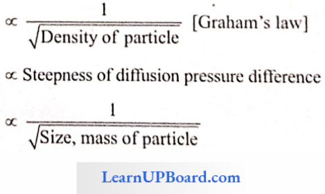
The rate and direction of a diffusing substance depend upon the concentration of that substance at different spots and is independent of the presence of other diffusing substances. The rate of diffusion is affected by many factors, for example,

Rate ∝ Temperature

NEET Biology Transport In Plant Diffusion Formula

Diffusion Pressure (DP): It is the pressure exerted by a substance due to the tendency of its particles to diffuse. It is also defined as the potential ability of a molecule or ion to diffuse. It is directly proportional to the diffusing particles.

DP ∝ Concentration

DP ∝ Temperature

The DP of pure water is maximum.

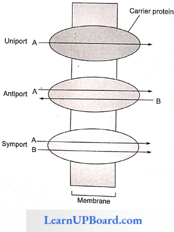
Facilitated Diffusion: Particles that are lipid soluble can easily pass directly through the cell membrane as it is mainly made of it. The hydrophilic solutes find it difficult to pass through the membrane. Their movement has to be facilitated. For this, the membranes possess aquaporins and ion channels.

NEET Biology Transport In Plant Facilitated Diffusion

Some carrier proteins allow transport only if two types of molecules move together. This is called cotransport. It is of two types. In the symport method of cotransport, both molecules cross the membrane in the same direction at the same time. In the antiport method of cotransport, both molecules move in opposite directions. When a molecule moves across a membrane independent of another molecule, the process is called uniport.

Read and Learn More NEET Biology Notes

NEET Biology Transport In Plant Types Of Transport Of Carrier Proteins

Osmosis (Diffusion Through Membrane)

Osmosis may be defined as “the passage of solvent molecules from a region of their higher concentration to a region of their lower concentration through a semipermeable membrane.” In all biological systems, the solvent is water.

Types Of Membranes

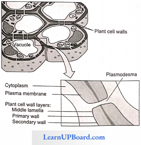
  • Permeable Membranes: Such membranes allow diffusion of both solvent and solute molecules or ions through them, for example, the cellulose wall of cells. Lignified cell walls are also quite freely permeable to water.
  • Impermeable Membranes: Such membranes prohibit the diffusion of both solvent and solute particles through them, for example, heavily cutinized or suberized cell walls in plants.
  • Semipermeable Membranes: Such membranes allow the diffusion of solvent molecules but do not allow the passage of solute molecules. Such membranes form a perfect partition between two osmometers, for example, membranes of collodion, parchment paper, and copper ferrocyanide membranes.

NEET Biology Transport In Plant Osmosis Types Of Membrances

Differentially Permeable Membranes: However, all membranes found in plants allow some solutes to pass through them along with the solvent molecules. Such membranes are called differentially permeable membranes, for example, all biological membranes.

Osmotic Pressure

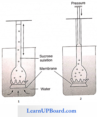
Osmotic pressure (OP) is the “maximum pressure which develops in a solution when it is separated from pure water by a semipermeable membrane.” It is also defined as “the pressure needed to prevent the passage of pure water into an aqueous solution through a semipermeable membrane thereby preventing an increase in the volume of the solution”.

NEET Biology Transport In Plant Osmotic Pressure

The osmotic pressure depends upon

  1. The concentration of solute particles,
  2. Ionization of solute particles,
  3. Hydration of solute particles, and
  4. Temperature.

An increase in the concentration of solutes in the solution increases the osmotic pressure. If the solute ionizes in the solution, the number of particles increases, thus raising the osmotic pressure. If the solute molecules are hydrated, the water molecules bound with the solute will be unable to diffuse and hence increase the osmotic pressure.

  • An increase in the temperature raises the osmotic pressure of the solution.
  • Plant cells exhibit a considerable range of variations in osmotic pressure. In land plants, it varies from 5 to 30 atm.
  • In aquatic plants, it varies from 1 to 3 atm. Plants of arid regions possess high osmotic pressure. The highest osmotic pressure is recorded in the halophytic plant, Atriplex confertifolia, i.e., 202.5 atm.
  • The osmotic pressure of an electrolyte is two to three times greater than a non-electrolyte.
  • Osmotic pressure can be calculated by OP = miRT where m is the molar concentration, i is the ionization constant, R is the gas constant, and T is the temperature (273 °C) (π). Osmotic pressure is numerically equal to osmotic/solute potential (ψs) but has a positive value ψs = π.

Measurement Of Osmotic Pressure: The methods for measuring osmotic pressure are

  1. Plasmolytic method by de Vries
  2. Pleffer osmometer
  3. Berkeley/Hartley osmometer
  4. Cryoscopic osmometer

Turgidity And Turgor Pressure

If a plant cell is placed in a hypotonic solution or pure water (i.e., a solution of higher water potential), the water starts moving into the cell by osmosis. As the volume of the protoplast increases, it begins to exert pressure against the cell wall and thus stretches it.

  • The pressure exerted by the protoplast against the cell wall is called turgor pressure (TP). The cell wall, being rigid, exerts an equal and opposite pressure on the protoplast, which is called wall pressure (WP).
  • The two pressures are equal and opposite in direction. As the turgor pressure of the cell increases, the cell becomes turgid. However, a stage is reached when osmotic pressure is exactly balanced by turgor pressure.
  • At this point, the amount of water leaving the cell equals that entering the cell. Hence, there is no further net movement of water and the cell is said to be in equilibrium with the exterior solution.

Diffusion Pressure Deficit (Suction Pressure)

The diffusion pressure of a pure solvent is maximum. The addition of solutes results in a decrease in the diffusion pressure. This deficit is termed diffusion pressure deficit (DPD). The greater the concentration of a solution, the greater is its DPD.

  • Such a solution will tend to take up water and become normal; therefore, water moves from low DPD to high DPD. The value of DPD for a cell is always positive. DPD for a cell is always positive and is calculated as (QP – TP).
  • DPD is an index of the water-absorbing capacity of cell. In a flaccid cell, TP = 0. Therefore, DPD = OP.
  • In a turgid cell, the entry of water into the cell causes the development of TP. At a point, TP = OP. Therefore, DPD = 0. Therefore, in a fully turgid cell, there is no net movement of water into the cell.

Chemical Potential

Chemical potential is the free energy of one mole of a particular chemical in a multi-component system. The lamer the chemical potential of a substance, the greater its tendency to undergo a chemical reaction. The chemical potential of water is called water potential and is given the value of zero at prevailing temperature and pressure.

Water Potential

Currently, the term water potential is used by biologists to describe the tendency of water molecules to move from one place to another. It is denoted by the symbol “ψ” (the Greek letter, psi).

Components Of Water Potential: Water potential is the difference in the free energy per unit molal volume of water in the pure state and in a system. For a solution, the value of water potential is always less than zero or negative. Water potential is determined by the following relation

Water Potential (ψ) = Solute potential (ψs) + Pressure potential (ψp)

Solute Potential (ψs): Also called osmotic potential, it is defined as the amount by which Tyr is reduced as a result of the addition of solute. It is always negative.

Pressure Potential (ψp): It is equal to TP and is positive except in plasmolyzed cell and in xylem vessels where it is negative. In terms of water potential, osmosis can be redefined as “the movement of water molecules from a region of higher water potential to the region of lower water potential through a differentially permeable membrane.”

Water potential may be regarded as the tendency of water to leave a system. The more the water potential, the greater is the tendency to leave. If two systems are in contact, water moves from the system with higher water potential to that with lower water potential or less negative ψw to more negative ψw.

Importance Of Osmosis: Osmosis is involved in

  1. Water absorption by roots
  2. Maintenance of turgidity
  3. Turgor movements in Mimosa and Desmodium.
  4. Stomatal movement
  5. Self-dispersal of fruits (autochory)
  6. Drought and frost resistance

Plasmolysis

When a cell is placed in a hypertonic solution, exosmosis occurs. Due to the loss of water to the external solution, TP of the cell decreases, hence the cell shape slightly changes. This stage is called limiting plasmolysis.

  • If the cell continues to be in a hypertonic solution, more and more water is lost, therefore protoplast starts shrinking. It first leaves the comers of the cell and this stage is called as incipient plasmolysis.
  • If more water is lost, the protoplast shrinks but remains in contact with the wall in one or two places. The space between the cell wall and the protoplast is occupied by the outer solution. This stage is called evident plasmolysis. The turgor pressure of a cell at limiting plasmolysis is equal to zero, whereas at incipient plasmolysis and evident plasmolysis, TP of the cell is negative.

Imbibition

The adsorption as well as absorption of a liquid by a solid without forming a solution is called as imbibition. The solid sub¬stance is called imbibing, whereas liquid is called as imbibe. The liquid particles are held in between the particles of solid by adsorption and capillarity.

The phenomenon depends upon the affinity of imbibant to imbibe. As a result of imbibition, the following events occur

Volume Change: The volume of the system increases, i.e., swelling. The total volume of water imbibed, plus the imbibing material is less after imbibition than before.

Heat Release: This is due to the compact arrangement of water molecules which liberate some of their kinetic energy.

Development Of Pressure: If the imbibing is confined, pressure may be developed. For example, in dry pea seeds, it is 1000 bars.

Imbibants present in plants are generally hydrophilic in nature. Among plant imbibing, hydrocolloids (for example, agar-agar, etc.) are the best imbibing followed by proteins, starch, and cellulose. Imbibition is affected by a number of factors such as temperature, pressure, etc., against the imbibing surface, the texture of imbibing, electrolytes, and pH.
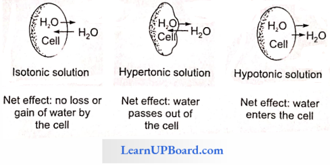
Hypotonic Hypertonic And Isotonic Solutions

A solution with a higher concentration of solutes is said to be the hypertonic solution. A lower concentration of solutes in a solution is called a hypotonic solution, and two solutions having the same concentration of solutes are called isotonic solutions.

NEET Biology Transport In Plant Hypertonic And Hypotonic Solutions

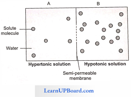
If a cell is placed in pure water or hypotonic solution, there is a net movement of water into the cell (endosmosis); if it is placed in a hypertonic solution, there is a net movement of water from the cell outwards (exosmosis); if the cell is placed in an isotonic solution, the amount of water leaving the cell equals that entering the cell and therefore, there is no net movement of water.

NEET Biology Transport In Plant Effect Of Isotonic Solution Hypertonic Solution And Hypotonic Solution

Classification Of Soil Moisture

The amount of water that can be held by soil depends upon the total pore space in the SCA. Water is present in the spaces between the soil. Various types of groundwater are

Run-off Water: Water that flows along the surface of the soil and reaches to the nearest water body constitutes run-off water. This water is not available to the plants.

Gravitational Water: Water that percolates down through the soil macropores (50 piM diameter) under the influence of gravity and reaches to the water table is called gravitational water. This type of water is also not available to the plants.

Capillary Water: Water retained by soil micropores (20 pm diameter) in the form of fine capillaries constitutes capillary water. This type of water is available to the plants.

Hygroscopic Water: Water held in the form of a very thin film around the soil particles due to adsorption constitutes hygroscopic water. This type of water is not available to the plant.

Chemically Combined Water: Water that combines with inorganic salts of the soil in the form of water of hydration constitutes chemically combined water. This type of water is also not available to the plants.

Water Vapors: They are present in soil air spaces. Normally, they are not available to the plants. Under certain conditions, they are useful in the phenomenon of night recovery.
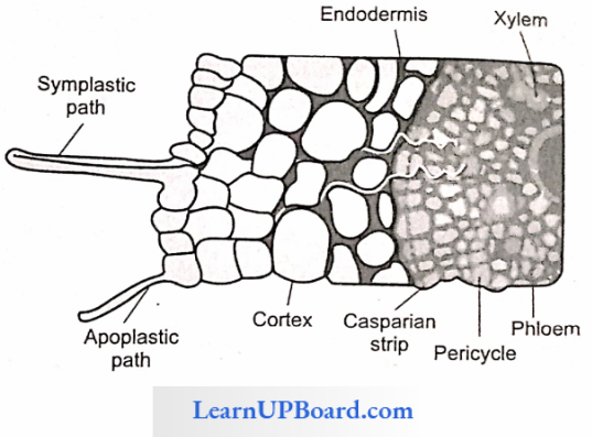
Absorption Of Water And Pathway Of Water Across The Root

Water is mainly absorbed by the root hair zone. Root hairs are tubular elongations of the external wall of epiblema cells. They range in length from less than a millimeter to about a centimeter and are usually 10 pm in diameter. They have a vacuole filled with salt, sugars, and organic acid.

Hence, the osmotic pressure of the root hair cell is high (approx. 3-8 atm) as compared to that of soil sap (approx, less than 1 atm). The movement of water from the root hair cell to the xylem may occur by two methods.

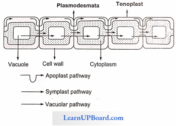
  1. Apoplast Pathway: In this method, water passes from the root hair cell to the xylem through the walls of intervening cells without crossing any membrane or cytoplasm. The apoplastic movement of water beyond the cortex is blocked due to the presence of Casparian strips in the endodermal cells. The major movement of water through cortical cells occurs by this method, as cortical cells offer the least resistance.
  2. Symplast Pathway: In this method, water passes from cell to cell by crossing the plasma membrane. Therefore, it is also known as a transmembrane pathway. This may occur by two methods
    • Non-Vacuolar Symplast Pathway: In this method, water passes between adjacent cells through plasmodesmata. It does not enter into the vacuoles.
    • Vacuolar Symplast Pathway: In this method, water crosses the tonoplast surrounding the vacuole. This pathway offers a lot of resistance. Beyond the cortex through the endodermis and pericycle water is forced to move through the symplast pathway.

NEET Biology Transport In Plant Apoplastic And Symplastic Pathways

NEET Biology Transport In Plant Three Pathways Of Water Movement

Mechanism Of Water Absorption: Water is mainly absorbed by two different mechanisms

  1. Passive (water is absorbed through roots)
  2. Active (water is absorbed by roots)

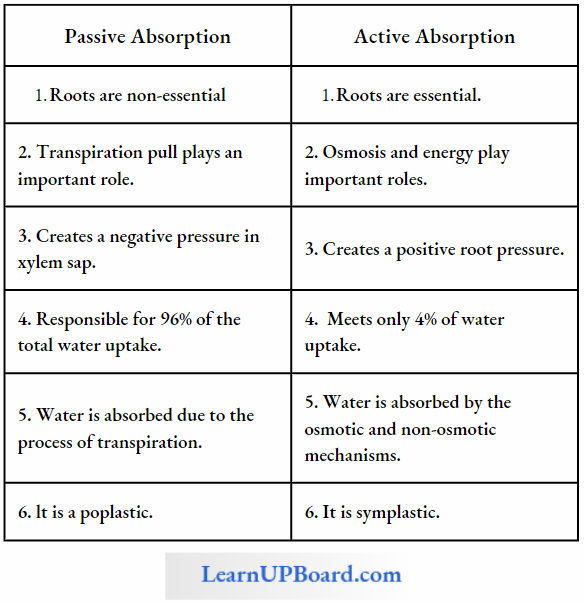
Water absorption by rapidly transpiring plants is called passive absorption because forces responsible for water uptake develop in shoots and is transmitted to roots through which water enters. Active absorption depends on forces developing in roots and is found in low-transpiring plants. The table describes the differences between passive and active absorption

Difference Between Passive Absorption And Active Absorption

NEET Biology Transport In Plant Differences Between Passive Absorption And Active Absorption

Factors Affecting Water Absorption: The rate of water absorption decreases with

  1. Increased salt concentration in soil
  2. Decreased soil temperature
  3. Decreased soil aeration (O2)
  4. Decreased soil moisture

Ascent Of Sap: The water absorbed by roots has to be conducted upwards so as to meet the needs of tissues there. This vertical conduction of water from the root to aerial parts of the plant is called the ascent of water or ascent of sap. Various theories explaining the ascent of sap have been put under three categories

  1. Vital Force Theories: These theories explain the ascent of sap by the force developing in living cells.
    • Relay Pump Theory Or Clambering Theory (By Godlewski): According to it, water rises in the stem by the rhythmic changes in osmotic pressure of the xylem parenchyma and medullary rays. The water is relayed in a staircase-like manner to the next higher cell. Now it has been established that water moves through tracheids and vessels. Hence, this theory has been rejected.
    • Pulsation Theory (By J.C. Bose): According to this theory, water rises in the stem due to pulsatory activity in the innermost layer of the cortex. This theory is not accepted nowadays, as neither these pulsations are universal in occurrence nor they are able to explain the magnitude of the ascent of sap.
    • Root Pressure Theory (By Priestley): Due to the movement of water from the soil into the root hairs and from there to xylem cells, hydrostatic pressure develops. This is called root pressure (the term given by Stephan Hales).
      • This pressure pushes the water up in the xylem vessels. The root pressure can be easily observed and measured when a freshly cut stump continues to exude water (bleeding) from its xylem vessels. The development of root pressure is an active process.
      • Stocking has defined root pressure as a pressure developing in tracheary elements of the xylem as a result of the metabolic activity of roots. Root pressure depends upon the active secretion of salts or other solutes into the xylem sap, thereby lowering its osmotic potential.
      • This involves the utilization of metabolically produced energy and is inhibited by respiratory inhibitors such as cyanide, lack of oxygen, and low temperatures. The positive hydrostatic pressure generated by root pressure (maximum 2 atm) is not sufficient to push up water to more than a few meters.
      • It cannot account for water movement up the xylem in tall trees. Also, actively transpiring plants and tall trees, especially conifers, do not generate root pressure. But it is a contributing factor in many plants. It may be sufficient for the transport of water in slowly transpiring herbaceous plants.
  2. Physical Force Theories: According to these theories, some physical force or dead cells are responsible for the ascent of sap.
    • Capillarity Theory (By Boehm): This theory explains the rise of the water column due to the joint force of capillarity and atmospheric pressure, as the xylem vessels have a narrow diameter and act as capillaries. This theory has been rejected as it does not explain the ascent of sap in tall trees because capillary force developed by a narrow vessel of 0.03 mm diameter would support a water column of about 1 m only.
    • Imbibition Theory (By Such): This theory explains the rise of the water column due to the force of imbibition. If it were true, water must rise through the walls of xylem vessels and not through their lumen as it always occurs. Hence, the concept was rejected.
  3. Cohesion-tension Or Transpiration Pull Theory: This is the most widely accepted theory proposed by Dixon and Joley (1894). The main features of the theory are as follows
    • Right from the root hairs to the leaves on top, water forms a continuous column in the plants.
    • Water molecules have high cohesive forces between them, i.e., these tend to stick to each other, because, being polar, they are electrically attracted to each other by hydrogen bonds. The high cohesive force means that a relatively large tension is required to break the water column.
    • In other words, the water column has a great tensile strength. The magnitude is generally 10-30 MPa.
    • The lignin cellulosic cell walls of xylem vessels have a strong affinity (adhesion) for water molecules. Therefore, a strong adhesive force exists between the walls of the xylem vessels and water, i.e., water tends to “stick” to the vessel wall.
    • As the water is lost from the leaf surface by transpiration, the DPD of the leaf cells increases.
    • As a result, the cells develop low water potential and water from the leaf veins (xylem) moves into leaf cells. The xylem vessels, in turn, draw water from the xylem of the main stem. A negative (pulling) pressure is thus exerted by all the leaves on the stem. The combined pressure, called transpiration pun, is strong enough to pull up the column of water to great heights.
    • The whole column of water moves. Water potential as low as -3 MPa or -30 bars has been measured in the leaves borne on tree tops.
    • Such a low water potential is sufficient to create pulling pres-sure which can overcome the gravitational pull and resistance offered by the capillaries of xylem vessels.
  4. Criticism Of Transpiration Pull Theory: The theory assumes tracheids to be more efficient than vessels. Dixon believed that partition walls of the tracheids confer stability on tensile stressed transpiration stream.

Transpiration

Less than 5% of absorbed water is used by plants and the rest is lost in transpiration.

Types Of Transpiration

  • Cuticular: Loss of water vapor from the general surface through the cuticle. Commonly, it is 3-10% of the total but in herbs and ferns it may be up to 50%.
  • Lenticular: Loss of water vapor from lenticels or aerating pores in the bark of trees or fruits, etc. It is hardly 0.1% of the total transpiration.
  • Bark: It is approximately 1% of the total transpiration.
  • Stomatal: Major form of transpiration constituting 50-97% of the total transpiration.

Number And Distribution Of Stomata: In most plants, there are 1000-60000 stomata per square centimeter of leaf surface. The total pore area is approximately 1-2% of the total leaf area. On the basis of their distribution, stomata are divided into the following categories

  • Apple Type (Mulberry Type): Stomata are present only on the lower surface of the leaf, for example, apple.
  • Potato Type: Stomata are present on both surfaces of the leaf but more on the lower surface, for example, potato, pea, tomato, and many other dicot plants.
  • Oat Type: The number of stomata is equal on both surfaces of the leaf, for example, oat and many other monocots.
  • Water Lily Type: The stomata are present only on the upper surface of the leaf, for example, water lily and most floating plants.
  • Potamogeton Type: Stomata arc cither absent or vestigial, for example, Potamogeton.

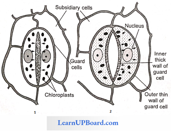
Structure Of Stomata: Each stoma is surrounded by two specialized cells called guard cells. They have chloroplasts and arc green and are surrounded by two or more specialized epidermal cells called accessory or subsidiary cells. Guard cells are of two types: kidney-shaped and dumbbell-shaped. Dumbbell-shaped guard cells are found in the family Gramineae and Cyperaceae. Kidney-shaped guard cells have an inner thick wall and an outer thin and elastic wall.

Dumbbell-shaped guard cells have thin-walled ends and thick-walled middle regions. The cell wall bordering the stomatal pore is thicker than that of next to the surrounding cells. Lotfield classified stomata as follows on the basis of their daily opening and closing movement

  • Alfalfa Type: Open the whole day, closed in night. For example, pea, radish, mustard, turnip, apple, grapes.
  • Potato Type: Open the whole day and night except for a few hours in the evening. For example, onion, potato, banana.
  • Barley Type: Open only for a few hours in day. For example, maize, wheat, and other cereals.
  • Equisetum Type: Always open.

Mechanism Of Opening And Closing Of Stomata: Stomata function as turgoroperated valves. When the osmotic concentration of guard cells increases, water comes in guard cells becomes turgid and stomata are open. Whenever the osmotic concentration of the guard cell decreases, water moves out, and guard cells become flaccid, and hence become closed. This increase and decrease in osmotic concentration is explained by a number of theories mentioned below.

NEET Biology Transport In Plant Structure Of Stomata

Photosynthetic Theory (von Mohl And Schwendener): This theory proposes that in the morning, as soon as light is available, the chloroplasts of guard cells start photosynthesis. As a result, sugars are produced, which increase the osmotic concentration of guard cells. Water comes in, guard cells become turgid, and stomata open. This theory was not accepted because the photosynthetic activity of guard cell chloroplasts seems to be negligible and sugar does not occur in detectable quantity in guard cells.

Starch ⇔ Sugar Interconversion Hypothesis (Classical Theory): The theory was proposed by Sayre and Scarth and was modified by Steward on the basis of the activity of the phosphorylase enzyme.

NEET Biology Transport In Plant Strach Sugar Interconversion Hypothesis Formula

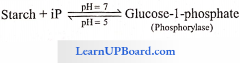
According to this theory, guard cells contain starch and phosphorylase enzyme, which causes the conversion of starch into glucose at higher pH which is developed by the utilization of CO2 in photosynthesis.

Glucose increases the osmotic concentration of guard cells, raising the turgor pressure and causing the opening of stomata. At night, CO2 accumulates in cell and intercellular spaces, thus lowering the pH at which phosphorylase causes the conversion of glucose to starch. The osmotic concentration of guard cells decreases. Hence, water moves out and, therefore, stomata are closed.

Objections: In some families, starch is absent, for example, onion. Glu¬cose does not appear in detectable quantity in the guard cells of open stomata, rather malic acid accumulates.

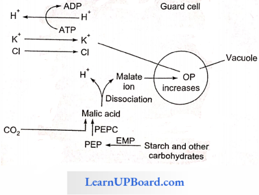
Active K+ Ion Uptake Theory (Levitt 1974): According to this theory, the pH of guard cells rises in the day due to the assimilation of CO2 in the photosynthesis and the uptake of H+ ions by chloroplast and mitochondria from cytoplasm. At this higher pH, starch is converted into phosphoenolpyruvate (PEP). PEP combines with CO2 with the help of PEPCase (phosphoenol pyruvate carboxylase) and forms oxaloacetic acid which gets changed into malic acid. Malic acid dissociates into malate and H+ ions.

  • There occurs an efflux of H+ ions and the influx of K+ ions, which forms potassium malate after reacting with malate. Potassium malate is a highly osmotically active substance which is stored in vacuoles. This raises the osmotic concentration of guard cells, water moves in, cells become turgid, and stomata are open. In the night, the process is reversed.
  • The potassium, chloride, and malate ions have a prominent role in stomatal opening. The ions accumulate in the vacuole of guard cells, lowering the water potential and thereby increasing water uptake and subsequently opening the stomata.

NEET Biology Transport In Plant Active K+ Ion Uptake Mechanism

Factors Affecting Transpiration: Blue light induces maximum opening of stomata. Red and blue lights constitute the action spectrum of transpiration.

A rise in the temperature of the leaf increases the vaporization of water within the leaves resulting in a greater DP gradient of vapors, which diffuses out into the air rapidly. The rate of transpiration is generally doubled with about every 10° = C rise in temperature. High CO2 closes stomata and cytokinins open stomata.

  • Wind Speed: If the wind velocity is high, the rate of transpiration is also high.
  • Relative Humidity: If relative humidity is low, there would be a high rate of transpiration and vice versa.
  • Root/shoot Ratio: An increase in the root/shoot ratio causes an increase in the rate of transpiration. Suolt plants have a high root/shoot ratio.
  • Plant Factors: Number and distribution of stomata, number of open stomata, water status of the plants, canopy structure, etc.

Significance Of Transpiration: The following roles have been given in relation to transpiration by different biologists

  • Since plants absorb far more amounts of water than is actually used by plants, transpiration helps in the removal of excess water.
  • Removal of water in the form of vapors has a cooling effect on the leaves. Transpiration, thus, does not allow the leaf temperature to rise to detrimental levels.
  • The transpiration pull created by transpiration is responsible for the ascent of water in tall trees.
  • Passive absorption of water through the roots takes place due to the negative pressure developing in the shoots as a result of transpiration.
  • Development of mechanical tissues, growth of root system, increasing ash and sugar content of fruits, and development of resistance are other beneficial effects of transpiration.
  • Many chemicals (antitranspirants) have been found to reduce the rate of transpiration without affecting CO2 uptake, for example, phenylmercuric acetate (PMA) abscisic acid (ABA), and CO2. Silicon emulsion and low viscosity wax cover stomata as a film, allowing CO2 and O2 but resisting diffusion of water.
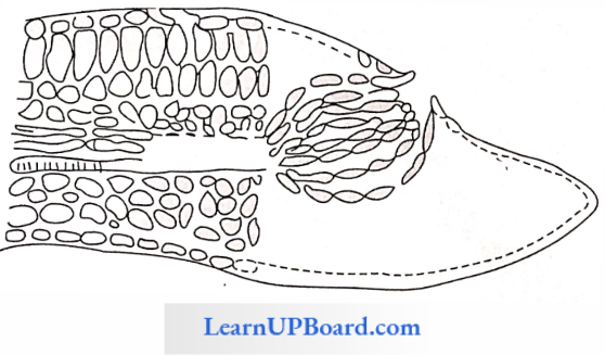
Guttation

Plants growing under humid conditions in moist warm soil often exhibit droplets of water along the margins of their leaves. This phenomenon is commonly seen in oats, tomatoes, cucumbers, garden nasturtium, saxifrage, etc. The loss of water in the form of liquid is called guttation.

In moist and humid conditions, the rate of absorption of water greatly exceeds transpiration. The root pressure is built up which pushes the water up in the xylem ducts, from where it comes out on the leaf surface through special structures called hydathodes.

NEET Biology Transport In Plant Guttation Process

Hydathodes are present at the tips of veins in the leaves. A hydathode consists of a pore in the epidermis followed by large intercellular spaces and loosely arranged parenchyma called epithem and blindly ending xylem elements. Guttated water contains inorganic and organic salts and is not pure.

Translocation Of Organic Solutes

Organic solutes such as glucose, and sucrose produced during photosynthesis are translocated through phloem tissue. The transport of photosynthates from the production centers (leaves) to the consumption centers (apices, roots, fruits, tubers) through the phloem is called translocation of organic solutes or long-distance transport. Translocation through phloem occurs in upward, downward, and radial directions from the source (leaves) to the sink (apices, roots, fruits, tubers, etc.)

NEET Biology Transport In Plant Translocation Of Organic Solutions

Chemical analysis of the phloem sap revealed the presence of sugars up to 90%. Sucrose constitutes 5-15% of the total sugars. Other sugars such as raffinose (triose). staehyose (tetrose), and verbascose (pentose) are also present in small quantities.

  • This analysis strongly suggests that the phloem is the tissue concerned with the translocation of organic solutes. Phloem sap may be collected by using sap-sucking aphids.
  • The tracer technique (Rabideau and Bun, 1945) supplied 14CO2 to a leaf during photosynthesis. Sugars synthesized in this leaf were labeled with 14C (tracer). The presence of labeled sugars (radioactivity) in the phloem showed that solutes are translocated through the phloem.

Theories Of Translocation Of Organic Solutes: Protoplasmic Streaming Hypothesis: This theory was proposed by a Dutch botanist, Hugo de Vries, in 18S5 and was supported by C.F. Curtis (1935). According to this theory, the protoplasm of the sieve tubes shows continuous streaming from one end to the other.

  • Organic solutes (sugars) that enter the sieve tube are passively carried by the streaming protoplasm from one end of the sieve tube to the other. Solutes move from one sieve tube to the next sieve tube by diffusion through the pores of the sieve plate.
  • Thus, the streaming protoplasm acts as a conveyor belt or two-way escalator. Different substances move in different directions at the same time in the same sieve tube.

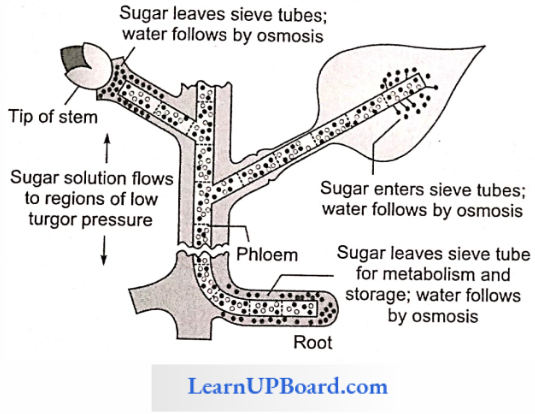
Pressure Flow Or Mass Flow Hypothesis: According to this theory proposed by Munch (1929) and elaborated by Crafts (1938), organic solutes are translocated “en masse’’ through the sieve tubes from the supplying end or source leaves to the consumption end or sink (roots, fruits, tubers).

  • Mesophyll cells synthesize sugars during photosynthesis. As these get dissolved in the cell sap, the osmotic concentration and DPD of mesophyll cells increase (ψw decreases). Water enters the mesophyll cells from the xylem. The turgor pressure or pressure potential (ψp) of the mesophyll cells increases.
  • Sugars dissolved in water move from mesophyll cells into the symplast system of sieve tubes. Solutes are carried “en masse” through the symplast to finally reach the consumption centers.
  • At the consumption end, food materials (solutes) are either used up (as in roots) or are stored in an insoluble form (as in fruits, or tubers). Hence, the osmotic concentration and. consequently, the turgor pressure in these cells will be low. Thus, a continuous turgor pressure gradient gets established across the symplast between the cells of the source (leaf) and the cells of the sink (root).
  • Water returns to the source (leaf) through the apoplast system.

Object To Mass Flow Hypothesis: The bidirectional transport of solution in the same sieve tube needs explanation. The slime content and other fibrils of the sieve tube reduce the speed of the flow of solutes even under high pressure.

Mass flow is not a purely physical process as described by Munch Phloem cells utilize 0.1 0.5% of sucrose translocated through them. This is evidence to show that phloem translocation is an active process and requires metabolic energy.

Factors Affecting Translocation Of Solutes

  • Temperature: The optimum temperature for translocation is between 20° and 30°C. The file rate of translocation increases with an increase in temperature. The temperature influences the root more than the shoot since it acts as a sink for the sugars.
  • Light: The root/shoot dry weight ratio increases with increased light intensities. This indicates that translocation to root increases as compared to shoot when light intensity is increased.
  • Metabolic Inhibitors: The metabolic inhibitors inhibit carbohydrate translocation. These include dinitrophenol (DNP), arsenite, azide, fluoride, and hydrogen cyanide.
  • Mineral Deficiencies: The absorption and translocation of sucrose by a leaf is facilitated by boron. It helps sucrose to move easily through the cell membranes in the form of the boron-sucrose complex.
  • Hormones: Sucrose is much more efficiently translocated when growth regulators are applied, such as kinetin, IAA, and gibberellic acid.

Transport In Plant Points To Remember

The unit for water potential is bar or Pascal (1 MPa = 10 bar) The osmotic pressure of 1 molar solution of a non-electrolytic would be 22.4 atm at 0°C. The equimolar concentrations of two solutions of non-ionizing substances will have the same osmotic pressure.

  • The value of the osmotic potential of an electrolyte will be greater by the degree of its dissociation into ions at a given temperature.
  • Bacteria do not survive in salted pickles because these get plasmolyzed.
  • Common salt kills weeds by plasmolysis.
  • Plasmolysis can be demonstrated in the epidermal peel of the Rhoeo discolor leaf.
  • A plasmolyzed cell regains normal condition if placed in a hypotonic solution. It is called deplasmolysis.
  • The government of H2O occurs from the high value of ψw to low value of ψw, i.e., from less negative value to more negative value of ψw.
  • The auxin-treated cells show an increase in their metabolism. Respiration in these cells increases and more energy is provided for the absorption of water.
  • At low temperatures, water in the intercellular spaces freezes into ice, thus having higher OP. It causes exosmosis of water from cells causing desiccation.
  • If a freshwater plant is transferred to marine water, it dies due to exosmosis.
  • The approximate water potential of root hairs is -3 to -43 bar.
  • Ringing experiments to prove the ascent of water through the xylem were first conducted by Malpighi (1672), Stephan Hales (1727), and Hartig.
  • Root pressure is measured by a manometer.
  • Stocking (1956) considered root pressure as an active process responsible for guttation and bleeding in plants.
  • Maximum root pressure recorded in plants is 2 bar which is sufficient to raise the water column to a height of 20 m.
  • Root pressure is absent in gymnosperms (some of the tallest trees are gymnosperms).
  • Photometers are used for measuring/comparing the rates of transpiration.

CoCl2 paper method is also used to compare the rates of transpiration. Moisture coming out of stomata turns blue CoCl2 paper to pink. Porometers are used for assessing the total pore area (stoma).

  • Generally, stomata are photoactive (open during the day and close at night). But in succulents such as Bryophyllum, Opimtia, and cacti, stomata close during the day and open at night (scotoactive).
  • Transpiration in old stems and fruits occurs through lenticels.
  • The fresh weight of a plant or leaf would be maximum in the morning and minimum in the afternoon.
  • If half of the total number of stomata on a leaf closes down, the rate of transpiration is not reduced by half.
  • Stomata are absent or non-functional in submerged hydrophytes.
  • Cytokinins help in the opening of stomata while ABA (abscisic acid) and low O2 close stomata.
  • Curtis (1926) considered transpiration “as a necessary evil” in plants.
  • Plants growing at high altitudes show xeromorphy (adaptation to minimize transpiration).

Transpiration Ratio: the amount of water lost per unit of dry matter produced during the growing season of a plant.

  • In Saxifraga, the rate of guttation is high during flowering.
  • In Colocasia antiquarian, the rate of guttation is very high.
  • Mechanical shock causes stomatal closure.

Stomatal index = \(\frac{E}{E + S}\) x 100 where E is the number of epidermal cells and S is the number of stomata.

Transpiration index = Leaf test tuneAVater test time.

A psychrometer is used to measure relative humidity and rate of transpiration.

The diameter of tree decreases during the day. It is due to the narrowing of tracheary elements due to the development of negative pressure. It is measured by dendrograph.

Matric Potential, ψm: It is used for surfaces that bind water. It is also negative. For example, soil particles, cell wall, cytoplasm, etc.

Gravity Potential, ψg: It denotes the effect of gravity on ψw. It depends on the height (h) of water above the reference state of water, the density of water, and acceleration due to gravity. The value of ψm negligible up to a height of 5 m from the reference level and also the value of ψm is ignored.

∴ ψw = ψs + ψp

Transport In Plant Multiple Choice Questions And Answers

Question 1. What determines the diffusion of water from one cell to another?

  1. OP
  2. WP
  3. DPD
  4. TP

Answer: 3. DPD

Question 2. The term “water potential” was used for the first time by

  1. Slatyer and Taylor
  2. Stocking
  3. Sachs
  4. Boehm

Answer: 1. Slatyer and Taylor

Question 3. In a hypertonic solution, the water potential of a cell

  1. Increases
  2. Decreases
  3. First increases and then decreases
  4. No chance occurs

Answer: 2. Decreases

Question 4. As a result of endosmosis, of a cell

  1. Increases
  2. Decreases
  3. Remains same
  4. Become zero

Answer: 1. Increases

Question 5. Which of the following equations is wrong?

  1. ψs = -π
  2. DPD = OP + TP
  3. ψw = ψs + ψp
  4. OP = CRT

Answer: 2. DPD = OP + TP

Question 6. When a cell is fully turgid, which of the following will be zero?

  1. Osmotic pressure
  2. Turgor pressure
  3. Wall pressure
  4. Suction pressure

Answer: 4. Suction pressure

Question 7. Osmotic potential is numerically equal to

  1. TP
  2. DPD
  3. OP
  4. WP

Answer: 3. OP

Question 8. The first sign of shrinkage of cell is detectable at

  1. Limiting plasmolysis
  2. Incipient plasmolysis
  3. Evident plasmolysis
  4. Permanent plasmolysis

Answer: 2. Incipient plasmolysis

Question 9. Osmotic pressure depends upon

  1. Concentration of solutes
  2. Temperature
  3. Ionization
  4. All of these

Answer: 4. All of these

Question 10. The water potential of a plasmolyzed cell will be

  1. ψw = -ψs + ψp
  2. ψs = ψp
  3. ψw = 0
  4. ψ= -ψs – ψp

Answer: 4. ψ= -ψs – ψp

Question 11. The water potential of soil at field capacity and wilting point are, respectively,

  1. —1.5 MPa and-0.01 MPa
  2. -0.1 MPa and -0.01 MPa
  3. -0.01 MPa and-1.5 MPa
  4. -0.01 MPa and -0.1 MPa

Answer: 3. -0.01 MPa and-1.5 MPa

Question 12. Which is not a characteristic of imbibition?

  1. It is a reversible phenomenon.
  2. Heat is generated.
  3. Involve capillarity and adsorption.
  4. It is a property of hydrophobic and lyophobic colloids.

Answer: 4. It is a property of hydrophobic and lyophobic colloids.

Question 13. The concept of apoplast and symplast imbibition was given by

  1. Munch
  2. Kramer
  3. Renner
  4. Dixon

Answer: 1. Munch

Question 14. The type of soil water that is available to roots is

  1. Gravitational water
  2. Hygroscopic water
  3. Capillary water
  4. Chemically combined water

Answer: 3. Capillary water

Question 15. Passive absorption is controlled by

  1. Transpiration
  2. Capillarity
  3. Presence of solutes in soil
  4. Temperature of atmosphere

Answer: 1. Transpiration

Question 16. If the texture of soil becomes fine, then the rate of movement of water through it will be

  1. Faster
  2. Slower
  3. Nil
  4. Same

Answer: 2. Slower

Question 17. At low temperatures, the rate of water absorption decreases due to

  1. Decreased viscosity of water
  2. Increased permeability
  3. Reduced rate of diffusion
  4. Increased root growth

Answer: 3. Reduced rate of diffusion

Question 18. Water absorption in plants is enhanced by

  1. Increased transpiration
  2. Decreased transpiration
  3. Decreased salt absorption
  4. Increased photosynthesis

Answer: 1. Increased transpiration

Question 19. The phenomenon of water uptake at the expense of energy by the cell and usually again osmotic phenomenon is known as

  1. Osmosis
  2. Active absorption
  3. Passive absorption
  4. Imbibition

Answer: 2. Active absorption

Question 20. Root pressure is measured by

  1. Osmometer
  2. Manometer
  3. Barometer
  4. Auxanometer

Answer: 2. Manometer

Question 21. When the temperature of soil is 0°C, then

  1. The absorption of water increases
  2. The absorption of water is not affected by temperature
  3. The absorption of water decreases
  4. The soil will lose capillary water

Answer: 3. The absorption of water decreases

Question 22. The osmotic theory for active water absorption was given by

  1. Bennet Clarks
  2. Thimann
  3. Atkin and Priestley
  4. Dixon

Answer: 3. Atkin and Priestley

Question 23. Root pressure is maximum when

  1. Transpiration is high and absorption is very low
  2. Transpiration is very low and absorption is high
  3. Transpiration is very high and absorption is also high
  4. Transpiration and absorption both are low

Answer: 2. Transpiration is very low and absorption is high

Question 24. The pulsation theory of the ascent of sap was given by

  1. Godlewski
  2. Dixon
  3. Tansley
  4. Sir J.C. Bose

Answer: 4. Sir J.C. Bose

Question 25. Which is not true for root pressure?

  1. Positive hydrostatic pressure
  2. Maximum during the day and minimum during night
  3. Magnitude is 1-2 bars
  4. Develops due to the metabolic activity of root.

Answer: 2. Maximum during the day and minimum during night

Question 26. A tension (transpiration pull) of 1 atm can pull water to a height of approx

  1. 10 feet
  2. 10 m
  3. 1 m
  4. 1 feet

Answer: 2. 10 m

Question 27. Amphistomatic leaves are generally found in

  1. Dicots
  2. Monocots
  3. CAM plants
  4. Aquatic plants

Answer: 2. Monocots

Question 28. The cutinized wall of epidermal cells is

  1. Permeable
  2. Semipermeable
  3. Impermeable
  4. Selective permeable

Answer: 3. Impermeable

Question 29. The stomata are widely open in

  1. Red light
  2. Blue light
  3. Greenlight
  4. Yellow light

Answer: 2. Blue light

Question 30. When transpiration is rapid

  1. ψw of epidermal cells decreases
  2. A negative pressure develops in the xylem vessel
  3. Water is absorbed through the root passively
  4. All of these

Answer: 4. All of these

Question 31. Which is not true regarding stomata?

  1. They are turgor-operated valves.
  2. They have differentially thickened walls in guard cells.
  3. They open when OP of the guard cell decreases.
  4. They show photoactive openings in CAM plants.

Answer: 3. They open when OP of the guard cell decreases.

Question 32. Cuticular transpiration is approx

  1. 50% in herbs and ferns
  2. 97% in most of the plants
  3. 1% of the total transpiration
  4. 50% in most of the plants

Answer: 1. 50% in herbs and ferns

Question 33. The rate of transpiration and stomatal movement are measured by, respectively,

  1. Porometer and photometer
  2. Potometer and pyrometer
  3. Potometer and tensiometer
  4. Hygrometer and porometer

Answer: 2. Potometer and pyrometer

Question 34. According to starch ⇔ sugar interconversion theory of stomatal opening, a stomatal opening is preceded by

  1. Increase in H+ concentration
  2. Decrease in the pH of the guard cell
  3. Increase in the pH of guard cell
  4. Inactivation of phosphorylase enzyme

Answer: 3. Increase in the pH of guard cell

Question 35. When stomata open in night only, they are called

  1. Photoactive stomata
  2. Scotoactive stomata
  3. Hydathodes
  4. All of these

Answer: 2. Scotoactive stomata

Question 36. Which of the following does not happen during stomatal opening?

  1. Accumulation of K+ ions in guard cell
  2. Lowering of osmotic pressure of guard cell
  3. Creation of water potential gradient between guard cell and subsidiary cell
  4. Increased thickening of the inner wall of the guard cell

Answer: 4. Increased thickening of the inner wall of the guard cell

Question 37. High amount of malate in guard cell during stomatal opening is due to

  1. Import from subsidiary cell
  2. Hydrolysis of starch
  3. Photosynthesis in guard cell
  4. Hydrolysis of proteins

Answer: 4. Hydrolysis of proteins

Question 38. Active K+ exchange mechanism for the opening and clos¬ing of stomata was given by

  1. Darwin
  2. Levitt
  3. Scarth
  4. Fujino

Answer: 2. Levitt

Question 39. Which of the following is a metabolic antitranspirant?

  1. PMA, CO2
  2. Colorless plastics and waxes
  3. Aspirin, ABA
  4. Both (1) and (3)

Answer: 4. Both (1) and (3)

Question 40. Which is not an advantage of transpiration?

  1. Development of mechanical tissues
  2. Development of root system
  3. Distribution of minerals
  4. Fixation of nitrogen

Answer: 4. Fixation of nitrogen

Question 41. The plant factor which affects the rate of transpiration is

  1. Leaf area
  2. Temperature
  3. Humidity
  4. Wind speed

Answer: 1. Leaf area

Question 42. In guttation, water is lost in the form of

  1. Water vapors
  2. A dilute solution of sugars
  3. Pure liquid water
  4. Dilute solution of salts and organic substances

Answer: 4. Dilute solution of salts and organic substances

Question 43. Which of the following is an effective adaptation for better gaseous exchange in plants?

  1. Presence of multiple epidermis
  2. Presence of hair on the lower epidermis
  3. The presence of a waxy cuticle covering the epidermis of the leaves
  4. Location of stomata on the lower surface of leaves and side turned away from direct sun rays

Answer: 4. Location of stomata on the lower surface of leaves and side turned away from direct sun rays

Question 44. The conditions under which transpiration would be most rapid are

  1. Excess of water in soil
  2. Low humidity, high temperature, turgid guard cells, and moist soil
  3. Low velocity of the wind
  4. High humidity

Answer: 2. Low humidity, high temperature, turgid guard cells, and moist soil

Question 45. The transpiration index is equal to

  1. \(\frac{\text{Leaf test time}}{\text{Water test time}}\)
  2. \(\frac{\text{Water test time}}{\text{Leaf test time}}\)
  3. The amount of water lost per unit of dry matter produced during the growing season
  4. \(\frac{\text{PWP}}{\text{Field capacity}}\)

Answer: 1. \(\frac{\text{Leaf test time}}{\text{Water test time}}\)

Question 46. Translocation of photosynthates occurs in the form of

  1. Starch
  2. Glucose
  3. Sucrose
  4. 3PGA

Answer: 3. Sucrose

Question 47. Although a girdled (up to bast) tree may survive for some time, it will eventually die because

  1. Water will not move upward
  2. Water will not move downward
  3. Sugars and other organic solutes will not move downward
  4. Sugars and other organic solutes will not move upward

Answer: 3. Sugars and other organic solutes will not move downward

Question 48. When stomata open, the pH of guard cells.

  1. Increases
  2. Decreases
  3. Remains same
  4. Both (1) and (2)

Answer: 1. Increases

Question 49. Water lost in pulsation is

  1. Pure water
  2. Impure water
  3. In vapor form
  4. Either (1) and (2)

Answer: 2. Impure water

Question 50. What will happen if plant cells are placed in a hypertonic solution

  1. Turgid
  2. Plasmolyzed
  3. Deplasmolyzed
  4. Lysed

Answer: 2. Plasmolyzed

Question 51. Loss of water from the tips of leaves is called

  1. Bleeding
  2. Gustation
  3. Respiration
  4. Transpiration

Answer: 2. Gustation

Question 52. Root pressure is measured by

  1. Manometer
  2. Potomcter
  3. Auxanomcter
  4. Osmometer

Answer: 1. Manometer

Question 53. Which of the following apparatus is commonly used to measure the rate of transpiration?

  1. Porometer
  2. Altimeter
  3. Potomcter
  4. Luxmeter

Answer: 3. Porometer

Question 54. Leaves of the Nehnnbo plant are

  1. Epistomatic
  2. Hypostomatic
  3. Amphistomatic
  4. None of these

Answer: 1. Epistomatic

Question 55. 0.1 M solution has the water potential of

  1. -2.3 bars
  2. 0 bar
  3. 22.4 bars
  4. +2.3 bars

Answer: 1. -2.3 bars

Question 56. A small mesophytic twig with green leaves is dipped into water in a big beaker under sunlight. It demonstrates

  1. Photosynthesis
  2. Respiration
  3. Transpiration
  4. None of the above

Answer: 3. Transpiration

Question 57. Which one is not related to transpiration?

  1. Regulation of plant body temperature
  2. Absorption and distribution of mineral salt
  3. Circulation of water
  4. Bleeding

Answer: 4. Bleeding

Question 58. Stomata can open at night also in

  1. Xerophyte
  2. Gamelophyte
  3. Hydrophyte
  4. None of these

Answer: 1. Xerophyte

Question 59. Who had said that “transpiration is a necessary evil”?

  1. Curtis
  2. Steward
  3. Andersen
  4. J.C. Bose

Answer: 1. Curtis

Question 60. Stomata open during day because the guard cells have

  1. Thin outer walls
  2. Kidney shape structure
  3. Chlorophyll
  4. Large nuclei

Answer: 1. Thin outer walls

Question 61. Stomata open and close due to

  1. Turgor pressure change
  2. Hormone change
  3. Temperature change
  4. All of the above

Answer: 1. Turgor pressure change

Question 62. In plasmolyzed cell, the space between cell wall and protoplasm is occupied by

  1. Hypotonic solution
  2. Hypertonic solution
  3. Isotonic solution
  4. Distil water

Answer: 2. Hypertonic solution

Question 63. In CAM plants, stomata are

  1. Closed at night and open during the day
  2. Closed during the day and open at night
  3. Never closes
  4. Never opens

Answer: 2. Closed during the day and open at night

Question 64. The real force responsible for the movement of water from cell to cell is

  1. OP
  2. TP
  3. DPD
  4. WP

Answer: 3. DPD

Question 65. Which of the following have sunken stomata?

  1. Nerium
  2. Mangifera
  3. Hydrilla
  4. Zea mays

Answer: 1. Nerium

Question 66. When a plasmolyzed cell is placed in a hypotonic solution then water will move inside the cell. Which force causes this?

  1. DPD
  2. OP
  3. WP
  4. None of these

Answer: 1. DPD

Question 67. Rate of transpiration is measured by

  1. Manometer
  2. Auxanometer
  3. Potometer
  4. Barometer

Answer: 3. Potometer

Question 68. If a cell shrinks when placed in a solution, the solution becomes

  1. Hypotonic
  2. Hypertonic
  3. Isotonic
  4. Pure solvent

Answer: 2. Hypertonic

Question 69. If cell A with DPD 4 bar is connected to cell B, C, and D whose osmotic pressure and turgor pressure are, respectively, 4 and 4, 10 and 5, 7 and 3 bar, the flow of water will be

  1. B to A, C, and D
  2. A to D, B, and C
  3. C to A, B, and D
  4. A to B, C, and D

Answer: 1. B to A, C, and D

Question 70. Guard cell controls

  1. The intensity of light entering
  2. Photosynthesis
  3. Closing and opening of stomata
  4. Change in green color

Answer: 3. Closing and opening of stomata

Question 71. Active transport

  1. Releases energy
  2. Requires energy
  3. Produces ATP
  4. Produces a toxic substance

Answer: 2. Requires energy

Question 72. Velamen tissues are associated within

  1. Haustorial function
  2. Assimilation
  3. Absorption of moisture
  4. Nutrition

Answer: 3. Absorption of moisture

Question 73. Cohesion tension theory regarding the ascent of sap was given by

  1. Dixon and Jolly
  2. J.C. Bose
  3. Cristian Wolf
  4. Godlewski

Answer: 1. Dixon and Jolly

Question 74. Velamen tissue is found in

  1. Mesophytes
  2. Epiphytes
  3. Hydrophytes
  4. Xerophytes

Answer: 2. Epiphytes

Question 75. In a fully turgid plant cell, which one is zero?

  1. Turgor pressure
  2. Wall pressure
  3. Suction pressure
  4. None of these

Answer: 3. Suction pressure

Question 76. Who proposed the cohesion theory of the ascent of sap?

  1. Strasburger
  2. Godlewski
  3. Western
  4. Oixon and Jolly

Answer: 4. Oixon and Jolly

Question 77. The most accepted theory for ascent of sap is

  1. Relay pump theory
  2. Pulsation theory
  3. Root pressure theory
  4. Transpiration pull cohesion theory

Answer: 4. Transpiration pull cohesion theory

Question 78. The transport of water and salt is mediated by

  1. Xylem
  2. Sieve tubes
  3. Sclerenchyma
  4. Phloem

Answer: 1. Xylem

Question 79. The removal of a ring of tissue outside the vascular cambi¬um from the tree trunk kills it because

  1. Water cannot move up
  2. Food does not travel down and roots become starved
  3. Shoots become starved
  4. Annual rings are not produced

Answer: 2. Food does not travel down and roots become starved

Question 80. Wilting of the plant is present in

  1. Moss
  2. Fern
  3. Algae
  4. Angiosperm

Answer: 4. Angiosperm

Question 81. Root hair absorbs water from the soil on account of

  1. Turgor pressure
  2. Osmotic pressure
  3. Suction pressure
  4. Root pressure

Answer: 3. Suction pressure

Question 82. Increased humidity in the atmosphere decreases the rate of

  1. Transpiration
  2. Photosynthesis
  3. Glycolysis
  4. Growth

Answer: 1. Transpiration

Question 83. In osmosis, there is movement of

  1. Solute only
  2. Solvte only
  3. Both (1) and (2)
  4. Neither solute nor solvent

Answer: 2. Solvte only

Question 84. Guttation takes place through

  1. Lenticles
  2. Pneumatophores
  3. Stomata
  4. Hydathodes

Answer: 4. Hydathodes

Question 85. Which of the following statements is correct?

  1. Cell membrane is involved only in exosmosis
  2. Cell membrane is involved only in endosmosis
  3. Cell membrane is involved both in exosmosis and endosmosis
  4. None of the above

Answer: 3. Cell membrane is involved both in exosmosis and endosmosis

Question 86. The root hairs absorb which of the following types of water?

  1. Capillary water
  2. Hygroscopic water
  3. Gravitational water
  4. All of the water

Answer: 1. Capillary water

Question 87. If flowers are cut and dipped in dilute NaCl solution, then

  1. Transpiration will become low
  2. Endoosmosis occurs
  3. No bacterial growth takes place
  4. Absorption of solute inside flower cell takes place

Answer: 1. Transpiration will become low

Question 88. A plant cell is plasmolyzed in a solution that is

  1. Hypotonic
  2. Hypertonic
  3. Isotonic
  4. Concentration no means

Answer: 2. Hypotonic

Question 89. Turgidity in guard cells is controlled by

  1. Chloride
  2. Malic acid
  3. Potassium
  4. Potassium, chloride, and malic acid

Answer: 4. Potassium, chloride, and malic acid

Question 90. Stomata are not found in

  1. Algae
  2. Mosses
  3. Ferns
  4. Liverworts

Answer: 1. Algae

Question 91. In which of the following, the rate of transpiration is high?

  1. CAM plant
  2. C3 plants
  3. C2 and C4 plants
  4. C4 plants

Answer: 1. CAM plant

Question 92. Cell sap is found in which cell organelle?

  1. Nucleolus
  2. Chloroplast
  3. Vacuole
  4. Golgi apparatus

Answer: 3. Vacuole

Question 93. Which one of the following fixes nitrogen?

  1. TMV
  2. Yeast
  3. Nostoc
  4. Denitrifying bacteria

Answer: 3. Nostoc

Question 94. Active transport of ions by the cell requires

  1. High temperature
  2. ATP
  3. Alkaline pH
  4. Salts

Answer: 2. ATP

Question 95. To initiate cell plasmolysis, the salt concentration must be

  1. Isotonic
  2. Hypotonic
  3. Hypertonic
  4. Atonic

Answer: 3. Hypertonic

Question 96. The basis of stomatal opening is

  1. Endosmosis
  2. Plasmolysis of guard cells
  3. Decrease in cell up concentration
  4. Exosmosis

Answer: 1. Endosmosis

Question 97. Plants absorb carbon dioxide from

  1. Vegetative
  2. Heterocyst
  3. Both vegetative and heterocyst
  4. None of these
  1. Millets
  2. Cereals
  3. Carbohydrates present in the soil
  4. Atmosphere

Answer: 4. Atmosphere

Question 98. Transpiration will increase with the increase of

  1. Humidity
  2. Temperature
  3. Carbon dioxide
  4. Sulfur dioxide

Answer: 2. Temperature

Question 99. If it is possible to drop a small particle through the stomata of a leaf, what will you conclude?

  1. It will fall on the earth surface.
  2. It will stop on the lower epidermis.
  3. It will stop on mesophyll cells.
  4. It will stop on vascular tissue.

Answer: 3. It will stop on mesophyll cells.

Question 100. During transpiration, turgidity in guard cells is controlled by

  1. Potassium
  2. Bromine
  3. Sodium
  4. Oxalic acid

Answer: 1. Potassium

Question 101. Apparatus used for measuring the transpiration

  1. Evapometer
  2. Potometer
  3. Osmometer
  4. Tensiometer

Answer: 2. Potometer

Question 102. Which of the following theories gives the latest explanation for the closure of stomata?

  1. ABA theory
  2. Munch theory
  3. Starch-glucose theory
  4. Active KT transport theory

Answer: 1. ABA theory

Question 103. The sugarcane plant has

  1. Dumbbell-shaped guard cells
  2. Pentamerous flowers
  3. Reticulate venation
  4. Capsular fruits

Answer: 1. Dumbbell-shaped guard cells

Transport In Plant Assertion Reasoning Questions And Answers

In the following questions, an Assertion (A) is followed by a corresponding Reason (R). Mark the correct answer.

  1. If both Assertion and Reason are true and the Reason is the correct explanation of the Assertion.
  2. If both Assertion and Reason are true, but the Reason is not the correct explanation of the Assertion.
  3. If Assertion is true, but Reason is false.
  4. If both Assertion and Reason are false.

Question 1. Assertion: Xerophytes have high water-retaining capacity.

Reason: They have high OR.

Answer: 1. If both Assertion and Reason are true and the Reason is the correct explanation of the Assertion.

Question 2. Assertion: There is an indirect relationship between the rate of respiration and water absorption.

Reason: Increased metabolism increases mineral uptake.

Answer: 1. If both Assertion and Reason are true and the Reason is the correct explanation of the Assertion.

Question 3. Assertion: Root pressure is a dynamic and always a positive hydrostatic pressure.

Reason: It is a universal phenomenon and develops under absorption lag.

Answer: 3. If Assertion is true, but Reason is false.

Question 4. Assertion: Stomata has delegated the task of providing food while preventing thirst.

Reason: They are made for gaseous exchange.

Answer: 2. If both Assertion and Reason are true, but the Reason is not the correct explanation of the Assertion.

Question 5. Assertion: During stomata opening, there is a relative change in TP of the guard cell and subsidiary cell.

Reason: TP of the subsidiary cell decreases during the opening.

Answer: 2. If both Assertion and Reason are true, but the Reason is not the correct explanation of the Assertion.


NEET Biology Notes – Mineral Nutrition

Mineral Nutrition Introduction

The sum total of various processes by which an organism with-draws and utilizes the substances required for the development and sustaining life-related processes is called nutrition, and these substances are called nutrients.

  • The absorption, distribution, and metabolism of various mineral elements by plants is called mineral nutrition. About 60 elements have been reported from plant ash (obtained after heating dry plant matter at 600°C). Out of these, 30 are present in all the plants.
  • Inorganic nutrients are classified as essential and non-essential elements. Seventeen elements have been placed in essential elements. These are the elements without which the reproduction and life cycle of a plant cannot be completed. The essential elements are C, H, O, N, P. K. S, Mg, Ca, Fe, Mo, Mn, Ni, Zn, B, Cl, and Cu.

Amon and Stout proposed the following criteria of the essentiality of an element:

  • The element must be absolutely necessary for supporting normal growth and reproduction.
  • The requirement of the element must be specific and not replaceable by another element.
  • The element must be directly involved in the nutrition of the plant.
  • A deficiency of minerals causes diseases in plants. Magnesium. for example, is a constituent of the chlorophyll molecule and is essential for photosynthesis. It cannot be replaced by any other element for the same function. It is also required as a cofactor by many enzymes involved in cellular respiration and metabolic pathways. Similarly, iron is a constituent of cytochromes.

Depending Upon The Quantity In Which These Elements Are Present In Cell, They Are Classified As:

Macronutrients: They are the elements that are present in a quantity of 1—10 mg/g dry wt. of the cell. They are C, H, O, N, P, S, K, Mg, and Ca.

Micronutrients: They are the elements that are present in a quantity equal to or less than 0.1 mg/g dry wt. of the cell (often less than 1 ppm). They are Fe, Mn, Mo, Ni, Zn, B, Cl, and Cu.

Mineral Nutrition Culture Medium

Soils normally contain sufficient quantities of essential minerals. However, three important elements need to be replenished in crop fields as they are depleted by repeated cultivation. These x fertilizer elements called critical elements are nitrogen, phosphorus, and potassium (NPK).

  • The common sources of these elements used in India are nitrate of sodium, ammonium sulfate, ammonium nitrate, ammonium chloride, urea, etc. The NPK fertilizers comprising bags of fertilizers are labeled 17-18-19 or 15-15-15 or other combinations. These numbers refer to the percentage by weight of nitrogen, phosphorus, and water-soluble potassium
  • To determine what elements are essential tor plant growth and deficiency symptoms of an essential element, a well-defined nutrient medium has to be used. Seeds are grown in highly washed pure sand in a glass, glazed porcelain, or plastic container and supplied with a carefully made-up nutrient solution.

Arnon And Hoagland’s Medium: Amon and Hoagland prescribed a medium containing micronutrients. Iron was earlier supplied as ferrous sulfate, but it often precipitated out. This problem has now been solved by dissolving ferrous sulfate along with a chelating agent Na-EDTA (disodium salt of ethylene diamine tetraacetic acid).

Read and Learn More NEET Biology Notes

Mineral Nutrition Solution Culture

Solution culture is performed in glass jars or polythene bottles. The container is covered with black paper after pouring the solution into them. Black paper has two functions

  1. Prevention of the growth of algae.
  2. Prevention of reaction of roots with light.

Seeds are allowed to germinate over the cork. Cotyledons are removed after seedling formation. The plant is properly sup¬ported with the help of split cork. The solution is aerated at regular intervals and is changed after two to three days.

Mineral Nutrition – Hydroponics

The commercial technique of soil-less culture is called hydroponics, which was first developed by Goerick (1940). The culture is performed in large tanks of metal or RCC tanks. The tanks are covered with wire mesh. Tanks are provided with aerating and circulating techniques. Seeds are suspended in the solution from the wire mesh with the help of threads. As the plant grows, additional support is provided. it depicts the diagrammatic views of the hydroponic technique.

NEET Biology Mineral Nutrition Hydroponics Technique

Liebig’s Law Of The Minimum

As per this law, “the yield of a crop plant is determined by the amount of the necessary element which is present in minimum quantity in proportion to the demand of the plant.”

General Functions Of Mineral Elements

Frame Work Elements: They form carbohydrates that form cell walls, for example, C, H, and O.

Protoplasmic Elements: They form protoplasm, for example, C, H, O, N, S, P.

Catalytic Elements: Fe, Cu, Zn, Mo, Mg, Mn, K (activator of over 40 enzymes)

Balancing Elements: Ca, Mg, and K counteract the toxic effect of other minerals.

Storage Elements: C, N, S, P.

Critical Elements: N, P, K.

Elements Affecting Cell Permeability: Monovalent cations (Na+, K+) increase the permeability of membranes while divalent and trivalent cations decrease it.

Toxic Elements: Al, As, Hg, Pb, Ag.

Non-mineral Elements: C, H, O, N. Nitrogen is a mineral as well as a non-mineral element.

Functional Elements: They are non-essential in most plants but have a definite activity in some species, for example, silicon in grasses.

Nitrogen Fixation

Atmosphere is the ultimate source of nitrogen. Nitrogen is a highly inert gas. It cannot be used directly but has to be combined with C, H, N, and O to form compounds before being used. Higher plants utilize nitrogen in the oxidized forms such as nitrate (NO3) and nitrite (NO2) or in the reduced form (NH4) made available to plants by the nitrogen fixers.

The best-known nitrogen-fixing symbiotic bacterium is Rhizobium. This bacterium lives in the soil to form root nodules in plants belonging to the family Leguminosae such as beans, gram, groundnut, and soya bean. Root nodules are little outgrowths on roots.

  • When a section of the root nodule is examined, it appears pinkish due to the presence of a pigment called leghemoglobin. Like hemoglobin, leghemoglobin is an oxygen scavenger. The enzyme that catalyses the fixation of nitrogen is nitrogenase which functions under anaerobic conditions. Leghemoglobin combines with oxygen and protects nitrogenase.
  • Free-living microorganisms such as the cyanobacteria can also fix nitrogen. Some cyanobacteria also have symbiotic as¬sociation with plants. They are found in lichens. Anthoccros. Azollcu and coralloid roots of Cycas.
  • In the process of biological nitrogen fixation, the dinitrogen molecule is progressively reduced by the addition of pairs of hydrogen atoms so that the three bonds between the two nitrogen atoms are cleaved and ammonia is formed.

These reactions occur only in the presence of a single enzyme nitrogenase. The process of nitrogen fixation is energy-intensive.

NEET Biology Mineral Nutrition Nitrogen Fixation

Requirements Of N2 Fixation

  • Nitrogenase enzyme complex (synthesized by nif genes of bacteria) is the seat of nitrogen reduction and contains Mo, Fe, and S.
  • It is a strong reducing agent, for example, NADPH2, FMNH2, ferredoxin, etc.
  • It takes place under anaerobic conditions.
  • The energy source is ATP.
  • Cofactors included are TPP, CoA, Mg+2.
  • Electron and H+ donor (generally glucose-6-phosphate)

Process Of Nodule Formation

Nodules are little out growths on the roots. When a root hair of a legume comes in contact with Rhizobium, there occurs an ex-change of plant and bacterial signals. Bacteria secrete nod fac-tors which result in the curling of root hair tips.

The plant responds by forming an infection thread that grows inward and carries the bacteria to the cortical cells of the root. Some of bacteria enlarge to become membrane-bound structures called bacteroids which are the seat of N2 fixation. Plant flavones act as the inducers of nod genes which specify early events of nodulation.

NEET Biology Mineral Nutrition Nodule Formation

Cortical cells are stimulated to divide rapidly. It is due to auxin secreted by plants and cytokinin contributed by bacteria. Nodules are pink in color due to leghemoglobin which is an oxygen scavenger and protects nitrogenase. Its heme comes from bacteria and globin from legumes.

Process Of N2 Fixation Can Be Summarized As: N2 + 8e + 8H+ + 16 ATP → 2NH3 + H2 + 16ADP + 16Pi

Ammonium ions can be taken up by higher plants but plants are more adapted to absorb nitrate (NO3) than ammonium ions (NH4+) from the soil. Soil bacteria such as Nitrosomonas and Nitrococcus convert ammonia to nitrite (NO2) ions. Nitrobacter oxidizes nitrite to nitrate. This process of converting ammonia into nitrate, a form of nitrogen more available to plants, is called nitrification.

NEET Biology Mineral Nutrition Nitrificication Formula

This process is an oxidation process and releases energy which is used by nitrifying microbes.

Nitrate Assimilation

The process of nitrate reduction to ammonia is called nitrate assimilation and is accomplished in two steps mediated by two specific enzymes. First, the nitrate is reduced to nitrite by an enzyme called nitrate reductase. This enzyme is a flavoprotein and contains molybdenum.

The nitrite ions are then reduced to ammonia by an enzyme called nitrite reductase. Ferredoxin is the most detect source of electrons for nitrite reduction and hence it occurs specifically in leaves. Therefore, nitrite ions formed in other parts of the plant are transported to leaves arid and further reduced to ammonia. Nitrite reductase does not require molybdenum but contains copper and iron.

Synthesis Of Amino Acids

There are two main processes for amino acid synthesis.

1. Reductive Animation: From glutamic acid all other amino acids can be synthesized by the process of transamination.

NEET Biology Mineral Nutrition Synthesis Of Amino Acids Reductive Amination

2. Transamination: It involves the transfer of amino groups from one amino acid to the keto group of the keto acid. The reactions are catalyzed by the enzyme transaminase.

Mineral Absorption

Mineral Absorption Occurs By Two Ways:

  1. Passive and
  2. Active.
  1. Passive Mineral Absorption: The main theories of passive mineral absorption. In most cases, the movement of mineral ions into the root occurs by diffusion. Molecules or ions diffuse from a region of their higher concentration to a region of their lower concentration. The movement of mineral ions into root cells as a result of diffusion is called passive absorption.
    • Donnan Equilibrium Theory: This theory was proposed by Donnan (1911). The entry of ions into the cell across the plasma membrane to maintain electrical equilibrium is called Donnan equilibrium. Some anions/cations get firmly attached to the inner surface of the plasma membrane (fixed and non-diffusible ions). To neutralize these, ions of opposite charges gain entrance in the cell passively against the concentration gradient without energy expenditure.
    • Ion Exchange Theory: It was proposed by Jenny and Overstreet (1938). The exchange of anions and cations absorbed on a colloidal fraction of the soil (clay and humus) with the ions adsorbed on the root surface is referred to as ion exchange. The small space in which the cations and anions attached to the surface of roots and particles oscillate is called oscillation volume. It is of two types
    • Contact Exchange: This includes the exchange of cation and anions from the root with similarly charged ions of soil solution.
    • Carbonic Acid Exchange: This includes the exchange of H+ and CHO3 ions from the root with similarly charged ions of the soil solution.
    • Bulk Flow/Mass Flow Theory: It was proposed by Hylmo (and supported by Kramer). According to it, the movement of ions occurs through roots along with the stream of water under the influence of transpiration.
  2. Active Mineral Absorption: The uptake of mineral ions against a concentration gradient is called active absorption. Such movement of minerals requires the expenditure of energy by the absorbing cell. This energy is derived from respiration and is supplied through ATP. Hence, when the roots are deprived of oxygen, they show a sudden drop in the active absorption of minerals. Mostly minerals are absorbed by active mechanisms. Various theories are given for the active uptake of minerals.
    • Carrier Concept (By Van den Honert): Carriers are specific proteins in membranes that form complexes with ions. This complex is capable of moving across the membrane and releasing ions on the inner side. There are separate carriers for cations and anions.
    • Cytochrome Pump (By Lundegardh And Burstrom): Cytochromes are iron-containing proteins in the membrane. They transport electrons from the inner surface to the outer surface of the membrane and also transport anions from the outer surface to the inner surface. Anions are transported actively while cation transport is passive. Anion uptake increases the rate of respiration called salt respiration.
    • Protein Lecithin Carrier (by Bennet and Clark): Carrier is a protein associated with a phosphatide called lecithin. It is an amphoteric earner and transports both cations and anions.

Mobility

The elements that cannot move freely in the plants (for example, Ca, B, S, Fe, etc.) are called immobile elements. Deficiency symptoms of such elements first appear in young leaves. Those elements that can move from old leaves to young leaves and growing tips (N, P, K, Cl, Mg) are called mobile elements. Their deficiency affects old leaves.

Mineral Nutrition Points To Remember

Sodium (Na) is required by a desert shrub Atriplex and not by most of the other species.

  1. Efforts are being made to develop varieties of plants that can mine metals from the soil so that that soil can be reclaimed for agriculture practice, called phytore¬mediation.
  2. Foliar application of Fe, Mn, and Cu is more efficient than application through the soil.
  3. Aeroponics (by Soifer Hillel and David Durger) is a system where roots are suspended into some plastic vessel and misted with oxygenated, nutrient-en-riched water.
  4. Winogradsky (1891) discovered biological nitrogen fixation.
  5. Cobalt is found mostly in hydathodes and is required in N2 fixation.
  6. Selenium is found in Astragalus.
  7. Excess of manganese may, in fact, induce the deficiency symptom of iron, magnesium, and calcium (toxicity of micronutrients).
  8. Gallium accumulates in Lcinnci and Aspergillus.
  9. Phytotron: When a plant is grown in controlled conditions of temperature, and light. pH, etc.
  10. Asparagine and glutamine are two amides formed from aspartic acid and glutamic acid, respectively, in a plant. Amides are the storage form of nitrogen.
  11. Stem nodules are found in Sesbania.
  12. Leaf nodules are found in Pavetta. Dioscorea.
  13. Associative symbiosis (loose symbiosis) is found in tropical grass (with Azotobacter) and maize and sorghum (with Azospirillum).
  14. True humus plant is Wullschleigelia aphylla.
  15. Single ion channels (discovered by Neher and Sakmann) are transmembrane proteins meant for the entry of specific ions.
  16. Aquaporins are water-filled pores in membranes.

Mineral Nutrition Multiple Choice Questions And Answers

Question 1. Which is not a criterion for the essentiality of a mineral?

  1. Direct role in metabolism
  2. Requirement is specific
  3. Deficiency causes hunger signs
  4. Dispensable for growth

Answer: 4. Dispensable for growth

Question 2. Essential elements are

  1. Only macronutrients
  2. Only micronutrients
  3. Both macro and micronutrients
  4. C, H, O, and N only

Answer: 3. Both macro and micronutrients

Question 3. Which is not a trace element?

  1. Mn
  2. Cu
  3. Mo
  4. K

Answer: 4. K

Question 4. Which is not a true statement regarding macronutrients?

  1. Macronutrients form plant structure.
  2. Macronutrients become toxic when present in excess.
  3. Macronutrients have no role in electron transfer.
  4. Macronutrients develop osmotic potential.

Answer: 2. Macronutrients become toxic when present in excess.

Question 5. Choose the false statement regarding micronutrients.

  1. Micronutrients become toxic in excess.
  2. Micronutrients do not cause osmotic potential.
  3. Micronutrients have little role in protoplasmic structure.
  4. Micronutrients play a secondary role in enzyme activation.

Answer: 4. Micronutrients play a secondary role in enzyme activation.

Question 6. Deficiency in plant growth and disorders caused by the reduced availability of a critical element is called

  1. Critical deficiency
  2. Secondary deficiency
  3. Primary deficiency
  4. Complete deficiency

Answer: 3. Primary deficiency

Question 7. Who prescribed a medium containing microelements for the first time?

  1. Gericke
  2. Arnon Ploagland
  3. Knop
  4. Stout

Answer: 2. Arnon Ploagland

Question 8. Excess of manganese may induce the deficiencies of

  1. Iron
  2. Calcium
  3. Magnesium
  4. All of these

Answer: 4. All of these

Question 9. A partial mineral element is

  1. N
  2. P
  3. K
  4. S

Answer: 1. N

Question 10.The deficiency of which element causes the deficiency of nitrogen?

  1. Mo
  2. K
  3. Mn
  4. S

Answer: 1. Mo

Question 11. Minerals associated with redox reactions are

  1. N, Cu
  2. Fe, Cu
  3. Fe, K
  4. Mn, Mo

Answer: 2. Fe, Cu

Question 12. Minerals that maintain cation-anion balance in cells are

  1. Cl, K
  2. Fe, Cu
  3. K, P
  4. Ca, Fe

Answer: 1. Cl, K

Question 13. Interveinal chlorosis is due to the deficiency of

  1. Fe
  2. Mn
  3. N
  4. B

Answer: 1. Fe

Question 14. Match columns A and B correctly.

NEET Biology Mineral Nutrition Match The Column

  1. (1) → (C), (2) → (A), (3) → (D), (4) → (B)
  2. (1) → (C), (2) → (A), (3) → (B), (4) → (D)
  3. (1) → (C), (2) → (B), (3)→(D), (4)→ (A)
  4. (1) → (A), (2) → (B), (3) → (C), (4) → (D)

Answer: 2. (1) → (C), (2) → (A), (3) → (B), (4) → (D)

Question 15. Which of the following groups of elements are mobile?

  1. Fe, Ca, B
  2. N, P, K
  3. B, K, Ca
  4. Ca, Mg, K

Answer: 2. Fe, Ca, B

Question 16. Which of the following elements are required for chlorophyll synthesis?

  1. Fe and Mg
  2. Mo and Ca
  3. Cu and Ca
  4. Ca and K

Answer: 1. Fe and Mg

Question 17. If chloroplast is burnt, then which of the following is left?

  1. Magnesium
  2. Manganese
  3. Iron
  4. Sulfur

Answer: 1. Magnesium

Question 18. Which one of the following is a sulfur-containing amino acid?

  1. Valine
  2. Methionine
  3. Tryptophan
  4. Phenylalanine

Answer: 2. Methionine

Question 19. Copper deficiency leads to

  1. Exanthema
  2. Whiptail of cauliflower
  3. Little leaf condition
  4. Interveinal chlorosis

Answer: 1. Exanthema

Question 20. Phosphorus is found maximum in

  1. Roots
  2. Fruits
  3. Flowers
  4. None of these

Answer: 2. Fruits

Question 21. Which of the following is required for auxin synthesis?

  1. Calcium
  2. Zinc
  3. Sugars
  4. Proteins

Answer: 2. Zinc

Question 22. Reversible binding of cations, a property possessed by clay particles, is known as

  1. Retentive capacity
  2. Cation exchange
  3. Adsorption
  4. Chelation

Answer: 2. Cation exchange

Question 23. A characteristic of ion channels is/are

  1. They are transmembrane proteins functioning a selective pores
  2. They were discovered by Neher and Sakman
  3. They are gated canners
  4. All of these

Answer: 4. All of these

Question 24. The theory of Donnan equilibrium explains the process of some

  1. Fixed diffusible cations on the inner side
  2. Fixed non-diffusible anions on the inner side
  3. Non-fixed diffusible anions on the inner side
  4. Non-fixed diffusible cations on the inner side

Answer: 2. Fixed non-diffusible anions on the inner side

Question 25. Mineral salts which are absorbed by the roots from the soil, are in the from of

  1. Very dilute solution
  2. Dilute solution
  3. Concentrated solution
  4. Very concentrated solution

Answer: 1. Very dilute solution

Question 26. The movement of electrolytes through the roots is a general

  1. Against the electrochemical gradient and requires energy
  2. Along the electrochemical gradient and does not require energy
  3. A passive process
  4. Dependent on aquaporins

Answer: 1. Against the electrochemical gradient and requires energy

Question 27. Ionic uptake against electrochemical gradient without the expenditure of metabolic energy can be explained by

  1. Ion exchange
  2. Donnan equilibrium
  3. Carrier proteins
  4. Both (1) and (2)

Answer: 4. Both (1) and (2)

Question 28. Transpiration pull or water tension in leaf is responsible for which one of the following methods of absorption of minerals by the plants from soil?

  1. Active absorption of minerals
  2. Mass flow
  3. Donnan equilibrium
  4. Ionic exchange

Answer: 2. Mass flow

Question 29. If nitrogen is bubbled in the rooting medium, active absorption of minerals will

  1. Increase
  2. Decrease
  3. Remain same
  4. Stop immediately

Answer: 2. Decrease

Question 30. During ionic flux, the uptake of ions into inner space is

  1. Active
  2. Passive
  3. Energy-dependent
  4. Both (1) and (3)

Answer: 4. Both (1) and (3)

Question 31. Carrier proteins for active salt uptake

  1. Have pores
  2. Form complex with ions
  3. Function under transpiration pull
  4. All of these

Answer: 2. Form complex with ions

Question 32. The translocation of solute is

  1. Equal to the rate of translocation of water
  2. Dependent on transpiration pull
  3. Through xylem vessel
  4. None of these

Answer: 4. None of these

Question 33. Find the odd one (with respect to the critical element).

  1. Nitrogen
  2. Potassium
  3. Nickel
  4. Phosphorus

Answer: 3. Nickel

Question 34. The process of conversion of NH4 → NO2  → NO3 is called

  1. Ammonification
  2. Nitrification
  3. N2 fixation
  4. Denitrification

Answer: 2. Nitrification

Question 35. Which of the following is/are diazotrophs?

  1. Rhizobitnn and Azotobacter
  2. Frankia and Klebsiella
  3. Anabaena and Nos toe
  4. All of these

Answer: 4. All of these

Question 36. Which is not true for nitrogenase enzyme in root nodules in legumes?

  1. Synthesized by nit genes of Rhizobium
  2. Site of reduction of N2 into NH3
  3. It is a Mo-Fe protein
  4. Resistant to O2 concentration

Answer: 4. Resistant to O2 concentration

Question 37. Cell division in root nodules is promoted by se creted by plants and secreted by bacteria.

  1. Auxin, Cytokinin
  2. Cytokinin, Auxin
  3. Auxin, Leghemoglobin
  4. Nitrogenase, Leg hemoglobin

Answer: 1. Auxin, Cytokinin

Question 38. Conversion of NO3 → NO2 → NH4 is called is catalysed by

  1. Nitrate assimilation; nitrate and nitrite reductase
  2. Nitrification; nitrate and nitrite reductase
  3. Ammonification; glutamate dehydrogenase
  4. Denitrification; transaminase

Answer: 1. Nitrate assimilation; nitrate and nitrite reductase

Question 39. Transported and storage forms of nitrogen in plants are

  1. Amides
  2. Polypeptides
  3. Amino acids
  4. α-ketoglutaric acids

Answer: 1. Amides

Question 40. The amino acid which plays a central role in nitrogen metabolism is/are

  1. Glutamic acid
  2. α-ketoglutaric acid
  3. Aspartic acid
  4. Double-aminated keto acids

Answer: 1. Glutamic acid

Question 41. The amino acid which plays a central role in nitrogen metabolism is/are

  1. Anthoceros
  2. Aulosira
  3. Nostoc
  4. Groundnut

Answer: 4. Groundnut

Question 42. Nitrite reductase enzyme is used to convert

  1. Nitrate into nitrite ion
  2. Nitrogen of the atmosphere into ammonia
  3. Ammonia into nitrates
  4. Nitrite to ammonium ion

Answer: 4. Ammonia into nitrates

Question 43. What do hemi parasites absorb from the host?

  1. Water and minerals
  2. Sugar
  3. Both (1) and (3)
  4. Nothing

Answer: 2. Sugar

Question 44. The small rootless aquatic herb in which a portion of the leaf forms a tiny sac or bladder that traps water insects is

  1. Dionaea
  2. Utricularia
  3. Sarracenia
  4. Drosera

Answer: 2. Utricularia

Question 45. The process of conversion of NO2, NO3, NH3 to N2 is called and is done by

  1. Nitrification, Nitrosomouas
  2. Denitrification, Pseudomonas
  3. Nitrate assimilation, Nitrogenase
  4. Ammonification, Bacillus

Answer: 4. Ammonification, Bacillus

Question 46. Which of the following elements are essential for the photolysis of water?

  1. Ca and Cl
  2. Mn and Cl
  3. Zn and I
  4. Cu and Fe

Answer: 2. Mn and Cl

Question 47. Which of the following is related with the transfer of food material?

  1. Xylem
  2. Collenchyma
  3. Phloem
  4. Parenchyma

Answer: 3. Phloem

Question 48. Which of the following elements is most mobile in plant metabolism?

  1. Calcium
  2. Phosphorus
  3. Carbon
  4. Magnesium

Answer: 2. Phosphorus

Question 49. The process of converting ammonia to nitrate by bacteria is known as

  1. Ammonification
  2. Nitrification
  3. Nitrogen fixation
  4. Denitrification

Answer: 2. Nitrification

Question 50. Root nodules that are present in plants are meant for fertilizers and are found in/on

  1. Certain leguminous plants
  2. Casuarina
  3. Ainas
  4. All of the above

Answer: 4. All of the above

Question 51. Agriculturists have reported about 40-50% higher yields of rice by applying

  1. Azolla
  2. Cyanophycean members
  3. Mycorrhizae
  4. Thom forest

Answer: 1. Azolla

Question 52. A nutrient element essential for the formation of microtubules of the mitotic spindle apparatus during cell division is

  1. Phosphorus
  2. Sulfur
  3. Calcium
  4. Zinc

Answer: 3. Calcium

Question 53. The non-symbiotic N2 fixer is

  1. Anabaena
  2. Rhizobium
  3. Azotobactor
  4. Azolla

Answer: 3. Azotobactor

Question 54. The N2 fixing bacterium associated with root nodules of legumes is known as

  1. Azotobacter
  2. Nitrobacter
  3. Lactobacillus
  4. Rhizobium

Answer: 4. Rhizobium

Question 55. The bacteria that converts nitrate into molecular nitrogen is called

  1. Nitrifying bacteria
  2. Methanobactcria
  3. Diazotrophic bacteria
  4. Denitrifying bacteria

Answer: 4. Denitrifying bacteria

Question 56. The bacterium capable of anaerobic N2 fixation is known as

  1. Clostridium
  2. Bacillus
  3. Azotobacter
  4. Rhizobium

Answer: 1. Clostridium

Question 57. Which element is essential for the photolysis of water?

  1. Nitrogen
  2. Manganese
  3. Carbon
  4. Oxygen

Answer: 2. Manganese

Question 58. Which of the following can utilise molecular nitrogen (N2) as a nutrient for growth?

  1. Rhizobium
  2. Spirogyra
  3. Mucor
  4. Methancoccus

Answer: 1. Rhizobium

Question 59. Sinks are related to

  1. Transport of organic solutes
  2. Stomata
  3. Enzymes
  4. Phytochrome

Answer: 1. Transport of organic solutes

Question 60. Supply ends in the transport of solute are

  1. Green leaves and storage organs
  2. Root and stem
  3. Xylem and phloem
  4. Hormones and enzyme

Answer: 1. Green leaves and storage organs

Question 61. Which of the following is a biofertilizer?

  1. Funaria
  2. Fern
  3. Anabaena
  4. Fungus

Answer: 3. Anabaena

Question 62. Mo is related with

  1. N2 fixation
  2. Flower induction
  3. Chromosome contraction
  4. Carbon collection

Answer: 1. N2 fixation

Question 63. Which one of the following elements is present in chlorophylls?

  1. Manganese
  2. Magnesium
  3. Copper
  4. Iron

Answer: 2. Magnesium

Question 64. Which one of the following bacteria has the potential for nitrogen fixation?

  1. Nitrosomonas
  2. Nitrobacter
  3. Nitrosococcus
  4. Rhizobium

Answer: 4. Rhizobium

Question 65. For nitrogen fixation, the pigment useful is

  1. Nitrogenase
  2. Hemoglobin
  3. Myoglobin
  4. Leghemoglobin

Answer: 4. Leghemoglobin

Question 66. Which of the following is a symbiotic bacteria?

  1. Rhizobium
  2. Azotobacter
  3. Clostridium
  4. Streptomyces

Answer: 1. Rhizobium

Question 67. The metal ion involved in stomatal regulation is

  1. Fe
  2. Mg
  3. Zn
  4. K

Answer: 4. K

Question 68. Legume plants are important for crop production because they

  1. Help in NO2 fixation
  2. Do not help in NO2 fixation
  3. Increase soil fertility
  4. All of these

Answer: 3. Increase soil fertility

Question 69. Which of the following is a nitrogen-fixing organism?

  1. Some BGA
  2. Rhizobium
  3. Both (1) and (2)
  4. Aspergillus

Answer: 3. Both (1) and (2)

Question 70. Which of the following bacteria is involved in the two-step conversion of NH3 into nitrate?

  1. Azotobacter and Nitrosomonas
  2. Nitrosomonas and Nitrobacter
  3. Azotobacter and Achromobacter
  4. Pseudomonas and Nitrobacter

Answer: 2. Nitrosomonas and Nitrobacter

Question 71. A metal ion involved in stomatal regulation is

  1. Iron
  2. Potassium
  3. Zinc
  4. Magnesium

Answer: 2. Potassium

Question 72. The plant ash is an indication of

  1. Organic matter of plant
  2. Waste product
  3. Mineral salts absorbed by plants
  4. None of these

Answer: 3. Mineral salts absorbed by plants

Question 73. Plant ash has a maximum content of

  1. Mg
  2. Fe
  3. K
  4. B

Answer: 1. Mg

Question 74. Which of the following is a part of cytochrome?

  1. Mg
  2. Zn
  3. Fe
  4. Ca

Answer: 3. Fe

Question 75. Food in plants is translocated in the form of

  1. Glucose
  2. Starch
  3. Sucrose
  4. Fructose

Answer: 3. Sucrose

Question 76. Which of the following is not related to N2 fixation?

  1. Rhizobium
  2. Anabaena
  3. Pseudomonas
  4. Azotobacter

Answer: 3. Pseudomonas

Question 77. Which of the following is not caused by a deficiency of minerals?

  1. Chlorosis
  2. Etiolation
  3. Shortening of internodes
  4. Necrosis

Answer: 2. Etiolation

Question 78. The mineral present in cell walls is

  1. Na
  2. Ca
  3. K
  4. Mg

Answer: 2. Ca

Question 79. What happens when we inoculate Rhizobium in wheat fields?

  1. No increase in production (nitrogen content of soil remains same)
  2. A lot of increase in production (nitrogen content of soil increase)
  3. Fertility of soil decreases
  4. Fertility of soil increases

Answer: 1. No increase in production (nitrogen content of soil remains same)

Question 80. Nitrifying bacteria are able to

  1. Convert atmospheric nitrogen into soluble form
  2. Convert ammonia to nitrate
  3. Ammonia to nitrogen
  4. Nitrate to nitrogen

Answer: 2. Convert ammonia to nitrate

Question 81. Magnesium is found in

  1. Chlorophyll
  2. Carotenoid
  3. Phycobilin
  4. Cytochrome

Answer: 1. Chlorophyll

Question 82. Which of the following is a trace element?

  1. S
  2. Mg
  3. Cu
  4. P

Answer: 3. Cu

Question 83. Which one of the following organisms may respire in the absence of oxygen?

  1. Azotobacter
  2. Clostridium
  3. Rhizobium
  4. Lactobacillus

Answer: 2. Clostridium

Question 84. Which of the following is not a trace element?

  1. Zn
  2. Mn
  3. Mg
  4. Cu

Answer: 3. Mg

Question 85. Symbiotic microorganism is

  1. Clostridium
  2. Azotobacter
  3. Rhizobium
  4. Chromatium

Answer: 3. Rhizobium

Question 86. Essential mineral nutrients are the elements

  1. In the absence of this plants cannot complete their life cycle
  2. Which cannot be replaced by another element in its function
  3. Which are directly associated with the plant metabolism
  4. All of the above

Answer: 4. All of the above

Question 87. Stomatal movement is controlled by

  1. Na
  2. Mg
  3. K
  4. P

Answer: 3. K

Question 88. Which of the following enzymes fixes nitrogen?

  1. Nitrate reductase
  2. Nitrogenase
  3. PEP carboxylase
  4. RuBisCo

Answer: 2. Nitrogenase

Question 89. The bacterium capable of anaerobic nitrogen fixation is

  1. Azatobacter
  2. Rhizobium
  3. Bacillus
  4. Clostridium

Answer: 4. Clostridium

Question 90. In plant metabolism, phosphorus plays a major role to

  1. Evolve oxygen during photosynthesis
  2. Create aerobic condition
  3. Generate metabolic energy
  4. Evolve carbon dioxide during respiration

Answer: 3. Generate metabolic energy

Question 91. Photosynthetic food material is transported in the form of

  1. Glucose
  2. Sucrose
  3. Starch
  4. Fructose

Answer: 2. Sucrose

Question 92. Chlorosis is caused due to the deficiency of

  1. Mg
  2. Ca
  3. B
  4. Mn

Answer: 1. Mg

Question 93. The major portion of the dry weight of plants comprises

  1. Nitrogen, phosphorus, and potassium
  2. Calcium, magnesium, and sulfur
  3. Carbon, nitrogen, and hydrogen
  4. Carbon, hydrogen, and oxygen

Answer: 4. Carbon, hydrogen, and oxygen

Question 94. Which one of the following mineral elements plays an important role in biological nitrogen fixation?

  1. Copper
  2. Manganese
  3. Zinc
  4. Molybdenum

Answer: 4. Molybdenum

Question 95. Stomata of CAM plants

  1. Are always open
  2. Open during the day and close at night
  3. Open at night and close during the day
  4. Never open

Answer: 3. Open at night and close during the day

Question 96. Stomata of a plant open due to

  1. The influx of potassium ions
  2. Efflux of potassium ions
  3. Influx of hydrogen ions
  4. The influx of calcium ions

Answer: 1. Influx of potassium ions

Question 97. Plants deficient of the element zinc show its effect on the biosynthesis of plant growth hormone

  1. Auxin
  2. Cytokinin
  3. Ethylene
  4. Abscisic acid

Answer: 1. Auxin

Question 98. In which one of the following is nitrogen not a constituent?

  1. Idioblast
  2. Bacteriochlorophyll
  3. Invertase
  4. Pepsin

Answer: 1. Idioblast

Question 99. Gray spots of oats are caused by the deficiency of

  1. Cu
  2. Zn
  3. Mn
  4. Fe

Answer: 3. Mn

Question 100. The most abundant element present in the plants is

  1. Iron
  2. Carbon
  3. Nitrogen
  4. Manganese

Answer: 2. Carbon

Question 101. The ability of Venus flytrap to capture insects is due to

  1. Chemical stimulation by the prey
  2. A passive process requiring no special ability on the part of the plant
  3. Specialized muscle-like cells
  4. Rapid turgor pressure changes

Answer: 4. Rapid turgor pressure changes

Question 102. The deficiencies of micronutrients not only affect the growth of plants but also vital functions such as photosynthetic and mitochondrial electron flow. Among the list given below, which group of three elements shall affect both photosynthetic and mitochondrial electron transport the most?

  1. Cu, Mn, Fe
  2. Co, Ni, Mo
  3. Mn, Co, Ca
  4. Ca, K, Na

Answer: 1. Cu, Mn, Fe

Question 103. Potometer works on the principle of

  1. The amount of water absorbed equals the amount that transpired
  2. Osmotic pressure
  3. Root pressure
  4. The potential difference between the tip of the tube and that of the plant

Answer: 1. Amount of water absorbed equals the amount that transpired

Question 104. Farmers in a particular region were concerned that the premature yellowing of leaves of a pulse crop might cause a decrease in the yield. Which treatment could be the most beneficial to obtain maximum seed yield?

  1. Removal of all yellow leaves and spraying the remaining green leaves with 2,4,5-trichloro phenoxy acetic acid
  2. Application of iron and magnesium to promote the synthesis of chlorophyll
  3. Frequent irrigation of the crop
  4. Treatment of plants with cytokinins along with a small dose of nitrogenous fertilizer

Answer: 2. Application of iron and magnesium to promote the synthesis of chlorophyll

Question 105. Sulfur is an important nutrient for optimum growth and productivity in

  1. Fiber crops
  2. Oil seed crops
  3. Pulse crops
  4. Cereals

Answer: 2. Oil seed crops

Question 106. A plant requires magnesium for

  1. Cell wall development
  2. Holding cells together
  3. Protein synthesis
  4. Chlorophyll synthesis

Answer: 4. Chlorophyll synthesis

Question 107. Which of the following is a flowering plant with nodules containing filamentous nitrogen-fixing microorganisms?

  1. Cicer arietinum
  2. Casuarina equisetifolia
  3. Crotalaria juncaea
  4. Cycas revolute

Answer: 2. Casuarina equisetifolia

Question 108. About 98% of the mass of every living organism is composed of just six elements including carbon, hydrogen, nitrogen, and oxygen.

  1. Calcium and phosphorus
  2. Phosphorus and sulfur
  3. Sulfur and magnesium
  4. Magnesium and sodium

Answer: 2. Phosphorus and sulfur

Question 109. Which one of the following elements is not an essential micronutrient for plant growth?

  1. Ca
  2. Mn
  3. Zn
  4. Cu

Answer: 1. Ca

Question 110. Carbohydrates are commonly found as starch in plant storage organs. Which of the following five properties of starch (1-5) make it useful as a storage material?

  1. Easily translocated
  2. Chemically non-reactive
  3. Easily digested by animals
  4. Osmotically inactive
  5. Synthesized during photosynthesis
  1. (1), (3), and (5)
  2. (1) and (5)
  3. (2) and (3)
  4. (2) and (4)

Answer: 4. (2) and (4)

Question 111. Nitrogen fixation in the root nodules of Anlus is brought about by

  1. Frankia
  2. Azorhizobium
  3. Bradyrhizobium
  4. Clostridium

Answer: 1. Frankia

Question 112. Guard cells help in

  1. Fighting against infection
  2. Protection against grazing
  3. Transpiration
  4. Guttation

Answer: 3. Transpiration

Question 113. Manganese is required in

  1. Chlorophyll synthesis
  2. Nucleic acid synthesis
  3. Plant cell wall formation
  4. Photolysis of water during photosynthesis

Answer: 4. Photolysis of water during photosynthesis

Question 114. An element playing an important role in nitrogen fixation is

  1. Manganese
  2. Zinc
  3. Molybdenum
  4. Copper

Answer: 3. Molybdenum

Question 115. Which one of the following is not a micronutrient?

  1. Zinc
  2. Boron
  3. Molybdenum
  4. Magnesium

Answer: 4. Magnesium

Question 116. The chief water-conducting elements of xylem in gymnosperms are

  1. Transfusion tissue
  2. Tracheids
  3. Vessels
  4. Fibers

Answer: 2. Tracheids

Question 117. Which one of the following structures between two adjacent cells is an effective transport pathway?

  1. Endoplasmic reticulum
  2. Plasmalemma
  3. Plasmodesmata
  4. Plastoquinones

Answer: 3. Plasmodesmata

Question 118. One of the free-living, anaerobic nitrogen fixers is

  1. Rhizobium
  2. Azotobacter
  3. Beijerinckia
  4. Rhodospirilium

Answer: 4. Rhodospirilium

Question 119. The common nitrogen fixer in paddy fields is

  1. Oscillatoria
  2. Frankia
  3. Rhizobium
  4. Azospirillum

Answer: 4. Azospirillum

Question 120. The transport of food material in higher plants takes place through

  1. Transfusion tissue
  2. Tracheids
  3. Sieve elements
  4. Companion cells

Answer: 3. Sieve elements

Question 121. Study the cycle and select the option that gives the correct words for all four blanks A, B, C, and D.

NEET Biology Mineral Nutrition Atmospheric Cycle

NEET Biology Mineral Nutrition Atmospheric Cycle

Answer: 1

Question 122. Leguminous plants are able to fix atmospheric nitrogen through the process of symbiotic nitrogen fixation. Which one of the following statements is not correct during this process of nitrogen fixation?

  1. Nodules act as sites for nitrogen fixation.
  2. The enzyme nitrogenase catalyzes the conversion of atmospheric N2 to NH3.
  3. Nitrogenase is insensitive to oxygen.
  4. Leghemoglobin scavenges oxygen and is pinkish in color.

Answer: 3. Nitrogenase is insensitive to oxygen.

Question 123. Which one of the following is correctly matched?

  1. Apoplast—Plasmodesmata
  2. Potassium—Readily immobilization
  3. Balance of rice seedlings—F. Skoog
  4. Passive transport of nutrients—ATP

Answer: 2. Potassium—Readily immobilization

Question 124. Which one of the following is a wrong statement?

  1. Root nodule-forming nitrogen fixers live as aerobes under free-living conditions.
  2. Phosphorus is a constituent of cell membranes, certain nucleic acids, and all proteins.
  3. Nitrosomonas and Nitrobacter are chemoautotrophs.
  4. Anabaena and Nostoc are capable of fixing nitrogen in a free-living state also.

Answer: 2. Phosphorus is a constituent of cell membranes, certain nucleic acids, and all proteins.

Question 125. The nitrogen-fixing microbe associated with Azolla in rice fields is

  1. Anabaena
  2. Frankia
  3. Tolypothrix
  4. Spimlina

Answer: 1. Anabaena

Question 126. Best defined function of Manganese in green plants is

  1. Calvin cycle
  2. Nitrogen fixation
  3. Water absorption
  4. Photolysis of water

Answer: 4. Photolysis of water

Question 127. For its activity, carboxypeptidase requires

  1. Zinc
  2. Iron
  3. Niacin
  4. Copper

Answer: 1. Zinc

Question 128. For its action, nitrogenase requires

  1. High input of energy
  2. Light
  3. Mn2+
  4. Super oxygen radicals

Answer: 1. High input of energy

Mineral Nutrition Assertion Reasoning Questions And Answers

In the following questions, an Assertion (A) is followed by a corresponding Reason (R). Mark the correct answer.

  1. If both Assertion and Reason are true and the Reason is the correct explanation of the Assertion.
  2. If both Assertion and Reason are true, but the Reason is not the correct explanation of the Assertion.
  3. If Assertion is true, but Reason is false.
  4. If both Assertion and Reason are false.

Question 1. Assertion: Some mineral nutrients are essential.

Reasoning: They can be synthesized by the plants.

Answer: 2. If both Assertion and Reason are true, but the Reason is not the correct explanation of the Assertion.

Question 2. Assertion: Ca++ cannot replace H+ adsorbed on clay or humus particles.

Reasoning: The retentive capacity of Ca+2 is more than that of H+.

Answer: 2. If both Assertion and Reason are true, but the Reason is not the correct explanation of the Assertion.

Question 3. Assertion: Chelating agents used in improving the availability of some minerals in soil are actually electron acceptors.

Reasoning: They increase the solubility of some minerals in acidic soils.

Answer: 2. If both Assertion and Reason are true, but the Reason is not the correct explanation of the Assertion.

Question 4. Assertion: N, P, and K are called critical elements.

Reasoning: They become deficient easily in soil due to leaching and higher requirements.

Answer: 4. If both Assertion and Reason are false.

Question 5. Assertion: When cation uptake exceeds anion uptake, certain changes occur in the ionic composition of the cell.

Reasoning: H2O and organic acids produced within cell dissociate and H+ moves outside the cell.

Answer: 1. If both Assertion and Reason are true and the Reason is the correct explanation of the Assertion.


NEET Biology Notes – Chemical Coordination And Integration

NEET Biology Notes – Chemical Coordination And Integration

Endocrine System

The branch of biology that deals with the study of the endocrine system and its physiology is known as endocrinology. Thomas Addison is known as the father of endocrinology.

Glands of Body

A cell, a tissue, or an organ that secretes certain useful chemical compounds is called a gland. Animals have three types of glands:

  1. Exocrine gland (Gr., ex = out + krinein – to secrete): These glands have ducts for discharging their secretions. Therefore, they are called duct glands. For example, the liver, sweat glands, sebaceous glands, gastric glands, and some intestinal glands.
  2. Endocrine glands (Gr., endo – within + kinetin = to secrete): These glands lack ducts and pass secretions into the surrounding blood directly. Therefore, they are called ductless glands. For example, thyroid, parathyroid, adrenal, pituitary, pineal body, and thymus.
  3. Heterocrine glands: These glands consist of both exocrine and endocrine tissues. The exocrine tissues discharge their secretion by a duct and the endocrine tissues discharge their secretion into the blood. Pancreas and gonads are heterocrine glands. These are also called mixed glands.

Coordination in the body of almost all the higher vertebrates is controlled by two systems: the nervous system and the endocrine system. The nervous system and endocrine system are called an integrative system of the body. The nervous system carries information in the form of impulses to different parts of the body. High-speed services are offered by this system, whereas the work of coordination by the endocrine system is slower due to the secretion of some chemical substances. Substances secreted by these glands are known as hormones. The meaning of the term “hormone” in Greek is to excite. Basic differences between nervous and endocrine coordination.

Differences Between Nervous And Endocrine Coordination:

NEET Biology Chemical Coordination And Integration Differences Between Nervous And Endocrine Coordinations

Hormone

The term “hormone” was coined by Starling. Hormones are also called primary messengers or chemical messengers.

  • The first discovered hormone is secretin. It was discovered by Bayliss and Starling in 1902.
  • Source and chemical nature: Hormones are chemical messengers that are secreted by one part of the body and are poured directly into the bloodstream, and they reach their target place with the help of blood. A small amount of hormone affects some specific cells or the physiology of the cells of an organ according to atmospheric conditions.

Read and Learn More NEET Biology Notes

Properties Of Hormones

The hormones have the following properties:

  1. They have low molecular weight.
  2. They are soluble in water and blood.
  3. They have no cumulative effect.
  4. They can act in low concentration.
  5. They are non-antigenic.
  6. They are organic catalysts.

Kinds Of Hormones

Based on their influence on physiological activities and control, the hormones may be divided into the following five categories.

  1. Hormones concerned with metabolism, e.g., insulin, etc.
  2. Hormones for growth and development, e.g., somatotropin, etc.
  3. Hormones of digestion, e.g., gastrin, secretin, etc.
  4. Hormones for reproduction, e.g., gonadotropin and sex hormones.
  5. Hormones that control other endocrine glands, e.g., thyrotropin, etc.

Hormone Points To Remember

Differences Between Nervous System And Endocrine System

NEET Biology Chemical Coordination And Integration Differences Between Nervous System And Endocrine System

Biochemical Classification of Hormones

All hormones, depending on their chemical structure, may be classified under the following categories:

  • Phenolic hormones: These are derived from an amino acid, tyrosine, e.g., thyroxine, adrenaline, nor-adrenaline, etc.
  • Proteinaceous or polypeptide hormones: Examples are oxytocin, vasopressin, parathormone, prolactin, somatotropin, insulin, glucagon, secretin, relaxin, etc.
  • Glycoproteinaceous hormones: Examples are thyrotropin, follicle-stimulating hormone, luteinizing hormone, etc.
  • Steroid hormones: Pass directly through the plasma membrane because of lipid affinity and bind to receptors in the cytoplasm which in turn act on DNA. Examples are estrogen, aldosterone, cortisol, estradiol, progesterone, testosterone, and cortisone.
  • Radioimmunoassay: Radioimmunoassay (RIA) is a technique to measure hormones, their precursors, and their metabolic end products quantitatively in a living body.

Types Of Endocrine Glands In Human

In man, the following major endocrine glands are present:

  1. Pituitary gland or hypophysis
  2. Thyroid gland
  3. Parathyroid gland
  4. Adrenal gland
  5. Islets of Langerhans in the pancreas
  6. Gastrointestinal lining
  7. Pineal gland
  8. Thymus gland
  9. Gonads
  10. Placenta

NEET Biology Chemical Coordination And Integration Location Of Various endocrine Glands

Hypothalamus

It is the major integrating link between the nervous and endocrine systems. It is the basal part of the diencephalon and contains several groups of neurosecretory cells called nuclei which produce hormones.

  • The hormones are of two types, releasing hormones (which stimulate secretion of pituitary hormones) and inhibiting hormones (which inhibit secretions of pituitary hormones).
  • These hormones reach the anterior pituitary lobe through the hypophysial portal system.
  • The posterior pituitary is under the direct neural regulation of the hypothalamus.

NEET Biology Chemical Coordination And Integration Diagrammatic Representation Of Pituitary And Its Relationship With Hypothalamus

Releasing or inhibiting hormones of the hypothalamus and their roles, factors, and specific hormones they control:

NEET Biology Chemical Coordination And Integration Releasing Or Inhibiting Hormones

Hypothalamus Points To Remember

Supra optic nuclei of the hypothalamus secrete the hormone vasopressin whereas paraventricular nuclei secrete the hormone oxytocin.

Pituitary Gland

It is a pink-colored pea-sized gland about 1.3 cm in diameter and weighs only 0.5 g.

  • It is located in a bony cavity called sella turcica of the sphenoid bone and is attached to the hypothalamus via the infundibulum.
  • The pituitary gland has two anatomically and functionally separate lobes—the much larger anterior lobe or adenohypophysis and the posterior lobe or neurohypophysis.
  • Adenohypophysis consists of two portions—pars dis-tails and pars intermedia.
  • Pars distalis produces a cluster of hormones whereas pars intermedia secretes only one hormone called melanocyte-stimulating hormone (MSH). However, in humans, pars intermedia atrophies and merges with pars distalis during fetal development.

Hormones Of Pituitary Gland And Their Action On Target Organs:

NEET Biology Chemical Coordination And Integration Hormones Of Pituitary Gland And Their Action On Target Organs.

Pituitary Gland Points To Remember

GH is the only adenohypophysis hormone that is linked directly to the body whereas other adenohypophysis hormones mostly control other glands.

  • GH stimulates hepatocytes to release glucose into the blood. In this respect, GH is an insulin antagonist and, thus, can be related to having a diabetogenic effect.
  • Pitocin is a synthetic oxytocin which is often given to induce labor.

Disorders Related to GH

Dwarfism: The failure of secretion of growth hormone from an early age stops the growth of long bones and the body prematurely; this makes the patient dwarf.

Gigantism: On the other hand, excessive secretion of this hormone from childhood turns the patient into a giant with abnormal elongation of all long bones.

Acromegaly: Over secretion of the growth hormone after adolescence causes abnormal elongation of long bones of arms, hands, legs, and lower jaw, and a gorilla-like appearance, but no giant stature.

Hormones of Posterior Pituitary: The posterior pituitary releases two hormones—vasopressin and oxytocin.

  • They are synthesized in some hypothalamic neurons and remain stored in their axon terminals inside the posterior lobe called herring bodies.
  • Nerve impulses that propagate along the axon and reach axon terminals trigger exocytosis of the secretory vesicles storing these hormones.

NEET Biology Chemical Coordination And Integration Diagrammatic Representation Of Posterior Pituitary

Vasopressin: Whenever the blood osmotic pressure rises due to the loss of water from the body, these neurons are stimulated to release vasopressin into the blood in the posterior lobe.

  • Vasopressin is also known as ADH because it reduces the volume of urine by increasing the reabsorption of water from the urine in the distal convoluted tubules, collecting tubules, and collection ducts in the kidney. This is done by rendering the walls of those tubules permeable to water.
  • Failure of secretion of vasopressin leads to reduced renal reabsorption of water and consequent elimination of a large volume of dilute (hypotonic) urine; this disease is known as diabetes insipidus. Although the volume of wine is increased, no glucose appears in the urine of such patients.
  • Besides its antidiuretic effect of reducing the urinary volume, vasopressin also enhances arterial blood pressure by causing constriction or narrowing of arteries.

Oxytocin (Pitocin): The other posterior lobe hormone, viz., oxytocin is secreted into the blood when the hypothalamic neurons are stimulated either due to the distension of the uterus by the full-term fetus or due to the sucking of the breast by the infant.

  • Oxytocin contracts the smooth muscles of the uterus and mammary glands. Uterine contractions, stimulated by oxytocin at the end of pregnancy, help in childbirth.
  • The oxytocin-induced contractions of the mammary gland muscles help in the flow of stored milk from the mammary gland to the mouth of a suckling infant.
  • Even the sight and sound of a baby can cause a nursing mother to secrete this hormone. That is why oxytocin is also called “milk ejection hormone” and “birth hormone.”

Parathyroids

These are four small pea-sized glands situated very close to the thyroid. They secrete a hormone called parathormone (Collip’s hormone).

  • They are under the feedback control of blood calcium levels.
  • A fall in blood calcium stimulates them to secrete parathormone and vice versa.
  • Parathormone increases the concentration of calcium ions in the blood plasma because it mobilizes more calcium from the bones to the plasma and reduces urinary elimination of calcium.
  • It is secreted whenever the plasma Ca2+ concentration falls and restores the Ca2+ concentration to normal in the plasma. On the other hand, it increases phosphate elimination in the urine and consequently lowers the phosphate concentration in the plasma. Thus, parathormone regulates the metabolism of calcium and phosphorus.

Calcium Homeostasis: A higher-than-normal level of calcium ions (Ca2+ ) in the blood stimulates the parafollicular cells of the thyroid gland. They release more calcitonin as the blood Ca2+ level rises.

  • Calcitonin promotes the deposition of blood Ca2t into the matrix of bone tissue. This decreases blood Ca2+ levels.
  • A lower-than-normal level of Ca2+ in the blood stimulates principal cells of the parathyroid gland.
  • They release more parathyroid hormone (PTH) as the blood Ca2+ level falls.
  • PTH promotes the release of Ca2+ from the bone matrix into the blood and retards the loss of Ca2+ in the urine. These actions help raise the blood level of Ca2+.
  • PTH also stimulates kidneys to release another hormone called calcitriol.
  • Calcitriol stimulates increased absorption of Ca2+ from foods in the gastrointestinal tract, which helps increase the blood level of Ca2+.

Disorders Related to Parathyroid

Hypoparathyroidism (parathyroid tetany): If the parathyroids fail to secrete a sufficient amount of parathormone, the concentration of calcium ions falls abnormally in the plasma.

  • This increases the excitability of nerves and muscles due to the deficiency of Ca2+ which causes depolarization without usual stimulus. Consequently, sustained contractions (tetany) of the muscles of the larynx, face, hands, and feet are produced.
  • This disease is called parathyroid tetany. It can also develop due to accidental damage to the parathyroid or the blood supply during thyroidectomy surgery.

Hyperparathyroidism: The parathyroid tumors secrete excessive amount of parathormone, which causes increased mobilization of bone minerals into the blood, softening of bones, rise in the concentration of calcium ions in the plasma, and deposition of calcium in kidney tubules and other soft tissues. It may cause osteitis fibrosa cystica.

Adrenal Gland

Adrenals arc two conical pyramid-shaped glands, one immediately above each kidney. Each adrenal is made up of an outer layer mesodermal in origin called the adrenal cortex and a central portion ectodermal in origin called the adrenal medulla.

NEET Biology Chemical Coordination And Integration Kidney And Adrenal Medulla

Adrenal Cortex

This part of the adrenal is vitally important for life, and its destruction or removal kills the animal. It secretes three groups of hormones, viz., mineralocorticoids, glucocorticoids, and sex corticoids.

  1. Mineralocorticoids are secreted from the outermost cellular layer zona glomerulosa of the adrenal cortex. Aldosterone is the principal mineralocorticoid in man, other mammals, and birds.
    • Mineralocorticoids regulate the metabolisms of sodium and potassium. Then- secretion is stimulated by a fall in plasma Na+ concentration or a fall in the circulating volume of blood.
    • Aldosterone reduces the elimination of Na+ in the urine, sweat, saliva, and bile by enhancing the active reabsorption of this ion from those fluids. It also increases the elimination of K+ in those fluids in exchange for the reabsorbed Na+.
    • By retaining more Na+ in the blood, it increases the reabsorption of water from the urine by the osmotic effect of Na+. Due to the same reason, it increases the volumes of blood and other extracellular fluids.
  2. Glucocorticoids such as cortisol are secreted from the middle cellular layer (zona fasciculata) of the adrenal cortex. Glucocorticoids stimulate gluconeogenesis, ipolysis, and proteolysis and inhibit cellular uptake and utilization of amino acids.
    • Cortisol is also involved in maintaining the cardiovascular system as well as kidney functions. The anterior pituitary hormone called corticotropin stimulates glucocorticoid secretion; glucocorticoids, on the other hand, exert a feedback inhibitory effect on corticotropin secretion.
    • Glucocorticoids are also anti-inflammatory as they act as the stabilizer of lysosomes of phagocytic cells. Prolonged use of glucocorticoids suppresses the immune response.
  3. Sex corticoids are secreted from both the middle and inner layers (zona reticularis) of the adrenal cortex. Their secretion is believed to be stimulated by corticotropin of the anterior pituitary.
    • They include steroids which may stimulate the development of external sex characters of the male type such as the male pattern and distribution of body hair (role in the growth of axial hair, pubic hair, and facial hair during puberty).
    • Examples of sex corticoids are arc androstenedione, dehydroepiandroster, and estrogens.

Adrenal Gland Points To Remember

  1. Glucocorticoids include three main hormones: cortisol, corticosterone, and cortisone. Of the three, cortisol is the most abundant (about 95%).
  2. Glucocorticoids are immunosuppressive, hence also used in transplantation surgery to avoid tissue rejection by the immune system of the recipient.
  3. Zona fasciculata is the widest layer of the adrenal cortex.

Disorders Related to Adrenal Cortex

  • Addison’s disease: A destruction of the adrenal cortex by diseases such as tuberculosis produces Addison’s disease due to the deficiency of both glucocorticoids and mineralocorticoids. Symptoms include a bronze-like pigmentation of the skin, low blood sugar, low plasma Na+, high plasma K+, increased urinary Na+, nausea, vomiting, and diarrhea.
  • Cushing’s syndrome: A tumor of the adrenal cortex may secrete too much cortisol to produce Cushing’s syndrome. High blood sugar, appearance of sugar in the urine, obesity, wasting of limb muscle, rise in plasma Na+, fall in plasma K+ rise in blood volume, and high blood pressure are observed in the patient.
  • Aldosteronism (Conn’s syndrome): An excessive secretion of aldosterone from an adrenal cortical tumor produces aldosteronism. This disease is characterized by high plasma Na+, low plasma K+, rise in blood volume, and high blood pressure.
  • Adrenal virilism: An excessive secretion of sex corticoids produces the male-type external sex characters such as beard, mustache, and male voice in women. The disease is called adrenal virilism.

Adrenal Medulla: The adrenal medulla helps the body to combat stress or emergency conditions.

  • But it is not vital for survival and may be removed without causing death.
  • The adrenal medulla secretes two hormones, viz. adrenaline and nor-adrenaline.
  • The proportion of the two hormones varies from species to species; in man, much more adrenaline is secreted than nor-adrenaline.
  • The secretion of these hormones is stimulated when nerve impulses reach the adrenal medulla through sympathetic nerve fibers.
  • These hormones act on organs and tissues supplied by sympathetic fibers and produce effects like those of sympathetic stimulation.
  • Nor-adrenaline is also released at sympathetic nerve terminals to transmit nerve impulses from them to smooth muscles and glands.
  • Both sympathetic nerves and adrenal medulla are stimulated by physical stress such as a fall in blood pressure or blood sugar, pain, cold, or injury; both are also stimulated by emotional stress such as anger, fear, and grief.
  • All these indicate that the adrenal medulla and sympathetic nervous system function as a closely integrated system; this may be called the sympathetic-adrenal system and is another instance of close coordination between nerves and hormones.

Hormones Of Adrenal Medulla And Their Action:

NEET Biology Chemical Coordination And Integration Hormones Of Adrenal Medulla And Their Action

Pancreas

The pancreas comprises both exocrine and endocrine parts. The endocrine part consists of small masses of hormone-secreting cells called islets of Langerhans.

Different Types Of Pancreatic Cells And Their Action:

NEET Biology Chemical Coordination And Integration Different Types Of Pancreatic Cells And Their Action

Disorders related to irregular insulin secretion: Failure of insulin secretion produces diabetes mellitus. In this disease, the Hood sugar remains abnormally high and exceeds the renal threshold for vorticose. Consequently, glucose appears in the urine (glucosuria).

  • The utilization of glucose is decreased: instead, the catabolism of fats and proteins arc enhanced. Increased oxidation of fat produces ketone bodies such as acetoacetate and acetone. Also, the blood cholesterol rises.
  • The osmotic effect of glucose in the urine considerably increases the volume of urine (polyuria). Thirst is enhanced due to urinary loss of water.
  • Injuries take a long tune to heal and may turn into gangrenes. In extreme cases, the patient suffers from coma and may die. Administration of insulin reduces the blood sugar and checks oilier symptoms of diabetes.

Pancreas Points To Remember

Diabetes mellitus (type 1): It is known as insulin-dependent diabetes mellitus (IDDM) and is also known as juvenile-onset diabetes because it most commonly develops in people younger than 20. It is an autoimmune disorder in which the immune system destroys β-cells.

Diabetes mellitus (type 2): It is known as non-insulin-dependent diabetes mellitus (NIDDM). It is also known as maturity-onset diabetes because it occurs later in life. It arises not from the shortage of insulin but because of target cells which become less sensitive to insulin.

Pineal Gland And Its Hormones

The pineal gland is regarded as the vestige of the third eye as well as a functional endocrine gland.

  • It is attached to the roof of the third ventricle in the rear portion of the brain, ectodermal in origin, and is known as the pineal gland, named for its resemblance to a pine cone.
  • It has no direct connection with the central nervous system.
  • It is variable in size and weighs about 150 mg, but is richly vascularized and secretes several hormones, including melatonin.
  • In humans, it has no light-sensitive cells, but in lower vertebrates, the pineal gland is cye-like and responds to light.
  • The pineal gland functions as a biological clock and a neurosecretory transducer, converting neural information.
  • More melatonin is produced during darkness.
  • Its formation is interrupted when light enters the eyes and stimulates tire retinal neurons.
  • They transmit impulses to the hypothalamus, and finally to the pineal gland.
  • The result is inhibition of melatonin secretion.
  • In this way, the release of melatonin is governed bv the diurnal dark-light cycle.
  • Melatonin also influences body temperature and metabolism. pigmentation, menstrual cycle, and defense capability.

Thymus

It is a soft, bilobed structure, where the two lobes lie side by side and are joined in the middle by connective tissue.

  • It is pyramidal in children with a maximum size reaching about 15 years of age.
  • Its size is reduced somewhat later due to a decrease in its lymphoid content.
  • The weight of the thymus at birth is 15-20 g in children remaining at that level thereafter.
  • It is deep red at a young age. becoming thinner and greyer with age. and later yellowish due to infiltration of adipose tissue.
  • The thymus is covered on the outside by a capsule of loose connective tissue which also penetrates the interior of the gland forming septa and irregular lobules.
  • There is an outer cortex of densely packed thymocytes (or T-lymphocyte lineage) and an inner medulla having connective tissue with fewer lymphoid cells.
  • The balls of flattened epithelial cells called HassaTs corpuscles occur here and there in the medulla.
  • Thymocytes also occur along with some B-lympho- cytes.
  • The hormone produced by the thymus gland is called thymosin.
  • Thymosin released in the bloodstream has a stimulating effect on the entire immune system.
  • It promotes the proliferation and maturation of T-lym-phocytes. It is also called “the throne of immunity” or “training school of T-lymphocytes.”

Thymus Points To Remember

Thymosin plays a major role in the differentiation of T-lymphocytes which provide cell-mediated immunity. These also promote the production of antibodies to pros ide humoral immunity

Sex Hormones And Their Functions

Testes in males and ovaries in females secrete sex hormones at puberty.

Hypogonadism: Defects in. or injury to. the hypothalamus, pituitary, testes, or ovary result in hypogonadism.

  • Male hypogonadism can consist of deficient androgen production (hypofunction of Leydig cell) deficient sperm formation (hypofunction of Sertoli cell) or both before puberty.
  • It results in the lack of development of secondary sexual characteristics and male musculature.
  • Female hypogonadism results from the hyposecretion of estrogen, resulting in the cessation of reproductive cycles. Such hypogonadism can result from a shortage of pituitary gonadotropins (LH, FSH, or both) or can represent primary testicular/ovary failure.

Precocious Puberty: True sexual precocity, i.e., the early maturation of ovaries and testes with the production of ova before the age of nine years in girls or the production of sperm before 10 years in boys occurs without evident cause.

  • Sexual pseudoprecocity results from excesses of sex hormones from the adrenal cortex, testis, ovary, or from other sources, including extragonadal tumors.
  • Sexual pseduoprecocity in boys occurs as a consequence of excess testosterone produced by the tumors of the testis or adrenals. In such cases, enlargement of the penis, accelerated appearance of sexual characteristics such as pubic and axillary hair, masculinization, faster body growth, and ultimate stunting are present.
  • Sexual pseudo-precocity in girls arises from an increased supply of estrogen secreted by tumors of the ovaries or adrenals.
  • The external manifestations of sexual maturation, for example, breast formation and appearance of pubic hair, appear early, but the maturation and discharge of ova do not occur.

Hormones Regulating Reproduction:

NEET Biology Chemical Coordination And Integration Hormones Regulating Reproduction

Eunuchoidism: Eunuchoidism results from the failure of testosterone secretion.

  • For this disorder, secondary sex organs such as the prostate gland, seminal vesicles, and penis remain infantile and small in size and fail to function.
  • Spermatozoa fail to be produced.
  • External sex characters such as beards, mustaches, and low-pitched male voices fail to develop.

Gynecomastia

Gynecomastia is the development of breast tissue in males and is usually due to the perturbation of the gen-to-androgen ratio.

  • In the neonatal period and during puberty, gynecomastia is due to a temporary increase in circulating estrogen.
  • Decreased testosterone in later life may also lead to gynecomastia.
  • Removal of testes in males is called castration.
  • It will lead to a decline in the androgen level and secondary characters fail to appear.
  • It can lead to the retention of a high-pitched juvenile voice in a male.

Hormones Of Heart, Kidney, And Gastrointestinal Tract

In addition to endocrine glands, hormones are also secreted by some tissues which are not endocrine glands. For example,

  • The atrial wall of the heart secretes an important peptide hormone called atrial natriuretic factor (ANF) which decreases blood pressure. When blood pressure is increased. ANF is secreted which causes dilation of blood vessels. This reduces the blood pressure. Juxtaglomerular cells of the kidney produce a peptide hormone called erythropoietin which stimulates poiesis (formation of RBC).
  • Endocrine cells present in the different parts of the gastrointestinal tract secrete four major peptide hormones, viz., gastrin, secretin, cholecystokinin (CCK), and gastric inhibitory peptide (mentioned in animal nutrition).

Molecular Mechanism Of Hormone Action

Catecholamines, peptides, and protein hormones are not lipid-soluble, and so cannot enter their target cells through the bilipid layer of the plasma membrane.

  • Instead, these water-soluble hormones interact with a surface receptor, usually a glycoprotein, and, thus, initiate a chain of events within it.
  • The hormone insulin provides a well-studied example.

Extracellular receptor: The membrane-bound receptors of insulin are heterotetrameric proteins consisting of four subunits, two a-subunits protrude out from the surface of the cell and bind insulin, and two β-subunits that span the membrane and protrude into the cytoplasm.

NEET Biology Chemical Coordination And Integration Insulin Hormone Binding To Its Receptor

Binding to the receptor: The binding of insulin to the outer subunits of the receptor causes a conformational change in the membrane-spanning β-subunits, which is also an enzyme, a tyrosine kinase. The activated β-subunits add phosphate groups of specific tyrosine residues located in the cytoplasmic domain of the receptor as well as a variety of insulin receptor substrates.

Secondary messengers (mediator): As a result of β-subunit activity, a transducer G-protein activates the enzyme phosphodiesterase. This enzyme breaks phosphatidylinositol 4,5-bishops-phase (PIP2) into a pair of mediators: inositol triphosphate (IP3) and diacylglycerol (DG). In turn, IP3, which is water-soluble diffuses into cytoplasm and triggers the release of another messenger, Ca2+ ions, for intracellular calcium-mediated processes, while DG remains v/ithin the membrane where it activates-, an enzyme called protein kinase C. which in turn, activates many other enzymes such as pyruvate dehydrogenase, and so brine. about the physiological effects.

Antagonistic effect: Many cells use more than one secondary messenger.

  • In heart cells, cAMP serves as a secondary messenger, speeding up muscle cell contraction in response to adrenaline, while cyclic guanosine monophosphate (cGMP) serves as another secondary messenger, slowing muscle contraction in response to acetylcholine.
  • It is in this way that the sympathetic and parasympathetic nervous systems achieve antagonistic effects on the heartbeat.
  • Another example of an antagonistic effect is insulin, which lowers blood sugar levels, and glucagon, which raises it.

Synergistic effect: Another type of hormonal interaction is known as synergistic effect. Here, two or more hormones complement each other’s actions and both are needed for full expression of the hormone effects. For example, the production, secretion, and ejection of milk by mammary glands require the synergistic effect of estrogens, progesterone, prolactin, and oxytocin.

Intracellular receptors: Unlikely catecholamine and peptide hormones, steroid and thyroid hormones are lipid-soluble hormones and readily pass through the plasma membrane of a target cell into the cytoplasm.

  • There they bind to specific intracellular receptor proteins, forming a complex that enters the nucleus and binds to specific regulatory sites on chromosomes.
  • The binding alters the pattern of gene expression, initiating the transcription of some genes (DMA) while repressing the transcription of others. This results in the production of specific mRNA translation products, proteins, and usually enzymes.
  • The actions of lipid-soluble hormones are slower and last longer than the actions of water-soluble hormones. These cause physiological responses that are characteristic of the steroid hormones.

NEET Biology Chemical Coordination And Integration Protein Hormone

NEET Biology Chemical Coordination And Integration Steroid Hormone

Disease Caused By Hormonal Irregularities:

NEET Biology Chemical Coordination And Integration Disease Caused By Hormonal Irregularities

Chemical Coordination And Integration Assertion-Reasoning Questions

In the following questions, an Assertion (A) is followed by a corresponding Reason (R). Mark the correct answer.

  1. If both Assertion and Reason are true and the Reason is the correct explanation of the Assertion.
  2. If both Assertion and Reason are true, but the Reason is not the correct explanation of the
    Assertion.
  3. If Assertion is true, but Reason is false.
  4. If both Assertion and Reason are false.

Question 1.

Assertion: Diabetes insipidus is marked by excessive urination and too much thirst for water.

Reason: Anti-diuretic hormone (ADH) is released by the posterior lobe of the pituitary gland.

Answer: 2. If both Assertion and Reason are true, but the Reason is not the correct explanation of the
Assertion.

Question 2.

Assertion: Insulin is not given orally.

Reason: Insulin hormone is lipid-soluble and directly enters inside the cell membrane.

Answer: 3. If Assertion is true, but Reason is false.

Question 3.

Assertion: Chorionic gonadotrophin prevents corpus luteum from involuting.

Reason: It has a property similar to luteinizing hormone.

Answer: 1. If both Assertion and Reason are true and the Reason is the correct explanation of the Assertion.

Question 4.

Assertion: Thyroxine shows a calorigenic effect.

Reason: Thyroxine increases catabolism, produces energy, and increases body temperature.

Answer: 1. If both Assertion and Reason are true and the Reason is the correct explanation of the Assertion.

Question 5.Assertion: Inhibin is secreted by the corpus luteum.

Reason: They inhibit the FSH and GnRH production.

Answer: 2. If both Assertion and Reason are true, but the Reason is not the correct explanation of the Assertion.

Question 6.Assertion: Adrenal glands have dual origin.

Reason: The adrenal cortex develops from the endoderm while the adrenal medulla develops from the mesoderm.

Answer: 3. If Assertion is true, but Reason is false.

Question 7.

Assertion: Vasopressin is also called an antidiuretic hormone.

Reason: Vasopressin reduces the loss of water in urine by increasing water reabsorption in nephrons.

Answer: 1. If both Assertion and Reason are true and the Reason is the correct explanation of the Assertion.

Question 8.

Assertion: Oxytocin is also known as anti-diuretic hormone (ADH).

Reason: Oxytocin can cause an increase in the renal reabsorption of water.

Answer: 4. If both Assertion and Reason are false.

Question 9.

Assertion: The failure of secretion of the hormone vasopressin causes diabetes mellitus in the
patient.

Reason: Vasopressin reduces the volume of urine by increasing the reabsorption of water from the urine.

Answer: 4. If both Assertion and Reason are false.

Question 10.

Assertion: The adrenal cortex is called the gland for “fight, fright, and flight.”

Reason: The hormones adrenaline and nor-adrenaline help the body combat stress and emergency conditions.

Answer: 4. If both Assertion and Reason are false.

NEET Biology Notes – Cell Cycle And Cell Division

Cell Cycle And Cell Division

Cell Division: The process of formation of a new cell from the pre-existing cell is called cell division.

Why Cell Divides:

  1. A cell divides due to a change in the nuclear-cytoplasmic ratio. This ratio is also known as the karyoplasmic ratio (KPR).
  2. A cell divides due to a change in surface area volume ratio (S/V ratio).

In small cells, the S/V ratio and KPR are high. As a cell increases in size, these two ratios decrease. In order to maintain these two ratios, a cell has to divide. Cells also divide due to the following points:

  1. \(\frac{R N A}{D N A}\) ratio determines the type of division.
  2. If \(\frac{R N A}{D N A}\) > 1 → Mitosis (reported by Hotta)
  3. If \(\frac{R N A}{D N A}\) < 1 → Meiosis (reported by Hotta)

Types of Cell Division: Cell division is further divided into direct and indirect cell divisions. These are explained below.

NEET Biology Cell Cycle And Cell Division Types Of Cell Divison

Cell Cycle

Cell cycle was discovered by Howard in Vicia faba. Every dividing cell must pass through the cell cycle. The sequence of events through which a cell duplicates its content and divides into daughter cells is called the cell cycle.

Read and Learn More NEET Biology Notes

Stages Of Cell Cycle: The division of a cell includes the following sequence of stages

NEET Biology Cell Cycle And Cell Division Depiciting The Formation Of Daughter Cell From One Cell

The cell cycle depicts the formation of daughter cells from one cell.

NEET Biology Cell Cycle And Cell Division The Cell Cycle

Interphase: Interphase is a longer phase of the cell cycle than the 1-phase. It is commonly called a resting phase because no visible changes in the nucleus take place. Interphase represents generation time. It is metabolically the most active phase, hence called the energy phase.

Synthesis of DNA, RNA, protein, ATP, enzyme, etc., takes place, hence the size of the nucleus increases. Chromosomes are present in the form of a chromatin network in this phase. This phase covers more than 95% time of the cell cycle.

Steps Of Interphase

G1-phase: G1-phase is also called post-mitotic, pre-synthetic, first gap, first growth phase.

  • Longest phase of interphase.
  • Synthesis of RNA and non-histone protein starts here and continues up to G2 phase.
  • Cell increases in its size to the maximum (double).
  • No synthesis of DNA takes place, hence no change in DNA content.
  • The cell divides under stress conditions, so the synthesis of ATP and amino acids takes place at the last stage of G1-phase, hence called antiphase (end of G1-phase).
  • The decision of cell division takes place in this phase, hence it is called the restriction point or checkpoint.
  • It is the most variable phase in different plant species. It takes different time period in different organisms. G0-phase (quiescent phase) is a temporary arrest of cell division and the cell undergoes differentiation, for example, heart cell, root tip.

S-Phase (Synthetic Phase): The replication of DNA takes place, hence replication of chromosomes takes place. The DNA content becomes doubled, but no change in the number of chromosomes so the ploidy level remains the same in the cell.

NEET Biology Cell Cycle And Cell Division Synthetic Phase

The duplication of centriole and synthesis of histone protein takes place in animal cells in S-phase. It is called the invisible phase because replicated chromosomes are not visible.

G2-phase: This phase is also known as the post-synthetic or premitotic phase second gap or second growth phase.

  • G2 is called the period of cytoplasmic growth.
  • Duplication of cell organelles takes place except cen-triole.
  • Synthesis of RNA and non-histone protein is continued up to this stage.
  • Synthesis of tubulin protein required for spindle formation takes place.
  • Repairing of damaged DNA takes place.
  • Synthesis of cyclin-dependent protein kinase (CDC- kinase) takes place, which controls the cell cycle.
  • Kinase enzyme causes the phosphorylation of the nuclear membrane, hence it disappears in late prophase.
  • G1 to S-transition (major checkpoint) requires G2-cyclin + CDC-2 kinase.
  • G2– to M-transition (minor checkpoint) requires mitotic cyclin + CDC-2 kinase. It is called the maturation-promoting factor (MPF).

Mitosis (Equational Division)

The term mitosis was coined by W. Flemming. Mitosis was discovered by Strassburger in plant cell and by W. Flemming in an animal cell. Mitosis is a type of indirect cell division in which one mother cell divides into two daughter cells in which the number of chromosomes is similar to the number of mother cells and both daughter cells are also similar to each other.

Generally, mitosis takes place is somatic cells, but also takes place in reproductive cells to increase the number. In animals, mitosis only takes place in diploid somatic cells, while in plants, it takes place in both haploid and diploid cells. A brief outline of the mechanism of mitosis is given below.

NEET Biology Cell Cycle And Cell Division Mechanism Of Mitosis

Karyokinesis: Karyokinesis takes place in the following phases

1. Prophase

  • The longest phase of M-phase.
  • Condensation and dehydration of the chromatin network take place resulting in the formation of chromosomes.
  • Chromosomes are much elongated and ends are not distinct. Hence, early prophase is also called the ball of wool or spireme stage.
  • Chromosomes appear in the form of double strands.
  • It is composed of two chromatids, and both chromatids are attached to centromere.
  • All chromosomes become arranged at the periphery of the nucleus.
  • During the late prophase, the nuclear membrane and nucleolus disappear.

NEET Biology Cell Cycle And Cell Division Prophase Stage

  • During prophase, the viscosity and refractive index of cytoplasm increases.
  • In animal cells, the already-divided centriole reaches to poles from which astral ray develops. Hence, animal mitosis is called amphiastral.
  • In plant cells, mitosis is called anastral due to the absence of an astral ray.
  • At the end of this phase, the cell does not show a Golgi body, ER, nucleolus, or nuclear envelope.

2. Metaphase

  • During metaphase, maximum dehydration of chromosome takes place. Hence, the chromosome becomes much shorter and thicker.
  • Hence, the size of the chromosome is measured in this stage. The morphology of chromosomes can be most easily studied in this phase.
  • All chromosomes are arranged at the central part of the cell to form an equatorial or metaphase plate.

NEET Biology Cell Cycle And Cell Division Metaphase Stage

  • The formation of a single metaphase plate takes place in mitosis.
  • Appearance of spindle fiber takes place.
  • Spindle fiber is composed of tubulin protein (97%) and RNA (3%).
  • The two types of spindle fiber are (1) continuous fiber (these attach to both poles of the cell) and (2) chromosomal fiber (one end attaches to the pole and the other end attaches to the kinetochore of the chromosome).

The arrangement of chromosomes in metaphase is called congression. Centromeres of chromosomes are aligned toward the equatorial plate and arms are toward the pole.

3. Anaphase

  • It is the shortest phase of the M-phase.
  • During early anaphase, the division of the centromere takes place. Hence, both chromatids of a chromo¬some separate from each other, and the separated chromatids are called daughter chromosomes.
  • During late anaphase, daughter chromosomes move to their respective pole due to
    • Contraction of spindle fiber
    • Relaxation of interzonal fiber which forms between two daughter chromosomes.

NEET Biology Cell Cycle And Cell Division Anaphase Stage

  • The shape of the chromosome is observed in anaphase.
  • The movement of chromosomes toward the pole requires 30 ATPs.
  • Cytokinesis also starts during anaphase.

4. Telophase

  • In telophase, all events are reverse of prophase.
  • Chromosomes reach to their poles.
  • Spindle fibers disappear. The nuclear membrane and nucleolus reappear at both poles.
  • Hydration and de-condensation of chromosomes take place.
  • It results in the formation of two daughter nuclei.
  • At in this stage, the chromosomes lose their individuality.
  • The nucleolus, Golgi body, and ER are reformed.

NEET Biology Cell Cycle And Cell Division Early Telophase And Late Telophase

Cytokinesis: The division of cytoplasm is known as cytokinesis.

1. In Plants: Cytokinesis takes place by cell plate formation. Phragmoplast develops between two daughter nuclei by the deposition of spindle fiber and vesicles of GB. The growth of phragmoplast takes place centrifugally. These phragmoplasts form a cell plate.

NEET Biology Cell Cycle And Cell Division Formation Of Cell Plate In Plants

2. In Animals: Cytokinesis takes place by cell furrow method. Contraction in the cell membrane takes place between two daughter nuclei which move centripctally forming two daughter cells.

NEET Biology Cell Cycle And Cell Division Division Of A Mother Cell Into daughter Cells In Animals

Significance Of Mitosis

  • In unicellular organisms, it increases the number of individuals.
  • In multicellular organisms, it is responsible for the growth of the body and the repairing of body parts.
  • It maintains the number of chromosomes in all cells of organisms.
  • It also maintains S/V ratio and KPR.

Mitosis Points To Remember

Number of mitosis required for n number of cells is (n – 1), where n is the required number of cells, A number of cells formed in n generation is 2″, where n is the number of generations.

  1. Chemicals that induce mitosis are auxin, gibberellins, cytokinin, insulin, and lymphokines.
  2. Mitotic poison includes those chemicals that inhibit mitosis, for example, chalones, mustard gas (agglutinates the chromosomes), ribonuclease, colchicine, cyanide (inhibits prophase), azides (inhibits prophase), and X-ray.
  3. Eumitosis: It is extranuclear mitosis, i.e., nuclear membrane degenerates.
  4. Pre-mitosis: It is intranuclear mitosis, i.e., the nuclear membrane does not degenerate but intranuclear spindle formation takes place, for example, fungi, protozoa, some algae, etc.
  5. Free Nuclear Division: Karyokinesis is not followed by cytokinesis and multinucleate cell forms, for example, Rhizopus, Vaucheria, Mucor, slime mold, etc.
  6. Denomitosis: In this type of mitosis, the nuclear membrane does not disappear and it is only found in dinoflagellates.
  7. Endomitosis was discovered by Meyer. The division of chromosomes takes place, but no division of the nucleus. Hence, all divided chromosomes remain present in the same nucleus. That is why the number of chromosomes has doubled.
  8. Endomitosis produces polyploidy, for example, tapetum cells. Endomitosis can be artificially induced by colchicines. Such mitosis is called C-mitosis (colchicines- induced mitosis).
  9. Colchicines dissolve microtubules and hence split or stop spindle fiber formation.
  10. Colchicines are produced from Colichicum autums of Liliaceae.

Meiosis

Meiosis takes place in diploid cells called as meiocytes. This division occurs only in reproductive cells. The term meiosis was coined by Fanner and Moore. It was discovered by von Benden and Winiwarter.

  • It is a type of indirect cell division in which one diploid reproductive mother cell divides into four haploid daughter cells.
  • Daughter cells are quite different from each other. The number of chromosomes in a daughter cell reduces to half of the mother cell.
  • Two divisions of the nucleus and only one division of the chromosome take place in meiosis.
  • The structural division of chromosomes takes place in meiosis II and the numerical division of chromosomes takes place in meiosis I.
  • In angiosperms, in the bud stage, the anther and ovule show meiosis.
  • Anther and ovule is the most suitable structures for meiosis study.
  • In algae, the zygote undergoes meiosis.
  • In bryophyta, pteridophyta, and gymnosperms, SMC (spore mother cell) undergoes meiosis.

In Fungi:

NEET Biology Cell Cycle And Cell Division Meiosis

  • Brachymeiosis was discovered by Gynenvaughan in fungi. Double reduction takes place in chromosome number in a tetraploid cell to produce a haploid cell.
  • The S-phase of meiosis is much longer than mitosis. Replication of DNA takes place here.
  • G2-phase is either very short or absent in meiosis.
  • Interkinesis is the period in between meiosis-1 and meiosis-2. It may or may not be present. It is generally present in animal cells. There is no S-phase and no DNA synthesis. Hence, called interkinesis.
  • Only duplication of centrioles takes place during interkinesis.

Types Of Meiosis

  1. Zygotic Or Initial Meiosis: Thallophyta (algae, fungi).
  2. Sporic Or Intermediate Meiosis: Bryophta to angiosperm.
  3. Gametic Or Terminal Meiosis: Animal and some brown algae.

Mechanism Of Meiosis: Meiosis includes the following phases

NEET Biology Cell Cycle And Cell Division Mechanism Of Meiosis

Meiosis 1

1. Prophase 1: It is the longest phase of meiosis, hence divided into five phases.

Leptotene: In this phase, condensation and dehydration of the chromatin network initiate.

  • Chromosomes appear as elongated, single-stranded, as shown. Beaded structures represent the chromomere.
  • Chromosomes always appear in homologous pairs; hence, they are diploid.
  • Each chromosome is composed of two chroma¬tids but these chromatids are not visible.
  • Both ends of the chromosome become attached to the nuclear lamina of the nuclear membrane and form a loop-like structure. Hence, this is also called the bouquet stage.

NEET Biology Cell Cycle And Cell Division Leptotene Phase

Zygotene: Dehydration of chromosomes continues. Hence, it becomes shorter and thicker.

  • Both chromatids of a chromosome are called diad.
  • The formation of recombination nodules takes place at several points in divalent chromosomes.
  • Exchange of part of nonsister chromatid takes place. This process is called as crossing over.
  • Formation of the synaptonemal complex takes place between bivalent chromosomes composed of ribonucleic protein (RNA 5% + Protein 95%) reported by Montrose

NEET Biology Cell Cycle And Cell Division Zygotene Phase

Pachytene: Both chromatids of chromosomes become visible, hence each bivalent is composed of chromatids called tetrad.

  • The formation of recombination nodules takes place at several points in divalent chromosomes.
  • Exchange of part of non-sister chromatid takes place. This process is called as crossing over.

NEET Biology Cell Cycle And Cell Division Pachytene Phase

Diplotene: Crossing over is an enzyme-mediated process and the enzyme involved is called recombinase.

  • In the oocyte of the same vertebrate, the diplotene stage can last for months or years.
  • The breaking of the synaptonemal complex starts. Hence, both chromosomes of bivalent tend to separate from each other called terminalization.
  • Both chromosomes are not separated completely due to the formation of an X-like structure at the point of crossing over called chiasmata.
  • Charismata is the result of crossing over.
  • Terminalization starts here but does not complete here.
  • The process of degeneration of the synaptonemal complex is called desynapsis.

NEET Biology Cell Cycle And Cell Division Diplotene Phase

Diakinesis: Both chromosomes of bivalent become separate, i.e., the process of terminalization is complete here. The nuclear membrane and nucleolus disappear.

NEET Biology Cell Cycle And Cell Division Diakinesis

2. Metaphase 1: The formation of a double metaphase plate takes place at the equator. Spindle fibers appear which attach to the centromeres and pull the chromosomes.

NEET Biology Cell Cycle And Cell Division Metaphase 1

3. Anaphase 1: Both chromosomes of a bivalent move to opposite poles due to contraction of the spindle fiber. This is called disjunction. There is no division of centromere.

NEET Biology Cell Cycle And Cell Division Anaphase 1

4. Telophase 1: Chromosomes continuously move and reach to their respective poles. The nuclear membrane and nucleolus reappear. It results in the formation of two daughter cells containing a haploid number of chromosomes and each chromosome is composed of two chromatids.

NEET Biology Cell Cycle And Cell Division Telophase 1

Meiosis 2: Both daughter nuclei divide by mitosis to form four haploid daughter nuclei. Each daughter nucleus is haploid but the chromosome is composed of a single chromatid due to the division of centromere in anaphase 2.

NEET Biology Cell Cycle And Cell Division Meiosis 2

Different stages of meiosis are collectively shown.

Cytokinesis: Cytokinesis is mainly of two types

  1. Successive Type: Cytokinesis takes place after meiosis 1 and 2 resulting in the formation of the isobilateral tetrad, for example, a monocot. It is of advanced type. In successive types, three cytokines are involved, first after meiosis 1 and two after meiosis 2.
  2. Simultaneous Type: Cytokinesis only takes place after meiosis 2 resulting in the formation of tetrahedral tetrad, for example, dicot.

NEET Biology Cell Cycle And Cell Division Different Stages Of Meiosis

Significance Of Meiosts

  • Mciosis maintains the number of chromosomes in each generation of organisms during sexual reproduction.
  • Crossing over lakes places in meiosis which produces variation, which plays an important role in evolution.
  • It is also required to complete the sexual life cycle of organisms.
  • Number of meiosis required for:
  • Formation of n number of pollen grams = \(\frac{n}{4}\)
  • Formation of n number of eggs = n
  • Formation of n number of seeds = n + \(\frac{n}{4}\)
  • The formation of n number of seeds in Cypraceae is n + n, i.e., 2n.

Cell Cycle And Cell Division Assertion Reasoning Type Questions And Answers

In the following questions, an Assertion (A) is followed by a corresponding Reason (R). Mark the correct answer.

  1. If both Assertion and Reason are true and the Reason is the correct explanation of the Assertion.
  2. If both Assertion and Reason are true, but the Reason is not the correct explanation of the Assertion.
  3. If Assertion is true, but Reason is false.
  4. If both Assertion and Reason are false.

Question 1. Assertion: Endomitosis does not cause karyokinesis or cytokinesis.

Reason: In endomitosis, mitosis occurs within the nucleus.

Answer: 1. If both Assertion and Reason are true and the Reason is the correct explanation of the Assertion.

Question 2. Assertion: Synaptonemal complex develops between two synapsed homologous chromosomes.

Reason: Mitosis cannot be completed without the synaptonemal complex.

Answer: 3. If Assertion is true, but Reason is false.

Question 3. Assertion: During anaphase 2, the chromatids of a chromosome separate.

Reason: The Centromere of a mitotic chromosome divides during anaphase.

Answer: 2. If both Assertion and Reason are true, but the Reason is not the correct explanation of the Assertion

Question 4. Assertion: Dictyotene stage occurs in females only.

Reason: Gametogenesis rests for a long period at the diplotene stage in females.

Answer: 1. If both Assertion and Reason are true and the Reason is the correct explanation of the Assertion.

Question 5. Assertion: Each chromosome of bivalent attaches with two spindles in metaphase.

Reason: In metaphase, bivalents migrate toward metaphase plate.

Answer: 4. If both Assertion and Reason are false.

NEET Biology Notes – Biomolecules

Biomolecules Introduction

Biomolecules Introduction: Protoplasm is a complex mixture of both organic and inorganic compounds.

  • Molecules found in the protoplasm of cells are called biomolecules.
  • The collection of various types of molecules in a cell is called the cellular pool.
  • The cellular pool consists of various types of biomolecules such as water, inorganic materials, and organic compounds.
  • Small molecules of low molecular weight, simple molecular conformations, and higher solubilities are called macromolecules. These include minerals, water, amino acids, simple sugars, and nucleotides.

A List Of Representative Inorganic Constituents Of Living Tissues

NEET Biology Biomolecules A List Of Representative Inorganic Constituents Of Living Tissues

  • Various minerals found in cells have many uses.
  • Mitochondria are rich in manganese.
  • Molybdenum is necessary for the fixation of nitrogen catalyzed by the enzyme nitrogenase.
  • Copper occurs in cytochrome oxidase.
  • Magnesium is essential for a large number of enzymes, particularly those utilizing ATP.
  • Calcium and magnesium decrease the excitability of nerves and muscles.
  • Sodium and potassium are responsible for the maintenance of extracellular and intracellular fluids through the osmotic effects of their concentrations. These two ions are also responsible for the maintenance of membrane potential and transmission of electrical impulses in the nerve cells. Both in cells and in extracellular fluids, diabasic phosphate (HPO42-) and monobasic phosphate (H2PO42-) act as acid-base buffers to maintain the W ion concentration.
  • The most abundant element in cell/living matter is oxygen (O > C > N > H ).
  • Fe++ and Cu++ are found in cytochromes.
  • The concentration of cations inside the cell is K > Na > Ca.

Read and Learn More NEET Biology Notes

A Comparison Of Elements Present In Non-Living And Living Matter

NEET Biology Biomolecules A Comparison Of Elements Present In Non Living And Living Matter

Methods To Analyze Chemical Composition

In order to study the various biomolecules found in living tissues (a vegetable, a piece of liver, etc.), the tissue is ground in trichloroacetic acid (Cl3CCOOH) using a pestle and mortar.

  • The resultant slurry is strained through cheese cloth or cotton and we obtain two fractions.
  • The filtrate is called an acid-soluble pool while the retentate is called an acid-insoluble fraction.
  • The acid-soluble pool represents roughly the cytoplasmic composition.
  • The macromolecules from the cytoplasm and organelles become the acid-insoluble fraction.
  • Chemicals present in both fractions are further separated by various analytical techniques and identified.

Average Composition Of Cells

NEET Biology Biomolecules Average Composition Of Cells

The acid-soluble pool contains chemicals called biomicromolecules as they have a small molecular mass of 18-800 daltons approximately.

  • Biomacromolecules are large in size, high molecular weight, and complex molecules that are formed by the condensation of biomacromolecules.
  • Their molecular mass is in the range of 10,000 daltons and above.
  • Biomacromolecules are of three types—proteins, nucleic acids, and polysaccharides.
  • Polymers occur in the form of threads. They are folded variously to form three-dimensional shapes required for their functioning.
  • Depending upon the molecular weight and solubility, biomolecules are divided into two categories.
  • Micromolecules: These are small-sized, have low molecular weight, simple molecular structure, and high solubility in the intracellular fluid matrix. These include water, minerals, gases, carbohydrates, lipids, amino acids, and nucleotides.
  • Macromolecules: These are large-sized, have larger molecular weight, complex conformation, and low solubility in the intracellular fluid matrix. They are generally formed by the polymerization of macromolecules. These include polysaccharides, proteins, and nucleic acids.

NEET Biology Biomolecules Types Of Molecules

Analytical techniques, when applied to the compound, give us an idea of the molecular formula and the probable structure of the compound.

  • All the carbon compounds that we get from living tissue can be called biomolecules. However, living organisms also have inorganic elements and compounds.
  • When the tissue is fully burnt, all carbon compounds are oxidized to gaseous form (CO2, water vapor) and are removed.
  • The residue is called ash. This ash contains inorganic elements such as calcium, magnesium, etc.
  • Inorganic compounds such as sulfate, phosphate, etc., are also seen in the acid-soluble fraction. Therefore, elemental analysis gives the elemental composition of living tissues in the form of H, O, Cl, C, etc., while the analysis of compounds gives an idea of the kind of organic and inorganic constituents present in living tissues.
  • From a chemistry point of view, one can identify functional groups such as aldehydes, ketones, aromatic compounds, etc. But from a biological point of view, we shall classify them into amino acids, nucleotide bases, fatty acids, etc.

Primary And Secondary Metabolites

The most exciting aspect of chemistry deals with isolating thousands of compounds, small and big, from living organisms, determining their structure and if possible, synthesizing them.

  • If one were to make a list of biomolecules, such a list would have thousands of organic compounds including amino acids, sugars, etc. We can call these biomolecules as metabolites.
  • In animal tissues, one notices the presence of all such categories of compounds. For example, proteins, carbohydrates, fats, amino acids, and nucleic acids.
  • These are called primary metabolites. However, when one analyzes plant, fungal, and microbial cells, one would see thousands of compounds other than these primary metabolites which are called secondary metabolites such as alkaloids, flavonoids, rubber, essential oils, antibiotics, colored pigments, scents, gums, and spices. Secondary metabolites are differentiated from primary metabolites in Table.
  • Primary metabolites have identifiable functions and play known roles in normal physiological processes. Many of the secondary metabolites are useful to human welfare (for example, rubber, drugs, spices, scents, and pigments). Their physiological role is unknown.
  • Some secondary metabolites have ecological importance too, which are mentioned in Table.

Difference Between Primary And Secondary Metabolites

NEET Biology Biomolecules Differences Between Primary And Secondary Metabolites

Some Secondary Metabolites

NEET Biology Biomolecules Some Secondary Metabolites

Carbohydrates

Carbohydrates are mainly composed of carbon, hydrogen, and oxygen. Carbohydrates are so called because in most of them, the proportion of hydrogen and oxygen is the same as that in water (H2O), i.e., 2:1. These are also known as saccharides (compounds containing sugar).

  • Carbohydrates are produced by green plants during photosynthesis. These constitute about 80% of the dry weight of plants.
  • Carbohydrates Are Divided Into Three Main Classes: monosaccharides, oligosaccharides, and polysaccharides.

NEET Biology Biomolecules Classification Of Carbohydrates And Their Examples

Monosaccharides: These are single saccharide units with CnH2nOn general formula which cannot be hydrolyzed further into smaller carbohydrates. These are composed of three to seven carbon atoms and are classified according to the number of C atoms as trioses (3C), tetroses (4C), pen-toses (5C), hexoses (6C), and heptoses (7C). Of these, pentoses and hexoses are the most common. Monosaccharides are important as energy sources and as building blocks for the synthesis of large molecules.

  • All monosaccharides are either aldoses or ketoses. The simplest monosaccharides include trioses, for example, glyceraldehyde and dihydroxyacetone.
  • Tetroses (for example, erythrose) are rare. Erythrose takes part in the synthesis of lignin and anthocyanin pigments.
  • Ribose, ribulose, xylulose, and arabinose are pentoses. Xyluloses and arabinoses polymerize to form xylans and arabans which are cell wall materials.
  • Glucose, fructose, mannose, and galactose are hexoses. These are white, sweet-tasting, crystalline, and extremely soluble in water.
  • Glucose is called “universal sugar” and is also known as dextrose grape sugar or corn sugar.

Fructose is called fruit sugar and is also known as levulose. It is a naturally occurring sweetest sugar. Honey has two sugars: dextrose and levulose.

Heptoses have seven carbon atoms per molecule of sugar with the general formula C7H14O7, for example, sedoheptulose. It is an intermediate of respiratory and photosynthetic pathways.

  • Pentoses and hexoses of monosaccharides occur in solid forms, i.e., open chain and ring form. There are two types of ring chains, i.e., pyranose ring, which has a hexagonal shape with five carbon atoms and one oxygen atom, and furanose ring, which has a pentagonal shape with four carbon atoms and one oxygen atom.
  • Monosaccharides have a “tree” aldehyde or ketone group which can reduce Cu++ to Cu+ Hence, these are also called reducing sugars.
  • Monosaccharides have two important chemical properties: first, sugars having a free aldehyde or ketone group can reduce Cu++ to Cu+ These are called reducing sugars. This property is the basis of Benedict’s test and Fehling’s test to detect the presence of glucose in urine.
  • Second, the aldehyde or ketone group of monosaccharides can react and bind with an alcoholic group of another organic compound to join the two compounds together. This bond is called the glycosidic bond. This bond can be hydrolyzed to give the original reactants.

NEET Biology Biomolecules Open Chain And Ring Form Of Different Monosaccharides

Differences Between Reducing And Non-Reducing Sugars

NEET Biology Biomolecules Differences Between Reducing And Non Reducing Sugars

Carbohydrates Points To Remember

Derived Monosaccharides

  • Deoxysugar: Loss of oxygen atom at the second carbon of ribose; yields deoxyribose, a constituent of DNA.
  • Aminosugar: Monosaccharides having an amino group, for example, glucosamine, and galactosamine.
  • Sugaracid: Examples, ascorbic acid, glucuronic acid, galacturonic acid.
  • Sugaralcohol: Examples, glycerol, and mannitol (present in brown algae).

Oligosaccharides: They are the condensation product of two to nine monosaccharides. These include trisaccharides, tetrasaccharides, hexasaccharides, heptasaccharides, etc.

Disaccharides

  • Disaccharides are formed by the condensation reactions between two monosaccharides (usually hexoses).
  • The bond formed between two monosaccharides is called a glycosidic bond.
  • It normally forms between C-atoms 1 and 4 of the neighboring units (1,4 bond).
  • Once linked, the monosaccharide units are called residues.
  • A molecule of sucrose is formed from a molecule each of glucose and fructose.

NEET Biology Biomolecules Structural Formulae Of Common Disaccharides

  • Sucrose is the storage product of photosynthesis in sugarcane and sugarbeet.
  • Lactose or milk sugar is found in human milk and cow’s milk.
  • It is formed from one glucose molecule and one galactose molecule.
  • Maltose or malt sugar is formed from two molecules of glucose during the germination of starchy seeds.
  • Maltose and lactose are reducing disaccharides.
  • Sucrose does not reduce Cu++ to Cu+. Hence, sucrose is a non-reducing sugar.

Trisaccharides

  • Sugars composed of three monosaccharide units are called trisaccharides (for example, raffinose).
  • Raffinose is a common trisaccharide found in plants.
  • Upon hydrolysis, it yields one molecule each of glucose, fructose, and galactose.
  • Larger oligosaccharides are attached to the cell membrane and enable cell-cell recognition due to their presence.
  • Trisaccharides also take part in antigen specificity.

Polysaccharides

  • These are polymers of monosaccharides and are branched or unbranched linear molecular chains.
  • These are insoluble carbohydrates and are considered to be non-sugars.
  • Starch, glycogen, cellulose, pectin, hemicellulose, and inulin are examples of polysaccharides.
  • Body cells store carbohydrates as polysaccharides since these are easy to store and can be easily converted back into simple carbohydrates upon hydrolysis. These are in more condensed form and they have high molecular weight. These cannot pass through the plasma membrane.

Based on the types of structural components involved, polysaccharides are of two types:

  • Hoinopolysaccharides: These consist of only one type of monosaccharide monomer for example starch, glycogen and cellulose, fructan, xylan, araban, and gnlactan.
  • Heteropolysaccharides: These consist of more than one type of monosaccharide monomer for example chitin, agar, arabanogalactans, arabanoxylans etc.
  • Based On Their Functions, Polysaccharides Are Of Three Main Types: storage (for example, starch and glycogen), structural (for example, chitin, cellulose), and mucopolysaccharides (for example, keratan sulfate, chondroitin sulfate, hyaluronic acid, agar, alginic acid, carrageenin. and heparin).

Storage Polysaccharides: Starch is found abundantly in rice, wheat, and other cereal grains and legumes, potato, tapioca, and bananas.

  • It is formed during photosynthesis and serves as an energy-storing material.
  • Glycogen found in the liver and muscles stores energy in mammals.
  • Storing carbohydrates in the form of polysaccharides has two advantages: first, during their formation, many molecules of water are removed from monosaccharides. This helps in condensing the bulk to be stored. Second, unlike small carbohydrates, polysaccharides are relatively easy to store.
  • When necessary, polysaccharides are broken down by enzymes for the release of energy.

Starch

  • Starch is a polymer of a-D-glucose. It is the major reserve food in plants.
  • Starch has two components: amylose (unbranched polymer) and amylopectin (branched polymer).
  • Amylopectin: Consists of 2000-200,000 glucose molecules forming a straight chain and shows branching (after 25 glucose units). The branching point has a-1,6 glycosidic linkages.
  • Amylose: Consists of a-1,4 glycosidic linkages between a-d glucose molecules. It is a straight chain of 200-1000 glucose units. Starch forms helical secondary structures; each turn consists of six glucose units.

NEET Biology Biomolecules Amylopection

NEET Biology Biomolecules Structure Of Amylose Showing Alpha 1 4 Linkage

Starch Points To Remember: Starch molecules accumulate in the form of layers (stratifications) around a shifting organic center (hilum) to form starch grains.

  • Hilum is made up of protein. In eccentric starch grains, hilum lies on one side. These are found in potatoes.
  • In concentric starch grains, hilum is present in the center. These are found in wheat, maize, and pea.
  • Dumb-bell-shaped starch grains are found in the latex of Euphorbia.
  • Starch grains with a single hilum are called simple (for example, maize) but those with more than one hilum are called compound (for example, potato, rice).
  • Starch turns blue with iodine as the helices in starch hold I2.

NEET Biology Biomolecules Strach Grains From Different Sources

Glycogen: Glycogen is the animal equivalent of starch; many fungi also stoic it. Glycogen turns red-violet with iodine. It consists of 30,000 glucose units joined by α-1.4 bonds, much more branched than starch. Each branch point has α-1,6 linkages and branching occurs after 10-14 glucose units.

Inulin: It is an unusual polysaccharide and a polymer of fructose. It is stored particularly in roots and tubers of the family Compositae, for example, Dahlia tubers.

Structural Polysaccharides (Cellulose) (Hexosan Polysaccharide)

  • Cellulose is the main structural unbranched homopolysaccharide of plants.
  • One molecule of cellulose has about 6000 β-glucose residues.
  • Cotton fibers contain the largest amount (90%) of cellulose among natural materials.
  • Wood contains between 25 and 50% cellulose, the rest being hemicellulose and lignin.
  • Fibers of cotton, linen, and jute are used for textiles and ropes.
  • The artificial fiber rayon is manufactured by dissolving cellulosic materials in alkali and by extruding and coagulating the filaments.
  • By treatment with other chemicals, cellulose is converted into cellulose acetate (used in fabrics, cellulosic plastics, and shatter-proof glass), cellulose nitrate (used in propellant explosives), and carboxymethyl cellulose (added to ice creams, cosmetics, and medicines to emulsify and give a smooth texture).

NEET Biology Biomolecules Diagrammatic Representation Of A Portion Of Glycogen

Cellulose can be hydrolyzed to soluble sugars. Microbes can then convert these sugars to form ethanol, butanol, acetone, methane, and other useful chemicals.

  • Cellulose is an unbranched homopolysaccharide of β-glucose.
  • Cellulose is the most abundant carbohydrate in the biosphere.
  • Cellulose is produced by plants and is used for building cell walls. It is also the most abundant organic compound in the biosphere.
  • Wood and cotton contain large quantities of cellulose.
  • Chitin is a polysaccharide found in the exoskeleton of insects, crabs, and prawns.
  • Chitin is similar to cellulose in many ways except that its basic unit is not glucose, but a similar molecule that contains nitrogen (N-acetylglucosamine).
  • Although chitin is soft and leathery, it becomes hard when impregnated with calcium carbonate or certain proteins.
  • The insolubility of these polysaccharides in water helps to retain the form and strengthens the structure of organisms.
  • Pectin and hemicelluloses are structural polysaccharides.
  • Middle lamella which binds the cells together is composed of calcium pectate.
  • Due to calcium pectinate, the water absorption capacity of the cell wall is increased.
  • Fruit walls contain a high percentage of pectin.
  • During ripening, pectin breaks down into simple sugars resulting in the sweetening and loosening of fruits.
  • Hemicellulose is a mixture of n-xylose linked by β-1,4 glycosidic bond.
  • Xylans, Arabians, and Galatians are hemicelluloses. Food such as dates have hemicellulose as the reserve food.

Mucopolysaccharides

  • The slimy substances produced by plants are called mucilages.
  • When you soak the seeds of isabgol (Phuuago ovate) or cut the fruit of okra (bhindi), you will notice the presence of a slimy substance.
  • Mucilages are polysaccharides formed from galactose and mannose.
  • Many seaweeds yield mucilages of commercial value such as agar, alginic acid, and carrageenin.
  • Mucopolysaccharides are found in the cell walls of bacteria and in the connective tissues of animals as well as in body fluids.
  • These bind proteins in cell walls and connective tissue and water in interstitial spaces thereby providing lubrication in ligaments and tendons.
  • The vitreous humor of the eye and synovial fluid also contain mucopolysaccharides.
  • Hyaluronic acid is found in connective tissue and in cell walls.
  • Keratin sulfate and chondroitin sulfate occur in cartilage, cornea, and skin, and impart strength and flexibility to them.
  • Keratin sulfate consists of acctylglucosaminc, galactose, and sulphuric acid. It provides strength and flexibility to the skin and cornea.
  • Hyaluronic acid consists of d-glucuronic acid or iduronic acid and n-acetyl glucosamine, present in the vitreous humor of the eye, synovial fluid, cerebrospinal fluid, etc.
  • Heparin is a polymer of α-1,4 glucosamine and glucuronic acid.
  • It is an anticoagulant present in human blood.
  • The husk of Plantago ovata and the mucilage of Aloe barbadensis are medicinally used.
  • Agar, alginic acid, and carrageenin are obtained from marine algae.
  • Artificial silk is a polysaccharide prepared from rayon.

Differences Between Oligosaccharides And Polysaccharides

NEET Biology Biomolecules Differences Between Oligosaccharides And Polysaccharides

Amino Acids

Amino acids are small molecules made of carbon, hydrogen, oxygen, nitrogen, and in some cases, sulfur.

  • Each amino acid has a free amino group, a free carboxyl group, and R as the side chain as the same substituents on the same carbon atom.
  • The amino group lends basic character while the carboxylic group lends basic character to the molecule
  • Lysine and arginine are basic amino acids because they carry two amino groups and one carboxylic group.
  • Glutamic acid (glutamate) and aspartic acid (aspartate) contain one amino and two carboxyl groups each and are classified as acidic amino acids.

NEET Biology Biomolecules Side Chain Of A Basic And An Acidic Amino Acid

  • Alanine, glycine, and valine are neutral amino acids as these contain one amino and one carboxyl group each.
  • There are 20 different amino acids coded by our DMA that differ in the side chain.
  • Most amino acids are laevorotatory while glycine is optically inactive.
  • There are three important non-protein amino acids.
  • They are ornithine, citrulline (both are involved in the ornithine cycle to synthesize urea), and diaminopimelic acid.

NEET Biology Biomolecules Examples Of Polar And Nonpolar Amino Acids

  • A particular property of amino acids is the in/able nature of -NH2 and -COOH groups. Hence, in the solutions of different pH, the structure of amino acids changes.
  • There are two types of amino acids, viz., essential and non-essential amino acids.
  • There are seven essential amino acids in animals whereas eight essential amino acids in man.
  • Threonine is an additional essential amino acid in human beings.
  • Two amino acids, viz., arginine and histidine, are semi-indispensable amino acids as they can be synthesized by human beings but very slowly.

NEET Biology Biomolecules Essential And Non Essential Amino Acids

Differences Between Essential And Non-Essential Amino Acids

NEET Biology Biomolecules Differences Between Essential And Non Essential Amino Acids

Amino Acids Points To Remembers

Amino acids are classified into the following groups:

  • Neutral Amino Acid: With one -NH2 and one -COOH group, for example, glycine, and alanine (non-polar).
  • Acidic Amino Acid: Have an extra -COOH group (mono-amino dicarboxylic), for example, glutamic and aspartic acid.
  • Basic Amino Acid: Have additional NH2 group (diamino monocarboxylic), for example, arginine, and lysine.
  • Sulfur-Containing Amino Acid: Have sulfur, for example, cysteine, cystine, and methionine.
  • Alcoholic Amino Acid: Have —OH group, for example, serine, and threonine.
  • Aromatic Amino Acid: Has cyclic structure having a side chain with -COOH and -NH2 groups, for example, phenylalanine, tryptophan, and tyrosine.
  • Heterocyclic Amino Acid: N is present in the ring. for example. proline, histidine, hydroxyproline.
  • Semi-Essential Amino Acid: Arginine and histidine are semi-essential amino acids required by children.

Protein amino acids are levorotatory and α-type except glycine. Glycine: simplest amino acid, involved in the formation of heme.

Functions Of Amino Acids

  • Besides their principal function as building blocks lor proteins, specific amino acids are also converted into different types of biologically active compounds.
  • For example, tyrosine is converted into the hormones thyroxin and adrenaline, as well as the skin pigment melanin. Glycine is involved in the formation of heme and tryptophan in the formation of the vitamin nicotinamide as well as the plant hormone indole-3-acetic acid.
  • After the removal of the amino group, the carbon chain of many amino acids is converted into glucose.
  • On losing the carboxyl groups as carbon dioxide, amino acids form biologically active amines such as histamine. Histamine is required for the functioning of muscles, blood capillaries, and gastric juices.
  • Ornithine and citrulline are components of the urea cycle.
  • Antibiotics contain non-protein amino acids.
  • Amino acids form organic acids which form glucose by gluconeogenesis.
  • Lysine is an essential amino acid because it is not formed in the body and has to be provided through diet.

Proteins

Berzelius coined the term protein. Proteins are heteropolymers of amino acids. Two amino acids can join through the amino group of one and the carboxylic group of the other amino acid forming an anhydrous bond (CO-NH linkage) also known as a peptide bond by the loss of water molecule.

  • A protein is a heteropolymer and not a homopolymer.
  • Collagen is the most abundant protein in the animal world and RuBisCO (ribulose biphosphate. carboxylase oxygenase) is the most abundant protein in the whole biosphere.
  • N and C refer to the two termini of every protein. Single-letter codes and three-letter abbreviations of amino acids are also indicated.

NEET Biology Biomolecules Classification Of Amino Acids

Structure Of Proteins: The four levels of protein structure are

1. Primary Structure: The sequence of amino acids in the polypeptide chain gives the protein its primary structure.

  • The primary structure is important as it determines the specificity of a protein but does not make a protein functional.
  • To be functional, the protein must have a particular three-dimensional structure (conformation).
  • A functional protein contains one or more polypeptide chains.
  • The sequence of amino acids in the chain determines where the chain will bend or fold and where the various lengths will be attracted to each other.

NEET Biology Biomolecules Primary Struture Of A Potion Of A Hypothetical Protein

2. Secondary Structure: Through the formation of hydrogen bonds, peptide chains assume a secondary structure.

  • When a chain is arranged like a coil, it is called an a-Helix.
  • When two or more chains are joined together by intermolecular hydrogen bonds, the structure is called a pleated sheet.
  • Helical structure is found in the keratin of hair and pleated structure is found in silk fibers.
  • Each protein has a specific secondary structure also.
  • It generally takes the form of an extended spiral spring, the a-helix, whose structure is maintained by many hydrogen bonds that are formed between adjacent -CO and -NH groups. The H-atom of the NH group of one amino acid is bonded to the O-atom of the CO group of three amino acids away. A protein which is entirely helical is keratin.
  • The other type of secondary structure is called a β-pleated sheet. Here, two or more chains are joined together by intermolecular hydrogen bonds as in silk fibers.
  • A special secondary structure is observed in collagen or tropocollagen helix which has three strands or polypeptides coiled around one another. The coil is strengthened by the establishment of a hydrogen bond between the -NH group of the glycine residue of each strand with the -CO group of the other two strands. The locking effect is due to proline and hydroxyproline.

NEET Biology Biomolecules Secondary Structure Of Proteins

3. Tertiary Structure: Usually, the polypeptide chain bends and folds extensively and forms a compact “globular” shape to obtain functional conformation. This is termed as the tertiary structure.

  • In a large protein such as hemoglobin, or in the case of an enzyme, the molecule undergoes further folding and coiling to attain functional conformation.
  • The coils and folds of the protein molecule are so arranged as to hide non-polar amino acid side chains inside and expose the polar side chains.
  • The three-dimensional conformation of a protein brings distant amino acid side chains closer.
  • The active sites of proteins such as enzymes are thus formed.
  • The conformation of proteins is easily changed by pH, temperature, and chemical substances, and hence the function of proteins is liable and subject to regulation.

NEET Biology Biomolecules Various Types Of Bonds Interactions Found During The Coiling Of Polypeptide Chain

4. Quarternary Structure: Many complex proteins consist of an aggregation of polypeptide chains held together by hydrophobic interactions and hydrogen and ionic bonds.

  • Their precise arrangement constitutes the quaternary structure.
  • In aqueous media, proteins carry both cationic and anionic groups on the same molecule.
  • The ionic state of the protein depends on the pH of the medium.
  • A protein-rich in basic amino acids such as lysine and arginine exists as a cation and behaves as a base at the physiological pH of 7.4 (basic protein), for example, histones of nucleoproteins.
  • Similarly, a protein with acidic amino acids exists as an anion and behaves as an acid, for example, most blood proteins (acidic proteins).

NEET Biology Biomolecules Cartton Showing A Secondary Structure And A Teritary Structure Of Proteins

Types Of Proteins: On the basis of constitution, proteins are classified as simple, conjugated, and derived.

  1. Simple Proteins
    • Simple proteins me composed of amino acids only.
    • Some are small, globular molecules mostly soluble in water and not coagulated by heat (for example, histones).
    • As the size of the protein molecule increases, it becomes less soluble and its heat coagulability increases.
    • For example, larger globular proteins (such as egg albumin, scrum globulins, and glutelins of wheat or rice) are coagulated by heat.
    • Fibrous proteins have long molecules and are insoluble in water (for example, keratin of skin and hair, and collagen of connective tissues).
  2. Conjugated Proteins
    • Conjugated proteins are formed by binding of a simple protein with a non-protein called a prosthetic group, for example, nucleoproteins have nucleic acids as their prosthetic group.
    • The conjugated proteins are of the following types
    • Nucleoproteins: Prosthetic group is a nucleic acid, for example, protamines.
    • Metalloproteins: The prosthetic group is a metal, for example, hemoglobin.
    • Chromoproteins: Prosthetic group is a pigment, for example, cytochromes.
    • Phosphoprotcins: Prosthetic group is a phosphoric acid, for example. casein of milk.
    • Lipoproteins: Prosthetic group is lipids, for example, chylomicrons, HDL. LDL, etc.
    • Glycoproteins: Prosthetic group is carbohydrates, for example, mucins.
    • Glycoproteins (and glycolipids) play an important role in cell recognition.

NEET Biology Biomolecules Classification Of Proteins Based On Different Criteria

The specificity of this recognition depends upon the particular sequence of sugars in carbohydrate portions. Ribulose bisphosphate carboxylase (an enzyme) present in large amounts in chloroplast stroma is the world’s most common protein.

  • Storage proteins include albumin of egg and those that occur in seeds (glutelin of wheat). Prolamines are storage proteins.
  • Protamines are basic proteins associated with the DNA of chromosomes, they are rich in lysine and arginine.
  • Keratin and fibroin form protective structures.
  • Antibodies arc defense proteins.
  • Snake venom, ricin of castor, and bacterial toxins are proteinaceous in nature.
  • Actin and myosin are essential for muscle contraction.
  • Microtubules have tubulin protein.
  • Hemoglobin and myoglobin are transport proteins.

Ovalbumin and glutelin are storage proteins present in cereals. Ferretin is an iron-storing protein of animal tissues. The types of prolamines and glutelins found in wheat are gliadin and glutenin.

  • Insulin and parathormone are proteinaceous hormones.
  • Fibrinogen and thrombin are blood-clotting proteins.
  • Rhodopsin and iodopsin are photoreceptor pigments. These are present in rods and cones of the retina and are proteins.
  • Proteins having all essential amino acids are called first-class proteins.
  • Monellin, a protein, is the sweetest chemical obtained from an African berry.
  • Cheese is a denatured protein.
  • Resilin is a perfectly elastic protein found in the wings of some insects.

Some Proteins And Their Functions

NEET Biology Biomolecules Some Protein And Their Functions

Lipids

Lipids are made of carbon, hydrogen, and little oxygen. The number of oxygen atoms in a lipid molecule is always less as compared to the number of carbon atoms.

  • Sometimes small amounts of phosphorus, nitrogen, and sulfur are also present.
  • Lipids are insoluble in water, but soluble in non-polar solvents such as chloroform and benzene. Lipids contain fatty acids which may be saturated or unsaturated.
  • Fatty acids are organic acids with a hydrocarbon chain ending in a carboxyl group (COOH).

Fatty acids are called saturated if they do not have any double bonds between the carbons of the molecular chain, for example, palmitic acid (16 C) and stearic acid (18 C).

NEET Biology Biomolecules A Triglyceride Fat

CH3(CH2)14COOH Palmitic acid

CH3(CH2)16COOH Stearic acid

The general formula of saturated fatty acids is CnH2nO2.

  • Their melting point is high.
  • Unsaturated fatty acids have one or more double bonds between the carbons of the chain.
  • The 18-C unsaturated fatty acids oleic, linoleic, and linolenic acids have 1, 2, and 3 double bonds, respectively.

CH3(CH2)7CH = CH(CH2)7COOH Oleic acid

The general formula of unsaturated fatty acids is CnH2n-2xO2 (where,v = number of double bonds). The arachidonic fatty acid has four double bonds.

Differences Between Saturated Fally Acids And Unsaturated Fatty Acids

NEET Biology Biomolecules Differences Between Saturated fatty Acids And Unsaturated Fatty Acids

They have a bend at each double bond which keeps them in liquid form at ordinary temperature. They are called polyunsaturated fatty acids (PUFA) when they have more than one double bond in them.

  • They are also called drying oils because they have a tendency to solidify on exposure.
  • Oils of groundnut, mustard seed, sesame seed, and sunflower are rich in unsaturated fatty acids.
  • The unsaturated fatty acids have lower melting points than those of saturated fatty acids.
  • In lipids, fatty acids are usually in the form of esters.
  • Just as acids and bases react to form salts, similarly organic acids react with alcohol to form esters. Here alcohol is glycerol.
  • Plants can synthesize all fatty acids.
  • Animals cannot synthesize linoleic, linolenic, and arachidonic acid.
  • These are called essential fatty acids. Their deficiency causes sterility, kidney failure, and stunted growth. The classification of lipids is given in detail

NEET Biology Biomolecules Classification Of Lipids

1. Simple Lipids: Simple lipids are esters of fatty acids with alcohol. For example, fats. oils, and waves.

  • The simplest alcohol in fats is glycerol (trihydroxypropane).
  • Triglycerides are common in nature.
  • Fats are esters of fatty acids with glycerol (glycerine).
  • Each molecule of glycerol can react with three molecules of fatty acids.
  • Depending on the number of fatty acids that are attached to the glycerol molecule, the esters are called mono-, di-. or triglycerides.
  • Fats that are generally liquid at room temperature are called oils.
  • Oils are rich in unsaturated fatty acids and consequently have low melting points.
  • On hydrogenation, the unsaturated fatty acids become saturated and the oil becomes a solid fat (vanaspati and margarine).
  • Waxes are another class of simple lipids. They are formed by the combination of a long-chain fatty acid with a long-chain alcohol. Waxes play an important role in protection. They form water-insoluble coatings on the hair and skin of animals and stems, leaves, and fruits of plants.

Beeswax is formed from palmitic acid (C16H32O2) and medical alcohol (C30H61O2). Bee wax is also called as Hexacosyl palmitate. secreted by worker bees. Lanolin (wool fat), forms a waterproof coat around the animal fur.

  • Bacteria that cause tuberculosis and leprosy produce a wax (wax-D) that contributes to their pathogenicity.
  • Cutin is formed by the cross etherification and polymerization of hydroxy fatty acids and other fatty acids without esterification by alcohols other than glycerol. Cuticle has 50-90% cutin.
  • Suberin is the condensation product of glycerol and phellonic acid. It makes the cell wall impermeable to water.

2. Compound Lipids: These lipids contain an additional group along with fatty acids and alcohols, for example, phospholipids, glycolipids, lipoproteins, and chromolipids.

Phospholipids

  • These are straight-chain compounds of glycerol, fatty acids, and phosphoric acid.
  • In these, only two fatty acids are attached to the glycerol molecule, and the third hydroxyl group of glycerol is esterified to phosphoric acid instead of fatty acid.
  • Depending upon the type of phospholipid, this phosphate is also bound to a second alcohol molecule which can be choline, cthanolamine, inositol, or serine.
  • Common phospholipids are lecithin and cephalin.
  • Phospholipids are amphipathic molecules having hydrophilic (water-loving) polar regions and hydrophobic (water-repelling) non-polar regions.
  • They are the basic constituents of biomembranes.
  • Many phospholipids arrange themselves in a double-layered membrane in aqueous media (lipid bilayer).
  • Cephalin is found in the brain and acts as an insulation material for nerves and also participates in blood coagulation.
  • Lecithin takes part in cell permeability, osmotic tension, and surface conditioning of cells.

NEET Biology Biomolecules Struture Of A Phospholipid

  • The hydrocarbon chains of the fatty acids are the non-polar tails of the molecule.
  • The phosphate and nitrogenous/non-nitrogenous groups form the polar head group of the molecule.
  • Many phospholipid molecules may a nan go themselves in a double-layered membrane (lipid bilayer) in aqueous media. These have one or more simple sugars.

NEET Biology Biomolecules Molecular Structure Of A Phospholid Molecule

Glycolipids: They are lipids having sugar residues. Two common glycolipids are cerebrosides and gangliosides.

  • Glycolipids Composition: Glycolipids contain fatty acids, alcohol sphingosine, and sugar such as galactose, glucose, etc.
  • Glycolipids Function: The glycolipids are components of cell membranes, particularly in the myelin sheath of nerve fibers on outer surfaces of nerve cell, and in chloroplast membranes.

Lipoproteins: Lipoproteins contain lipids (mainly phospholipids) and proteins in their molecules.

Function: Membranes are composed of lipoproteins. Lipids are transported in the blood plasma and lymph as lipoproteins. Lipoproteins occur in milk and egg yolk.

Chromolipids: These contain pigments such as carot¬enoids, for example, carotene, and vitamin A.

3. Derived Lipids: These are isoprenoid structures, for example, steroids, terpenes, carotenoids, and prostaglandins.

  • Sterols
    • Sterols belong to a class of lipids that are not straight-chain compounds.
    • These are composed of fused hydrocarbon rings and a long hydrocarbon side chain. One of the examples is cholesterol.
    • Cholesterol is found in animals only.
    • It exists either free or as a cholesterol ester with a fatty acid.
    • Cholesterol is also the precursor of hormones such as progesterone, testosterone, estradiol, and cortisol.
    • Another steroid compound, diosgenin, produced by the yam plant (Oioscorea) is used in the manufacturing of antifertility pills.
  • Prostaglandins
    • It is a group of hormone-like unsaturated fatty acids that function as messenger substances between cells.
    • They are derived from arachidonic acid and related C20 fatty acids.
    • Prostaglandins occur in human seminal fluid, menstrual fluid, amniotic fluid, and a number of tissues.
    • They also circulate in blood.
    • They produce a variety of effects in different organs.
    • Prostaglandins regulate the production of acid in the stomach and stimulate the contraction of smooth muscles.
    • They are used to induce labor because they cause uterine contractions.
    • These can reduce the effects of asthma and gastric acidity. Analgesics such as aspirin inhibit prosta-glandin synthesis.
    • Cholesterol helps in the absorption of fatty acids and the formation of sex hormones, vitamin D, and bile salts. Potato is rich in cholesterol.
    • Terpenes are lipid-like carbohydrates formed of isoprene units (C5H8)n, for example, menthol, camphor, carotenoids, etc.

Nucleotides

Nucleotide is a group of small complex molecules forming a part of the information transfer system in cells. They are the basic units of nucleic acids. They also participate in energy transfer systems.

  • Nucleotides contain carbon, hydrogen, oxygen, nitrogen, and phosphorus.
  • Each nucleotide is made up of a cyclic nitrogenous base, a pentose, and one to three phosphate groups.
  • The nitrogenous bases occurring in nucleotides are either a purine or a pyrimidine.
  • Major purines are adenine and guanine. Thymine, uracil, and cytosine are pyrimidines.

NEET Biology Biomolecules Strutures Of Purines And Pyrimides

  • The sugar pentose is either ribose or deoxyribose.
  • The nucleotides are, thus, called ribonucleotides or deoxyribonucleotides.
  • Examples of ribonucleotides and deoxyribonucleotides are adenylic acid (AMP) and deoxyadenylic acid (dAMP), respectively.
  • Ribonucleotides arce the basic units of ribonucleic acids (RNA) and deoxyribonucleotides arc the basic units of deoxyribonucleic acids (DNA).
  • Nucleotides are mono-, dk, or triphosphate of nucleosides. For example, adenylic acid or adenosine monophosphate (AMP).
  • Adenosine diphosphate (ADP) and adenosine triphosphate (ATP) arc higher adenine nucleotides.
  • Nucleotides with more than one phosphate group arc are called higher nucleotides, for example, ATP and ADP. Likewise. other purines and pyrimidines can also form higher nucleotides.
  • Higher nucleotides of purines and pyrimidines occur in the free state, for example. ATP and ADP. Their third and second phosphate bonds can release about 8 kcal or more of tree energy per mole on hydrolysis. This far exceeds the energy released on the hydrolysis of most other covalent bonds. Therefore, these phosphate bonds of higher nucleotides are called high-energy bonds.
  • Nucleotides of the vitamins nicotinamide and riboflavin in occur either freely or in combination with specific proteins, thus working as coenzymes. They do not participate in the formation of nucleic acids. Instead, they act along with oxidizing enzymes and participate in oxidation reactions occurring in the cell.

Nicotinamide And Riboflavin Nucleotides

NEET Biology Biomolecules Nicotinamide And Riboflavin Nucleotides

Functions Of Nucleotides

  • Purine and pyrimidine nucleotides polymerize to form nucleic acids.
  • Higher purine and pyrimidine nucleotides, particularly ATP, store energy in their high-energy phosphate bonds.
  • They arc formed during photosynthesis and respiration.
  • Hydrolysis of the phosphate bonds of ATP releases their bond energy for driving energy-dependent reactions and processes.
  • Nicotinamide and riboflavin nucleotides act as coenzymes of oxidizing enzymes.

NEET Biology Biomolecules Molecular Structure Of Ribonucleic Acid

Nucleic Acids

First discovered by Meischer, nucleic acids are giant molecules having a variety of functions. There are two major types of nucleic acids deoxyribonucleic acid or DNA and ribonucleic acid or RNA.

  • DNA is found mainly in the nucleus but also occurs in chloroplasts and mitochondria.
  • It is the genetic material and contains all the information needed for the development and existence of an organism.
  • RNA occurs as genetic material in some viruses.
  • Nucleic acids are linear polymers of purine and pyrimidine nucleotides.
  • The nucleotides are linked serially by phosphate groups, each linking the C’5(5′ – C) and C’3(3′ – C) of the pentoses of the successive nucleotides.
  • A DNA molecule consists of a double chain of nucleo¬tides, whereas RNA consists of a single chain.
  • The nucleotides of DNA contain the bases adenine (A), thymine (T), guanine (G), and cytosine (C), while RNA contains A, G, C, and uracil (U) instead of T.
  • The backbone of the nucleic acid is uniformly made up of alternating pentose and phosphate groups.
  • The pentose in DNA is deoxyribose (C5H10O4) and that in RNA is ribose (C5H10O3).

Double Helix Structure Of DNA

  • In the double-stranded DNA, the bases of the opposite strands pair in a specific relationship by means of hydrogen bonds.
  • A always pairs with T and G always pairs with C. This complementarity is known as the base pairing rule.
  • Nucleic acids exhibit a wide variety of secondary structures. For example, one of the secondary structures exhibited by DNA is the famous Watson-Crick model.

NEET Biology Biomolecules A Polynucleotide Strand Of DNA And Watson Crick Model O DNA Double Helix

  • The double helix model says that DNA exists as a double helix.
  • The two strands of polynucleotides arc antiparallel i.e., run in the opposite direction.
  • The backbone is formed by the sugar-phosphate-sugar chain.
  • The nitrogen bases are projected more or less perpendicular to this backbone but face inside.
  • A and G of one strand compulsorily base pairs with T and C, respectively, on the other strand.
  • There are two hydrogen bonds between A and T.
  • There are three hydrogen bonds between G and C.
  • Each strand appears like a helical staircase.
  • Each step of the ascent is represented by a pair of bases.
  • At each step of ascent, the strand turns 36°.
  • One full turn of the helical strand would involve 10 steps or 10 base pairs.
  • Attempt drawing a line diagram.
  • The pitch would be 34 Å.
  • The rise per base pair would be 3.4 Å.
  • This form of DNA with the above-mentioned salient features is called B-DNA.

In 1950, Erwin Chargaff found that in any DNA molecule:

  1. The amount of purines and pyrimidines is equal, i.e., A+ G = T + C.
  2. The amount of adenine is always equal to that of thymine; and the amount of guanine is always equal to that of cytosine (i.e., A = T and G = C).
  3. The base ratio (A + T)/(G + C) may vary from one species to another but is constant for a given species.
  4. The deoxyribose sugar and phosphate components occur in equal proportions.

Structure Of RNA

  1. RNA is usually single-stranded, but sometimes (as in reovirus and rice dwarf virus) it is double-stranded.
  2. RNA does not follow Chargaff’s rules, i.e., 1: a 1 ratio does not exist between purines and pyrimidines bases due to its single-stranded nature and lack of complementarity.

NEET Biology Biomolecules Secondary Struture Of DNA

Types Of RNA: There are three types of non-genetic RNA.

  1. Messenger RNA (mRNA): It is produced in the nucleus and carries the information for the synthesis of proteins; it was discovered by Jacob and Monod (1961).
  2. Ribosomal RNA (rRNA): It is the largest RNA and constitutes about 80% of the total cellular RNA. It is found in the ribosomes where protein synthesis takes place.
  3. Transfer RNA or soluble RNA or adaptive RNA (s-RNA, t-RNA): It is the smallest type of RNA and constitutes about 10-15% of the total cellular RNA. These are found in the cytoplasm and are of different types (as many types as those of amino, acids, usually 20). Their function is to collect amino acids from the cytoplasm for protein synthesis. t-RNA molecule is folded to form a clover leaf-like structure. This structure was given by Holley.

NEET Biology Biomolecules A Polynucleotide Strand Of RNA

Nucleic Acids Points To Remember

The study of X-ray diffraction patterns of DNAs isolated from various organisms by Wilkins, Franklin, and Astbury revealed that DNA has a right-handed helical structure.

  1. Using all the available chemical and physical information, James Watson and F.C. Crick of Cambridge gave the double helix model of DNA for which they were awarded the Nobel Prize in 1962.
  2. The width between the two backbones is constant and equal to the width of a base pair (i.e., the width of a purine + a pyrimidine).
  3. Along the axis of the molecule, the base pairs are spaced at intervals of 0.34 nm. Therefore, one complete turn of the double helix comprises 3.4 nm (10 base pairs).
  4. There is no restriction on the sequence of bases in one chain. However, due to the rule of base pairing, the sequence of one chain determines the sequence in the other. The two chains are thus said to be complementary.
  5. As a result, the (purine) adenine in either chain is associated with (pyrimidine) thymine in the other. Similarly, the (purine) guanine in either chain is associated with the (pyrimidine) cytosine in the other.
  6. The two chains are held together by hydrogen bonding between the bases (joined together in pairs), a single base from one chain being hydrogen-bonded to the complementary base from the complementary chain.
  7. The adenine-thymine pair has two hydrogen bonds and the guanine-cytosine pair has three hydrogen bonds.
  8. The double helix has a diameter of 20 Å, i.e., the distance between two strands is 19.8 Å (or 20 Å).
  9. DNA with a higher percentage of G = C has more density than those with a higher percentage of A = T.
  10. Upon heating at temperatures above SO-90 C, the two strands of DNA uncoil and separate (denaturation). On cooling, the strands come closer and are rejoined together (denaturation/annealing). The low melting area of DNA is A = T base pairs.
  11. 1 μm of DNA contains about 3000 base pairs.

DNA is mostly right-handed. This type of DNA exists in four forms:

  1. B form: The usual DNA, having 10 base pairs per turn.
  2. A form: Having 11 base pairs (instead of 10 base pairs per turn), the base pairs are not perpendicular to the axis, but are tilted.
  3. C form: Like B form, but have nine base pairs per turn.
  4. D form: Like B form, but have eight base pairs per turn.

DNA with left-handed coiling is called Z-DNA. In this, the repeating unit is dinucleotide. In some cases, as in ∅ x 174 and S-13 viruses, the DNA is single-stranded.

NEET Biology Biomolecules Representation Of Small Molecular Weight Organic Compounds In Living Tissuess

Palindromic And Repetitive DNA: DNA duplex possessing areas of the same sequences of nucleotides is called palindromic DNA. Repetitive DNA has a sequence of nitrogen bases repeated several times in tandem.

Enzymes

Enzymes are proteinaceous, biocatalysts. The first enzyme was discovered by Buchner. The term enzyme was given by Kuhne.

  • Zymase (from yeast) was the first discovered enzyme (Buchner)
  • The first purified and crystalized enzyme was urease (by J.B. Sumner) from the Canavaliat/jack bean (labia plant).
  • The proteinaceous nature of the enzyme was established by Northrop and Sumner.
  • Enzymes are biocatalysts made up of proteins (except ribozyme), which increase the rate of biochemical reactions by lowering the activation energy.
  • The first discovered ribozyme was L19 RNAase by T. Cech from rRNA of a protozoan Tetrahymena thermophila and RNAase P or Ribonuclease P by Altman in a prokaryotic cell (Nobel prize).

General Properties Of Enzymes

  • Large-sized biomolecules, colloidal nature with high molecular weight.
  • The large size (equal to colloid particles) provides more surface area and, hence possesses a large number of active sites. A large number of substrates get converted into products by one molecule of enzyme at a time.
  • The highest molecular weight is of the enzyme pyruvate dehydrogenase complex (46 lakh), which participates in the link reaction of respiration.

Proteinous Nature Of Enzymes

  • The monomer unit of an enzyme is an amino acid.
  • Amino acids are linked together to form a polypeptide chain.
  • Most of the enzymes are arranged in the tertiary structure of protein or globular proteins except isoenzyme (quaternary structure).
  • The tertiary structure of proteins provides stability and water-soluble nature to enzymes.
  • The synthesis of enzymes occurs on ribosomes under the control of genes.

Enzymes Specificity

  • Enzymes are specific in the case of pH, temperature, and substrate.
  • The common pH range for enzyme activity is 6-8.
  • Every enzyme works on a specific pH, pepsin: 2.5 pH, hydrolase: 4-5.
  • RuBisCO, per case: 8.5 pH, trypsin: 8.5 pH.

NEET Biology Biomolecules Effect Of pH The Rate Of Reaction

Enzymes Temperature

  • The common range of temperature for enzyme activity is 20-40°C.
  • Enzymes work on the body temperature of an organism not on environmental temperature.
  • Plant enzymes are affected by environmental temperature change as plants do not show homeostasis.
  • At low temperatures, enzymes become functionally inactive and at high temperatures, denatured.

Enzymes Substrate

  • Every enzyme works on a specific substrate.
  • The substrate binds at the active site of the enzyme which is made up of a specific sequence of amino acids and recognizes its substrate.
  • An example is succinic dehydrogenase acts on succinic acid while pyruvate dehydrogenase acts on pyruvic acid.
  • Enzymes increase the rate of reaction by decreasing activation energy.
  • Activation Energy: Minimum amount of energy required more than the free energy of reactants to reach the transition state of a chemical reaction or to undergo the chemical reaction.

NEET Biology Biomolecules Activation Energy

Enzymes Turn Over Number (TON): The number of reactant molecules converted into products by one molecule of enzyme in a unit of time. The highest TON is of carbonic anhydrase (360 lakh/minute)

NEET Biology Biomolecules Turn Over Number

Enzymes KM Constant

  • Enzymes follow the Michaelis-Menten reaction kinetics.
  • It represents the substrate concentration at which the rate of enzymatic reaction becomes half of the maximum velocity or rate.
  • If an enzyme passes a high KM constant then its affinity towards the substrate is low and the rate of reaction is also low.
  • The energy required for a chemical reaction to proceed is called activation energy.
  • Enzymes lower the activation energy. (Remember that enzymes cannot start the chemical reaction.)

NEET Biology Biomolecules Substrate Concentration

  • With the increase in concentration of substrate, the enzymatic velocity also increases.
  • At a certain value, all the active sites of enzyme molecules are saturated, and an increase in the substrate concentration does not increase the velocity of the enzymatic reaction. (The concentration of substrate at which the velocity of enzymatic action reaches half of its maximum value is called KM value or Michaelis constant).

The higher is the affinity of an enzyme for a substrate, the lower is its KM value, i.e., KM value ∝ 1/Affinity

Ki Constant (Enzyme Inhibitor Complex Dissociation Constant): The substrate concentration at which the enzyme inhibitor complex dissociates and the reaction becomes normal. It is applicable only for competitive reversible inhibitions.

Structure Of Enzyme

  • Simple Enzymes: They are made up of proteins only, for example, pepsin, trypsin, etc.
  • Conjugated Enzymes: They are made up of protein as well as non-protein parts.
    • Co-enzymes: Co-enzymes are non-protein, organic groups, which are loosely attached to apoenzymes. They are generally made up of vitamins.
    • Prosthetic Group: When a non-protein part is tightly or firmly attached to apoenzymes, a prosthetic group is formed.
    • Metal Activators/Co-Factors/Metallic Factor: Loosely attached inorganic co-factor, for example, Mn, Fe, Co, Zn, Ca, Mg, Cu, etc

NEET Biology Biomolecules Concept Map Enzymes

Enzyme Active Site: The part of the polypeptide chain made up of a specific sequence of amino acids at which a specific substrate is to be bound and catalyzed is known as an active site. The specific sequence of amino acids at the active site is determined by genetic codes.

Enzyme Allosteric Site: Besides the active site, some enzymes possess additional sites, at which chemical other than the substrate (allosteric modulators) are bound. These sites are known as allosteric sites and enzymes with allosteric sites are called as allosteric enzymes, for example, hexokinase, and phosphofructokinase.

Enzymes Points To Remember

  • Endoenzymes: Enzymes that are functional only inside the cells. For example, enzymes of metabolism.
  • Exoenzymes: Enzymes catalyze the reactions outside the cell. For example, enzymes of digestion, some enzymes of insectivorous plants, zymase complex of fermentation, etc.
  • Proenzyme/zymogen: These are precursors of enzymes or inactive forms of enzymes. For example, pepsinogen, trypsinogen, etc.
  • Isoenzymes: Enzymes having Similar action, but little difference in their molecular configuration are called isoenzymes. Sixteen forms of α-amylase of wheat, five forms of LDH (lactate dehydrogenase), and three forms of per case are known. These isoenzyme forms are synthesized by different genes and are tissue- and organ-specific.
  • Inducible Enzymes: When the formation of the enzyme is induced by substrate availability. For example, lactase, nitrogenase, β-galactosidase, etc.
  • Extremozymes: Enzymes that may also function at extremely adverse conditions (very high temperature), for example, Taq polymerase.
  • Abzynics: When monoclonal antibodies (Mab) are used as enzymes.
  • Biodetergents: Enzymes used in washing powders are known as detergents, for example, amylase, lipase, and proteolytic enzymes.

House-keeping/constitutive Enzymes: These are always present in constant amounts and are also essential to cells. For example, enzymes of cell respiration.

NEET Biology Biomolecules Metallo Enzymes

Classification Of Enzymes

  • Enzyme names such as ptyalin (salivary amylase), pepsin, and trypsin give no indication of their action.
  • Other enzymes such as amylase, sucrase, protease, and lipase were named after the substrates on which they act: amylose (starch), sucrose, protein, and lipids, respectively.
  • Still others were named according to the source from which they were obtained papain from papaya, bromelain from pineapple (belongs to the family Bromeliaceae).
  • Some like DNA polymerase indicate its specific action, polymerization.
  • Duclaux (1883) provided a system for naming enzymes by adding the suffixase at the end of the enzyme name.
  • In this system, each enzyme ends with an -ase and consists of two parts, the first part indicates its substrate and the second the reaction catalyzed.
  • For example, glutamate pyruvate transaminase transfers an amino group from the substrate glutamate to another substrate pyruvate. However, arbitrary names such as ptyalin and trypsin still continue to be used because of their familiarity.

Enzymes Are Grouped Into Six Major Classes:

  1. Class 1 Oxidoreductases: These catalyze oxidation or reduction of their substrates and act by removing or adding electrons (and/or H+) from or to substrates, for example, cytochrome oxidase oxidizes cytochrome.
  2. Class 2 Transferases: These transfer specific groups from one substrate to another. The chemical group transferred in the process is not in a free state, for example, glutamate pyruvate transaminase.
  3. Class 3 Hydrolases: These break down large molecules into smaller ones by the introduction of water (hydrolysis) and the breaking of specific covalent bonds. Most digestive enzymes belong to this category, for example, amylase which hydrolyzes starch, and lipases.
  4. Class 4 Lyases: These catalyze the cleavage of specific covalent bonds and removal of groups without hydrolysis, for example, histidine decarboxylase cleaves the C-C bond in histidine to form carbon dioxide and histamine.
  5. Class 5 Isomerases: These catalyze the rearrangement of molecular structure to form isomers, for example, phosphohexose isomerase changes glucose-6-phosphate to fructose-6-phosphate (both are hexose phosphates).
  6. Class 6 Ligases: These catalyze the covalent bonding of two substrates to form a large molecule. The energy for the reaction is derived from the hydrolysis of ATP. Pyruvate carboxylase combines pyruvate and carbon dioxide to form oxaloacetate at the expense of ATP.

Factors Affecting Enzyme Activity: The activity of an enzyme can be affected by a change in the conditions which can alter the tertiary structure of the protein. These include temperature, pH, change in substrate concentration, or binding of specific chemicals that regulate enzyme activity.

  • Enzymes generally function in a narrow range of temperatures and pH.
  • Each enzyme shows its highest activity at a particular temperature and pH called the optimum temperature and optimum pH.
  • Activity declines both below and above the optimum value.
  • Low temperature preserves the enzyme in a temporarily inactive state, whereas high temperature destroys enzymatic activity because proteins are denatured by heat.

NEET Biology Biomolecules Effect Of Change In pH And Temperature And Concentrartion Of Substrate On Enzyme Activity

Enzyme Optimum Temperature

  • Enzymes generally work over a narrow range of temperatures.
  • Usually, it corresponds to the body temperature of the organism. For instance, human enzymes work at the normal body temperature.
  • Each enzyme shows its highest activity at a particular temperature called the optimum temperature
  • Activity declines both above and below the optimum temperature.
  • Every enzyme has a specific optimum temperature.
  • According to the general rule of thumb, Q10 (temperature coefficient) for enzymes is 2-3, i.e., in between minimum and optimum temperature (5-40°C), the rate of reaction increases 2-3 times with rise in 10°C temperature.

If the temperature is reduced to near or below freezing point, the enzymes are inactivated (not denatured).

NEET Biology Biomolecules Effect Of Temperature On The Velocity Of Enzyme Action

  • Most enzymes show maximum activity in a temperature range of 25-40°C.
  • Enzymes are thermolabile, i.e., they get denatured at high temperatures.
  • The loss of catalytic properties begins at 35°C and is almost complete around 60°C. However, dried enzyme extracts can endure temperatures of 100-120°C or even higher. That is why dry seeds can endure higher temperatures than germinating seeds.
  • Thermal stability is thus an important quality of some enzymes isolated from thermophilic organisms.

Enzyme Optimum pH

  • Each enzyme shows its highest activity at a specific pH. This is called optimum pH.
  • Activity declines both above and below the optimum pH.
  • Most intracellular enzymes function best around neutral pH.
  • Some digestive enzymes have their optimum pH in the acidic or alkaline range. For example, the protein-digesting enzyme pepsin found in the stomach has an optimum pH of 2.0.
  • Another protein-digesting enzyme, trypsin, found in the duodenum functions best at an alkaline pH of 8.0.

Enzyme Concentration Of Substrate: With an increase in substrate concentration, the velocity of the enzymatic reaction rises at first.

  • The reaction ultimately reaches a maximum velocity (Vmax) which is not exceeded by any further rise in the concentration of substrate.
  • This is because the enzyme molecules are fewer than the substrate molecules and after the saturation of these molecules, there are no free enzyme molecules to bind with the additional substrate molecules.

How Enzymes Speed Up Reactions: A certain amount of energy is necessary to initiate any chemical reaction. This is called activation energy or free energy of activation.

  • In a population of molecules of each substrate, the majority have average kinetic energy, some have higher and some have lower than the average energy.
  • Under normal temperature, only the molecules having relatively high energy are likely to react to form the product. Therefore, the reaction takes place very slowly.
  • One way to make the reaction go faster is to raise the temperature of the mixture.
  • Heat increases the kinetic energy of the molecules, causing their collisions and reactions.
  • The other method of quickening the reaction is by adding an enzyme.
  • The enzyme lowers the activation energy of the reaction and allows a large number of molecules to react at a time.
  • Exactly how the enzymes lower the activation energy is not clear. However, it is known that the enzymes combine with the substrate molecules and bring them close together which favors their collisions in the most suitable directions and locations for the reaction to occur.
  • The inorganic catalysts work in the same manner.
  • It is now held that conformational changes in the active sites of the enzymes actually “push” the substrate molecules toward an interaction.
  • The hydrolysis of starch into glucose is an organic chemical reaction.
  • The rate of a physical or chemical process refers to the amount of product formed per unit time. The rate can also be called velocity if the direction is specified for a given reaction.
  • Reactants absorb energy from surroundings to climb the hill of activation energy (EA) and reach the unstable, short-lived, transition state Enzyme speeds up the reaction by reducing the uphill climb to the transition state. In the transition state, the reactants are in an unstable condition and a reaction can occur.

Mechanism Of Enzyme Action: Two hypotheses have been put forward to explain the mode of enzyme action.

1. Lock And Key Hypothesis

  • This hypothesis was given by Emil Fischer (1894).
  • According to this hypothesis, both enzyme and substrate molecules have specific geometrical shapes.
  • It is similar to the system of lock and key, which have special geometrical shapes in the region of their activity.
  • The active sites contain special groups having -NH2, -COOH, -SH, etc., for establishing contact with the substrate molecules.

NEET Biology Biomolecules Activation Energy Requirements Of Uncatalyzed And Enzyme Catalyzed Reactions

  • Just as a lock can be opened by its specific key, a substrate molecule can be acted upon by a particular enzyme. This also explains the specificity of enzyme action.
  • After coming in contact with the active site of the enzyme, the substrate molecules or reactants form a complex called enzyme-substrate complex.

NEET Biology Biomolecules Lock And Key Hypothesis To Show The Specificity Of Enzymes

Enzyme + Substrate ⇔ Enzyme -Substrate Complex

Enzyme-Substrate Complex ⇔ Enzyme + End Products

  • In the enzyme-substrate complex, the molecules of the substrate undergo chemical change and form products.
  • The product no longer fits into the active site and escapes in the surrounding medium, leaving the active site free to receive more substrate molecules. This theory explains how a small concentration of an enzyme can act upon a large amount of the substrate. It also explains how the enzyme remains unaffected at the end of a chemical reaction.
  • The lock and key theory explains how a substance having a structure similar to the substrate can work as a competitive inhibitor.

2. Induced Fit Hypothesis

  • This hypothesis was proposed by Koshland (1960).
  • According to this hypothesis, the active site of the enzyme does not initially exist in a shape that is complementary to the substrate but is induced to assume the complementary shape as the substrate becomes bound to the enzyme.
  • According to Koshland, “the active site is induced to assume a complementary shape in much the same way as a hand induces a change in the shape of a glove.”
  • An active site of an enzyme is a crevice or a pocket into which the substrate fits. Thus, enzymes through their active site catalyze reactions at a high rate. Hence, according to this model, the enzyme (or its active site) is flexible.

NEET Biology Biomolecules Induced Fit Theory Of Enzyme Action

The active site of the enzyme contains two groups:

  1. The buttressing group is meant to support the substrate.
  2. The catalytic group is meant to catalyze the reaction.

When a substrate comes in contact with the buttressing group, the active site changes to bring the catalytic group opposite the substrate bonds to be broken.

Isoenzymes: Multiple molecular forms of an enzyme (synthesized by different genes) occurring in the same organism and having a similar substrate activity are called iso-enzymes. Over 100 enzymes are known to have iso-enzymes such as

  1. α-amylase of wheat endosperm has 16-iso-enzymes.
  2. Lactic acid dehydrogenase has five iso-enzymes.
  3. Alcohol dehydrogenase has four iso-enzymes.

Site of Enzyme Action: All enzymes are produced in the living cells. About 2000 enzymes have been recorded. These are of two types with regard to the site where they act: intracellular and extracellular.

  1. Intracellular Enzymes
    • Most of the enzymes remain and function inside the cells. They are called intracellular enzymes or endoenzyines.
    • Some are dissolved in the cytoplasmic matrix.
    • The water extract of ground-up liver cells contains all the 11 enzymes necessary to change glucose to lactic acid.
    • Certain enzymes are bound to particles such as ribosomes, mitochondria, and chloroplast.
    • The respiratory enzymes needed to convert lactic acid to carbon dioxide and water are found in mitochondria.
  2. Extracellular Enzymes
    • Certain enzymes leave the cells and function outside them.
    • They are called extracellular enzymes or exoenzymes.
    • They mainly include the digestive enzymes, for example, salivary amylase, gastric pepsin, and pancreatic lipase; secreted by the cells of the salivary glands, gastric glands, and pancreas, respectively.
    • Lysozyme present in tears and nasal secretion is also an exo-enzyme.
    • Rennet tablets, containing the enzyme rennin from the calf’s stomach, are used to coagulate milk protein carcinogen for cheese (casein) formation.

Inhibition of Enzyme Activity

  • Any substance that can diminish the velocity of an enzyme-catalyzed reaction is called an inhibitor.
  • Reversible inhibitors bind to enzymes through noncovalent bonds.
  • Dilution of the enzyme-inhibitor complex results in the dissociation of the reversibly-bound inhibitor and recovery of enzyme activity.
  • Irreversible inhibition occurs when an inhibited enzyme does not regain activity upon dilution of the en¬zyme-inhibitor complex.
  • Some irreversible inhibitors act by forming covalent bonds with specific groups of enzymes; for example, the neurotoxic effects of certain insecticides are due to their irreversible binding at the catalytic site of the enzyme acetylcholinesterase.
  • The two most commonly encountered types of inhibition are competitive and non-competitive.

NEET Biology Biomolecules Types Of Enzyme Inhibition

Competitive Inhibition: This type of inhibition occurs when the inhibitor binds reversibly to the same site that the substrate would normally occupy and, therefore, competes with the substrate for that site.

  • Effect On Vmax: The effect of a competitive inhibitor is reversed by increasing [S]. At a sufficiently high substrate concentration, the reaction velocity reaches the Vmax observed in the absence of an inhibitor.
  • Effect On Km: A competitive inhibitor increases the apparent for a given substrate. This means that in the presence of a competitive inhibitor, more substrate is needed to achieve (1/2) Vmax, for example, sulpha drugs for folic acid synthesis in bacteria and inhibition of succinic dehydrogenase by malonate.

NEET Biology Biomolecules Effect Of A Competitive Inhibitor On the Reaction Velocity Versus Substrate Plot

Non-competitive inhibition: This type of inhibition is recognized by its characteristic effect on Vmax. Non-competitive inhibition occurs when the inhibitor and substrate bind at different sites on the enzyme. The non-competitive inhibitor can bind cither free enzyme or the ES complex, thereby preventing the reaction from occurring.

NEET Biology Biomolecules A Non Competitive Inhibitor Binding To Free Enzyme Substrate Complex

  • Effect On Vmax: Noncompetitive inhibition cannot be overcome by increasing the concentration of substrate. Thus, non-competitive inhibitors decrease Vmax of the reaction.
  • Effect On Km: Non-competitive inhibitors do not interfere with the binding of substrate to enzyme. Thus, the enzyme shows the same Km in the presence or absence of the non-competitive inhibitor. For example, cyanide kills an animal by inhibiting cytochrome oxidase.


Biomolecules Assertion Reasoning Questions And Answers

In the following questions, an Assertion (A) is followed by a corresponding Reason (R). Mark the correct answer.

  1. If both Assertion and Reason are true and the Reason is the correct explanation of the Assertion.
  2. If both Assertion and Reason are true, but the Reason is not the correct explanation of the Assertion.
  3. If Assertion is true, but Reason is false.
  4. If both Assertion and Reason are false.

Question 1. Assertion: Heparin is a natural anticoagulant inside the blood vessels.

Reason: It is an example of homopolysaccharide.

Answer: 3. If Assertion is true, but Reason is false.

Question 2. Assertion: Hemoglobin is a monomeric protein.

Reason: It is made up of two polypeptide chains.

Answer: 4. If both Assertion and Reason are false.

Question 3. Assertion: Saturated fatty acids are non-essential fatty acids.

Reason: They can be synthesized in an animal body.

Answer: 1. If both Assertion and Reason are true and the Reason is the correct explanation of the Assertion.

Question 4. Assertion: Lipids provide more energy as compared to carbohydrates on oxidation.

Reason: Lipid is the first respiratory substance.

Answer: 3. If Assertion is true, but Reason is false.

Question 5. Assertion: In protoplasm, protein functions as a buffer.

Reason: The protein molecule is amphoteric.

Answer: 1. If both Assertion and Reason are true and the Reason is the correct explanation of the Assertion.

Question 6. Assertion: Phospholipids form a bimolecular layer in an aqueous medium.

Reason: Phospholipid molecules are amphipathic.

Answer: 1. If both Assertion and Reason are true and the Reason is the correct explanation of the Assertion.

Question 7. Assertion: Starch is the storage polysaccharide in plants.

Reason: Starch is a polymer of p-glucose.

Answer: 3. If Assertion is true, but Reason is false.

Question 8. Assertion: Lecithin is important in membranes.

Reason: It has an amphipathic nature.

Answer: 1. If both Assertion and Reason are true and the Reason is the correct explanation of the Assertion.

Question 9. Assertion: Glucose is dextrose.

Reason: The open chains of glucose have four asymmetrical carbons.

Answer: 1. If both Assertion and Reason are true and the Reason is the correct explanation of the Assertion.

Question 10. Assertion: Histones are acidic proteins.

Reason: These join all nucleic acids.

Answer: 4. If both Assertion and Reason are false.

Question 11. Assertion: Disaccharides show optical activity.

Reason: Cellobiose is an example of a disaccharide.

Answer: 4. If both Assertion and Reason are false.

Question 12. Assertion: Isabgol is used as a medicine.

Reason: The husk of isabgol contains mucilage.

Answer: 1. If both Assertion and Reason are true and the Reason is the correct explanation of the Assertion

Question 13. Assertion: Monellin is the sweetest chemical.

Reason: Monellin is a protein.

Answer: 2. If both Assertion and Reason are true, but the Reason is not the correct explanation of the Assertion.

Question 14. Assertion: Natural silk is made up of protein.

Reason: Artificial silk is a polysaccharide.

Answer: 2. If both Assertion and Reason are true, but the Reason is not the correct explanation of the Assertion.

Question 15. Assertion: Specific substrate binds at the active site of the enzyme.

Reason: Enzymes increase the activation energy of the substrate.

Answer: 3. If Assertion is true, but Reason is false.

Question 16. Assertion: Enzymes become denatured at high temperatures.

Reason: The tertiary structure of proteins gets damaged at high temperatures.

Answer: 1. If both Assertion and Reason are true and the Reason is the correct explanation of the Assertion.

Question 17. Assertion: All enzymes are proteins.

Reason: Ribozymes are enzymes without proteins.

Answer: 2. If both Assertion and Reason are true, but the Reason is not the correct explanation of the Assertion.

Question 18. Assertion: Allosteric modulators accelerate or retard the rate of catalysis of an allosteric enzyme.

Reason: Allosteric modulators modulate the configura¬tion of the active site of enzyme.

Answer: 1. If both Assertion and Reason are true and the Reason is the correct explanation of the Assertion.

NEET Biology Notes – Evolution

NEET Biology Notes – Evolution Introduction

  • The theory of evolution maintains that the different kinds of organisms that we see today have evolved from common ancestors over millions of years.
  • This theory is one of the most important concepts in biology.
  • The distinguished scientist Theodosius Dobzhansky has said, “Nothing in biology makes sense except in the light of evolution.”
  • For more than a century, the theory of evolution has exerted a very strong influence not only on our thinking about biology, but also on developments in other disciplines such as sociology, politics, economics, and religion.
  • Life originated on the earth between 3000 and 4000 million years ago in the form of unicellular organisms.
  • How did these simple cells lead to (or evolve into) organisms as large as a whale or a Sequoia (redwood) tree and structures as complex and delicate as the eye and the brain?
  • The theory of evolution by natural selection was put forward by Charles Darwin and Alfred Russel Wallace towards the middle of the 19th century.
  • It has provided us with a scientific framework for understanding the evolutionary changes that have occurred and continue to take place in the biological world.

From Origin Of Earth To Origin Of Life

  • Evolution is a slow, continuous, and irreversible process of change.
  • Origin of earth: The big bang theory proposes that the universe had an explosive beginning. The universe originated about 20 billion years ago by a big bang (thermonuclear explosion) of a dense entity. About 4.6 billion years ago, our solar system was probably created when the gaseous cloud called solar nebula started to collapse under the force of its own gravity, until it became a flattened spinning disc of atoms and particles. Its central region heated up and became a star.
  • The earth is about 4.6 billion years old, and the oldest rocks that have persisted in recognizable form are about 3.8 billion years old. For many years, scientists believed that such ancient rocks did not contain any fossils, but they knew that fossils were simply too small to be seen clearly without an electron microscope.
  • The oldest microfossils discovered so far are that of cyanobacteria that appeared 3.3-3.5 billion years ago.
  • Massive limestone deposits called stromatolites became frequent in the fossil record about 2.8 billion years ago. Produced by cyanobacteria, stromatolites were abundant in virtually all freshwater and marine communities until about 1.6 billion years ago.
  • The fossil records indicate that unicellular protists the first eukaryotes-appeared about 1.5 billion years ago.
  • The basic unit of evolution is population.
  • According to recent literature, the first non-cellular forms of life could have originated 3 billion years ago. These were giant molecules (RNA, protein, polysac- charides, etc.). These capsules reproduced their molecules perhaps. The first cellular form of life did not possibly originate till about 2000 million years ago.

Read and Learn More NEET Biology Notes

Theories On The Origin Of Life

  • Theory of special creation: It states that life was created by supernatural power in the form that has not undergone any change. It was given by Father Suarez. God created life in six days from materia prima and man was created by Him on the sixth day. According to this theory, the earth is about 4000 years old.
  • Theory of catastrophism: It was given by Cuvier. According to him, after a gap of certain period (called age), the world undergoes a catastrophe (sudden calamity) that kills almost all living organisms and then God creates a new generation or new life from inorganic matter.
  • Theory of biogenesis (i.e., life from life, omnis vivum ex vivo): This theory was proved by Redi, Spallanzani, and Pasteur independently. They disproved (refuted) the theory of spontaneous generation (abiogenesis). Francesco Redi (1668) proved that flies could not arise from putrefying meat without their eggs. Spallanzani (1767) demonstrated that the putrefaction of meat is due to microbes in the air and it can be prevented by boiling and sealing the meat in air-tight containers. Pasteur gave a definite proof of life arising from pre- existing life using microbes and sterilization methods. He performed “swan neck flask” experiment.
  • Cosmozoic theory (theory of panspermia): This theory was given by Richter (1865) and Helmholtz (1884) and supported by Arrhenius (1908). They suggested that life reached the earth from some heavenly body through meteorites. According to this theory, panspermia (primitive form of life, as suggested by Arrhenius) consisted of spores or seeds (sperms) and microbes that existed throughout the universe and produced different forms of life on this earth.
  • Abiogenic or Chemical Origin of Life
    Majority of scientists are of the opinion that life originated from inanimate matter. Since the theory of abiogenic origin or chemi- cal evolution of life is the only one that provides an explanation that can be tested, most scientists have tentatively accepted it.

Oparin-Haldane Hypothesis

  • Alexander I. Oparin (1894-1980), a Russian bio- chemist, and J.B.S. Haldane (1892-1964), a British scientist, put forward the concept that the first living organism evolved from non-living material. They also suggested the sequence of events that might have oc- curred. In 1923, Oparin postulated that life originated on the earth at some point of time in the remote past and under the conditions no longer observed. In his book, The Origin of Life (1938), Oparin submitted “abiogenesis first, but biogenesis ever since.” Oparin’s theory is known as primary abiogenesis.
  • According to Oparin and Haldane (1929), the sponta- neous generation of early molecules might have taken place if the earth once had more reducing atmosphere compared to the present oxidizing atmosphere. Oparin and Haldane agreed that the primeyal earth contained little, if at all, oxygen. Perhaps in the primitive atmos- phere, oxygen in the free gaseous state was virtually absent. Therefore, no degradation of any organic com- pound arising in the primeval earth could have taken place.
  • As there was no ozone layer in the atmosphere, any absorption of UV radiations, which is lethal to our pre- sent lives, was not possible in the primeval earth.
  • The early gas cloud was rich in hydrogen, being pre- sent in the combined form in methane (CH), ammonia (NH3), and water vapor (H2O).
  • Moreover, the atmospheric water vapor along with early gas cloud condensed into drops of water and fell as rain that rolled down the rock surfaces and accumu- lated to form liquid pools and oceans. In the process, the erosion of rocks and the washing of minerals (e.g., chlorides and phosphates) into the oceans were inevi- table. Thus, Haldane’s hot dilute soup was produced and the stage was set for the combination of various chemical elements.
  • Atmospheric chemicals and those in water produced small precursor molecules such as amino acids, sugars, and nitrogenous bases. These precursor molecules then combined resulting in the appearance of proteins, polysaccharides, and nucleic acids.
  • The energy sources for such reactions of organic synthesis were the UV radiations (solar radiation), cosmic rays, electrical discharges (lightning), intense dry heat (volcanic eruption), and radioactive decay of various elements on the earth’s surface. Once formed, the organic molecules accumulated in water because their degradation was extremely slow in the absence of any life or enzyme catalysts. Such transformation is not possible in the present oxidizing atmosphere because oxygen or microorganisms will decompose or destroy the living particle that may arise by mere chance.

Experimental Evidence for Abiogenic Molecular Evolution of Life

  • Harold C. Urey (1893-1981), an astronomer, accorded the first adequate recognition of Oparin-Haldane’s view on the origin of life in 1952.
  • Urey asked his student Stanley L. Miller, a biochemist, to replicate the primordial atmosphere as propounded by Oparin and Haldane.
  • Miller (1953) made the first successful simulated experiment to assess the validity of the claim for the origin of organic molecules in the primeval earth’s conditions.

NEET Biology Evolution Miller's experiment

  • The abiotic synthesis of biomolecules is studied under the following headings:
    • Chemogeny: It is the synthesis of organic molecules by chemical reactions.
    • Biogeny: It is the formation of self-replicating biomolecules in broth (primordial hot soup or warm little pond).
    • Cognogeny: It is the evolution of various forms of life or the diversification of existing groups.

Enclosing the Prebiotic Systems

  • The experiments of Miller and other scientists demonstrate that prebiotic molecules could have been formed under the conditions which most likely existed on the early earth.
  • Still, the formation of prebiotic soup of small molecules does not necessarily lead to the origin of life.
  • For the origin of life, at least three conditions needed to have been fulfilled:
    • There must have been a supply of self-replicators, i.e., self-producing molecules.
    • Copying of these replicators must have been subject to error via mutation.
    • The system of replicators must have required a perpetual supply of free energy and partial isolation from the general environment.
  • The high temperature prevailing in the early earth would have easily fulfilled the second condition, that is, the requirement of mutation. The thermal motion would have continually altered the prebiotic molecules.

Evidences Of Evolution

Evidences from Anatomy

  • Homologous organs
    • These organs have similar basic structure and developmental origin.
    • The organisms that possess such organs are said to have originated from common ancestor.
    • Consider, for example, the seal’s flipper, the bat’s wing, the horse’s foot, the cat’s paw, and the human’s hand.
    • In plants, a thorn of Bougainvillea differs from a tendril of Cucurbita in its function; both are located in a similar (auxiliary) position and have similar origin.
    • Thorns and tendrils are considered homologous.

NEET Biology Evolution Examples of homologous organs

    • Homologous organs show divergent evolution. It means that similar structures developed along different directions due to adaptions to different needs or adaptive radiation (which is the development of dissimilar functional structures in closely related group of organisms).

NEET Biology Evolution Hearts of different vertebrates

NEET Biology Evolution Homologous structures

  • Analogous organs
    • These are organs that are not anatomically similar though they perform similar functions.
    • For example, the wings of birds and butterfly look alike; they perform the similar function of flying but are not anatomically or structurally similar.
    • Even the wings of birds and bats are also analogous structures which have different origins.
    • Other examples are the flippers of penguin and dolphin (the former is a bird and the latter is a mammal), and the eyes of octopus and mammals (both differ in retinal position; still the function is the same).
    • In plants, sweet potato (root modification) and potato (stem modification) is another example of analogy. Both are meant for the storage of food but are modifications of different parts of plant.
    • Now, what is the reason of the development of analogous structures?
    • The possible explanation may be that it is the similar habitat that has resulted in the selection of similar adaptive features in different (distantly related) groups of organisms put toward the same function.
    • This phenomenon is termed as adaptive convergence or convergent evolution. It is the opposite of adaptive radiation as seen in homologous structures.
  • Vestigial organs
    • These are believed to be the remnants of organs that were complete and functional in the ancestors.
    • The study of vestigial organs offers an evolutionary explanation of such rudimentary vestiges by stating that adaptations to the new environment of the organism have made these structures redundant.
    • Such structures are called vestigial organs.

NEET Biology Evolution Some vestigial organs in human body

    • The rudiment of the reptilian jaw apparatus and the rudiments of the hind limbs of python and Greenland whales are some examples of vestigial organs.
    • In humans, many vestigial structures indicate a relationship with other mammals, including the primates.
    • For instance, the muscles of the external ear and scalp are rudimentary and often non-functional.
    • But these are common to many mammals where they are functional.
    • The reduced tailbone and nictitating membrane of the eye, the appendix of cecum, rudimentary body hair, and wisdom teeth are examples of vestigial organs.
    • The appendix of man is thought to be a remnant of the large cecum-the storage organ for cellulose digestion in herbivorous mammals.
    • Similarly, the non-functional vestiges of the pelvic girdle in python and porpoise show, for instance, that they originally evolved from four-footed ancestors.
  • Atavism
    • The sudden reappearance of an ancestral character is called atavism.
    • For example, tail in new born human baby.
    • The winged petiole of Citrus represents that the unifoliate condition is derived from the trifoliate leaf.

NEET Biology Evolution Atavism

Biogeographical Evidences

  • The study of the patterns of distribution of animals and plants in different parts of the earth is called biogeography.
  • Alfred Russel Wallace (1823-1913) divided the whole world into six major biogeographical regions or realms.

NEET Biology Evolution Biogeographical realms

    • Palaearctic: Europe and Asia, north of the tropics, north-western corner of Africa, including the Atlas Mountains.
    • Nearctic: North America exclusive of the tropics, Alaska, Canada, United States, and Mexico.
    • Neotropical: Central America including low lands of Mexico, islands of the Caribbean, and all of South America.
    • Ethiopian: Africa (with exception of the Atlas Mountains) and Madagascar and adjacent islands.
    • Oriental: Tropical part of Asia (including India); south of the Himalaya Mountains; and eastward through Sumatra, Java, Borneo, and Philippines.
    • Australian: Australia; Tasmania; New Guinea; and all islands of the Indonesian archipelago that lie to the east of Borneo; beginning with Celebes.
  • Biogeographic map of the world is that in which the six major biogeographic realms are present.
  • Geologists believe that millions of years ago, all continents we demarcate today were in the form of a single land mass.
  • On account of geological changes, especially the movements of crustal plates below the surface of the earth, huge land masses broke off and drifted apart from one another.
  • As these land masses (continents) moved away, the seas separated them and acted as barriers to the free movement of organisms among the continents.
  • Because of variable environmental conditions prevailing on the different continents, over centuries, plants and animals evolved independently in each biogeographical region.
  • Consider, for example, two instances that show similarities in the pattern of distribution of plants and animals between two land masses which were once the part of a larger land mass.
    • The flora and fauna on each of the Galapagos Islands a chain of 22 islands in the Pacific Ocean on the west coast of South America-resemble those of the South American mainland with which the Galapagos Islands were once connected.
    • Magnolias, tulips, and sassafras are found naturally growing in the eastern USA and in China. Hence, these show disjunct distribution. This means that these floras have different groups that are related but widely separated geographically.
  • The distribution patterns of the present-day animals and plants as well as fossils are best explained on the basis of the theory of evolution.
  • The birds on Galapagos Islands show differences in bills and feeding habits.
  • The bills of several of these species resemble those of different, distinct families of birds on the mainland.
  • All these birds are thought to have evolved from a single common ancestor.

What is Adaptive Radiation?

  • During his journey, Darwin went to Galapagos Islands. There he observed an amazing diversity among creatures.
  • Of particular interest were small black birds, later called Darwin’s finches, which amazed him.
  • He realized that there were many varieties of finches on the same island.
  • All the varieties, he conjectured, evolved on the island itself.
  • From the original seed-eating features, many other forms with different sizes of beaks arose, enabling them to become insectivorous and vegetarian finches.
  • These birds on the Galapagos Islands show difference in bills and feeding habits, but still resemble the birds present on the original mainland.
  • Hence, we have seen that different species have evolved from single common ancestor.

NEET Biology Evolution Varieties of beaks of finches that Darwin found in Galapagos Islands

NEET Biology Evolution Darwin's finches

  • This process of evolution of different species in a given geographical area starting from a point and literally radiating to other areas of geography (habitats) is called adaptive radiation, of which Larwin’s finches represent one of the best example.
  • The clusters of species that have been formed on the Galapagos Islands (Tasmanian wolf) are clear examples of species formation arising by microevolutionary divergence from an ancestral form occupying different habitats of microevolution leading to macroevolution.
  • Another example is Australian marsupials.

NEET Biology Evolution Adaptive radiation of marsupials of Australia

  • A number of marsupials, each different from the other, evolved from an ancestral stock but within the Australian continent.
  • When more than one adaptive radiation appears to have occurred in isolated geographical areas (representing different habitats), one can call this convergent evolution.
  • Placental mammals in Australia also exhibit adaptive radiation in evolving into varieties of such placental mammals each of which appears to be similar to a corresponding marsupial (e.g., placental wolf and Tasmanian wolf marsupial)

NEET Biology Evolution Picture showing convergent evolution of Australian marsupials and placental mammals

Embryological Evidences

  • The sequence of embryonic development in different vertebrates shows striking similarities.
  • Gill clefts and notochord appear in the embryonic development of all vertebrates from fishes to mammals.
  • The notochord is replaced by the vertebral column in all adult vertebrates.
  • Similarly, gills are replaced by lungs in adult amphibians, reptiles, and mammals.
  • Such similarities in embryonic development once again reinforce the idea of evolution from common ancestors.
  • Occasionally, embryonic features such as tail and gill slits persist in adults.

NEET Biology Evolution Similarity in the embryo of different vertebrates

  • According to Ernst Haeckel, ontogeny (development of embryo) is the recapitulation of phylogeny (the ancestral sequence).
  • This view was summarized by his biogenetic law: Ontogeny recapitulates phylogeny.
  • Developmental evidence for evolution is also available from plants.
  • It is generally believed that mosses and ferns are more evolved than algae.
  • Protonema of mosses resembles certain green algae.
  • This provides a clue to their evolutionary relationship.
  • Both bryophytes and pteridophytes have ciliated sperms and require water for fertilization.
  • Gymnosperms do not need water for fertilization.
  • But Cycads and Gingko, the primitive gymnosperms, have ciliated sperms like pteridophytes.
  • This suggests that gymnosperms have descended from pteridophyte-like ancestors.
  • The occurrence of ancestral traits in embryo is called palaeogenesis.

Palaeontological Evidences

  • Fossils are the remains and/or impressions of organisms that lived in the past few centuries. Palacontologists have painstakingly built up extensive collections of fossils from all over the world.
  • Fossil record has helped in building the broad historical sequence of biological evolution.
  • Phylogeny, the evolutionary history of the organism, can sometimes be reconstructed with the help of fossils.
  • Horse, elephant, and man are good examples of relatively complete reconstructions of phylogeny.
  • Besides form and structure, the habits and behavior of extinct species can be inferred from well-preserved fossils.
  • It is also possible to reconstruct the entire habitat of an organism from fossils.
  • Fossils also indicate the connecting links between two groups of organisms.
  • Archaeopteryx shows the features of both reptiles and birds.

NEET Biology Evolution Archaeopteryx

  • The reptilian characters of Archaeopteryx are as follows:
    • The body axis is more or less lizard-like.
    • A long tail is present.
    • The bones are not pneumatic.
    • The jaws are provided with similar teeth.
    • A weak sternum is present.
    • Free caudal vertebrae are present as found in lizards.
    • The hand bears typical reptilian plan and each finger terminates in a claw.
  • The avian characters of Archaeopteryx are as follows:
    • Feathers are present on the body.
    • The two jaws are modified into a beak.
    • The forelimbs are modified into wings.
    • The hindlimbs are built on the typical avian plan.
    • There is an intimate fusion of the skull bones as seen in the birds.
  • By careful analysis of the distribution of fossils in different strata of rocks, the time in history when different species were formed or became extinct can be inferred.

Timeline of Evolution

  • When scientists first began to study and date fossils, they had to find some way to organize the different time periods from which the fossils came.
  • They divided the earth’s past into large blocks of time called eras.
  • Eras were further subdivided into smaller blocks of time called periods, and some periods, in turn, were subdivided into epochs.
  • The major geological eras, with their approximate dates in millions of years.

Fossil Parks

  • Our country has rich deposits of fossil plants spanning a gap of 3500 million years.
  • 20 million years old fossil forests have been discovered and studied by the Birbal Sahni Institute of Palaeobot any, Lucknow. These forests need to be systematically studied and conserved for scientific understanding and enlightenment. Some excellent localities that can be raised to the status of national fossil parks are as follows:
    • 50 million years old fossil forests preserved in the sediments between the streaming lava flow that poured out into the Deccan country at Mandla district, Madhya Pradesh.
    • 100 million years old fossil forest located in Raj-mahal Hills, Bihar.
    • 260 million years old coal-forming forests in Orissa.

Microfossils and Fossil Fuel Exploration

  1. Paleobiological study helps in understanding and locating coal and hydrocarbon sources.
  2. Palynofossils-tiny microscopic spores, pollen, and other vegetal remains of the past-assist us in inter- preting ancient environmental conditions favorable for organic matter accumulation and its conversion to fossil fuels by transformation and subsequent thermal alteration.
  3. By the quantitative analysis of microfossils, it is possible to determine the approximate location and con- figuration of nearshore marine deposits, which are in turn responsible for the formation and accumulation of hydrocarbons.
  4. The main sources of hydrocarbons are phytoplankton, marine, and terrestrial algae as well as lipid-rich plant remains.
  5. Thus, the study of fossil plants offers an effective tool in stratigraphic geology and can be exploited in tapping organic fuel resources.

NEET Biology Evolution Important Fossils

Evolution Of Modern Horse

Eohippus (Hyracotherium)

  • The evolution of modern horse began in Eocene epoch.
  • The first fossil named Eohippus (dawn horse) was in North America.
  • This horse was about the size of a fox or terrier dog (a type of small dog for unearthing foxes), only 40 cm high at the shoulders.
  • It had short head and neck.
  • The forelimbs were with four complete fingers (2, 3, 4, and 5) and one splint of the first finger and the hindlimbs with three functional toes (2, 3, and 4) and one splint of the fifth toe. (Splints are non-functional reduced fingers and toes of horse.)
  • Teeth were with incomplete cement.
  • Molar teeth had no serrations.
  • Low-crowned molar teeth were adapted to browse soft lush vegetation.

Mesohippus

  • Mesohippus, the intermediate horse, evolved from Hyracotherium about three crore years ago during Oligocene epoch.
  • It was of the size of modern sheep, about 60 cm high at the shoulders.
  • Forefeet had three fingers and one splint of the fifth finger and hind feet possessed three toes, but the middle one was longer than the others and supported most of the body weight.
  • Molar teeth had some serrations.

Merychippus

  • Merychippus, the ruminating horse, arose from Mesohippus in Miocene epoch about two crore years ago.
  • It was of the size of a small pony, about 100 cm high at the shoulders.
  • It had a longer neck.
  • Its forelimbs and hindlimbs had three fingers and three toes, the middle finger and toe being longer than the others and supported the entire body weight.
  • There was no splint.
  • Teeth were longer with cement.
  • Molar teeth had well-developed serrations.

Pliohippus

  • Pliohippus, the Pliocene horse, evolved from Merychippus in Pliocene epoch about one crore years ago. It was the size of modern pony, about 120 cm high at the shoulders.
  • Its every forelimb and hindlimb had one complete finger and one complete toe and two splints hidden beneath the skin.
  • Pliohippus is, therefore, referred to be the first one toed horse.
  • The molar teeth were long with well-developed cement and serrations.
  • Teeth were adapted for eating grass.

Equus

  • Equus is the modern horse that arose from Pliohippus in Pleistocene epoch about nine to ten lakh years ago in North America and later spread throughout the world except Australia.
  • It is about 150 cm high at the shoulders.
  • It has a long head and a long neck.
  • Each forelimb and hindlimb of the modern horse has one finger and one toe and two splints.
  • The crowns of molar teeth are elongated with enameled ridges and are highly suitable for grinding.
  • During the evolution of horse, there was
    • General increase (with occasional decrease) in size,
    • Progressive loss of toes,
    • Lengthening of toes that were retained,
    • Lengthening of limbs in general,
    • Enlargement of brain, especially cerebral hemi- spheres,
    • Increase in height, and
    • Increase in the complexity of molar teeth and an enlargement of the last three premolars until they came to resemble molars.

NEET Biology Evolution Evolution of horses

Evolution of Vertebrates and Major Groups of Plants

  • The patterns of evolution of vertebrates and major groups of plants are conspicuously different.
  • The major groups of vascular plants have left relatively small number of fossils which even show gaps (fossil-less dark periods).
  • There are relatively a few major lineages, and all lineages are very distinct from one another.
  • Instead of showing gradual and continuous change through time, the major lineages appeared suddenly in the fossil record.
  • After that, they persisted with little fundamental change for hundreds of millions of years.
  • The existence of many major subdivisions of vascular plants living today can be recognized about 345 mil- lion years ago on the basis of their distinctive reproductive structure.
  • All primitive land plants reproduce via tiny spores contained in the sporangia. The major taxonomic groups are distinguished by the position of sporangia on the plant.
  • The sporangia are terminal, located at the tip of the plant in the most primitive Psilopsida.
  • These are placed at the base of the leaves in Lycopsida (represented in the modern flora by Lycopodium and Selaginella).

NEET Biology Evolution Evolution of vertebrates

  • The sporangia are arranged in whorls at the top of the plant in Sphenopsida (horsetails).
  • Fossil evidences document that these basic patterns have been maintained for more than 350 million years. A few, if any, intermediates are known between these patterns.
  • The origin of seeds in the land plants was achieved about 345 million years ago in lineages recognized as ancestral to all more advanced vascular plants.
  • The last major evolutionary advancement among vascular plants was the emergence of flowering plants (the angiosperms) about 140 million years ago.
  • But the fossils left no clue as to their ancestors.
  • The fossil records also indicate that nearly all living orders of angiosperms and most of the characters of their modern-day representatives evolved from them.
  • The continuous change of a character within an evolying lineage is termed as evolutionary trend.
  • A lineage is an evolutionary sequence arranged in linear order from an ancestral group to a descendant group.
  • The number of trends in any lineage is, therefore, the same as the number of characters evolving.
  • A trend may be progressive (simple to complex, from unicellular to multicellular) or retrogressive (complex to simple from bacteria to virus).

NEET Biology Evolution Evolution of plants

Brief Accout Of Evolution

  • About 2000 million years ago (mya), the first cellular forms of life appeared on earth.
  • The mechanism of how non-cellular aggregates of giant macromolecules could evolve into cells with membranous envelop is not known.
  • Some of these cells had the ability to release O2.
  • The reaction could have been similar to the light re- action in photosynthesis where water is split with the help of solar energy captured and channelized by appropriate light harvesting pigments.
  • Slowly, single-celled organisms became multi-cellular life forms.
  • By about 500 mya, invertebrates were formed and active.
  • Jawless fishes probably evolved around 350 mya.
  • Sea weeds and a few plants were existent probably around 320 mya.
  • We are told that the first organisms that invaded land were plants.
  • They were widespread on land when animals invaded land.
  • Fishes with stout and strong fins could move on land and go back to water.
  • There are no specimens of these left with us.
  • However, these were ancestors of modern day frogs and salamanders.
  • The amphibians evolved into reptiles.
  • These lay thick-shelled eggs that do not dry up in the sun unlike those of amphibians.
  • Again, we only see their modern day descendentsturtles, tortoises, and crocodiles.
  • This was about 350 mya.
  • In 1938, a fish caught in South Africa happened to be a Coelacanth which was earlier thought to be extinct.

NEET Biology Evolution Representative evolutionary history of vertebrates through geological period

  • These animals called lobefins evolved into the first amphibians that lived on both land and water.
  • In the next 200 million years or so, reptiles of different shapes and sizes dominated the earth.
  • Giant ferns (pteridophytes) were present but they all fell to form coal deposits slowly.
  • Some of these land reptiles went back into water to evolve into fish-like reptiles probably 200 mya (e.g., Ichthyosaur).
  • The land reptiles were, of course, the dinosaurs.
  • The biggest of them-Tyrannosaurus rex-was about 20 ft in height and had huge fearsome dagger-like teeth.
  • About 65 mya, dinosaurs suddenly disappeared from the earth.
  • We do not know the true reason.
  • Some say climatic changes killed them. Some say most of them evolved into birds.

NEET Biology Evolution A family tree of dinosaurs and their living modern day counterpart organisms

  • The truth may lie in between.
  • Small-sized reptiles of that era still exist today.
  • The first mammals were like shrews.
  • Their fossils are small sized.
  • Mammals were viviparous and protected their unborn young inside the mother’s body.
  • Mammals were more intelligent in sensing and avoiding danger at least.
  • When reptiles came down, mammals took over this earth.
  • In South America, there were mammals resembling horse, hippopotamus, bear, rabbit, etc.
  • Due to continental drift, when South America joined North America, these animals were overridden by North American fauna.
  • Due to the same continental drift, the pouched mammals of Australia survived because of lack of competition from any other mammal.
  • Lest we forget, some mammals live wholly in water. Whales, dolphins, seals, and sea cows are some examples.
  • Evolutions of horse, elephant, dog, etc., are special stories of evolution.
  • The most successful story is the evolution of man with language skills and self-consciousness.

Theories Of Evolution

Lamarck’s Theory of Evolution

  • Lamarck’s theory is often called as the theory of inheritance of acquired characters or the theory of use and disuse of organs.
  • The first attempt to explain the origin of species and their adaptation to the environment was done by Jean Baptiste Lamarck (1744-1829).
  • He was the greatest French naturalist.
  • Lamarck’s theory was published in 1809 (year of Darwin’s birth) in his book “Philosophie Zoologique.”
  • According to this theory, organisms undergo changes to adapt themselves to the environment.
  • The changes acquired by organisms during their lifetime are passed on to the next generation.
  • He took the example of long neck of Giraffe. They continuously stretched their neck to reach the vegetation on trees.
  • This acquired change was passed on to the next generation.
  • He also gave the principle of use and disuse.
  • The use of an organ will lead to strengthening of the organ, and disuse will lead to weakening of the organ.
  • Lamarck arranged his theory in the form of four postulates.
    • Internal forces tend to increase the size of the body.
    • The formation of new organs is the result of the need or want continuously felt by organisms Doctrine of appetency/desires.
    • The development and power of action of an organ is directly proportional to its use.
    • All changes acquired by the organism during its life are transmitted to the offsprings by the process of inheritance.

Darwin’s Theory of Evolution

  • Charles Robert Darwin put forward the concept of natural selection as the mechanism of evolution.
  • The theory was put forward along with Alfred Russell Wallace.
  • Darwin had written the book “Origin of Species.”
  • Darwin was greatly influenced by “An Essay on Population” written by Thomas Rev Malthus. He was also influenced by Charles Lyell’s essays on “Principles of Geology.”
  • Darwin was a British naturalist.
  • In 1831, at the age of 22, he was appointed on a world survey ship of British government, HMS Beagle.
  • For five years on this ship, Darwin explored the fauna and flora of continents and islands.
  • Branching descent and natural selection are the two key concepts of the Darwinian theory of evolution.
  • According to Wallace’s chart, the main points of Dar- win’s theory of natural selection were as follows:
    • High rate of reproduction
    • Total number almost constant
    • Struggle for existence
    • Variations
    • Survival of fittest
    • Natural selection
  • All successful organisms have a high biotic potential or reproductive rate.
  • The organisms produce a large number of offsprings that can possibly survive.
  • For example, a mouse produces a dozen mice at a time. A rabbit produces six young ones in a litter (there are four litters in a year). A rabbit starts reproducing at the age of 6 months.
  • Not all but only some individuals that survive reach adulthood, and those which reach adulthood, reproduce at different rates. This is called differential reproduction.
  • The success in survival and reproduction depends on the characteristic traits of an organism. For example, only those rabbits will survive which are the fastest. There is “struggle for existence” and, in this, there will be “survival of the fittest.” The phrase “survival of the fittest” was first used by Herbert Spencer. The same context was asserted by Darwin as “natural selection.”
  • So, evolution is the change in the genetic composition of the population which is brought about by natural selection (which acts upon the variability in population).

Causes of Variations

  • Mutation is the ultimate source of variations.
  • At the next level is recombination.
  • Intermingling of two widely separated populations also causes variation.

Weakness of Darwinism

  • He was not able to explain the cause of discontinuous variations observed by him in nature and the mode of transmission of variants to the next generation.
  • In 1868, Darwin put forward the theory of pangenesis. According to this theory, every organ of the body pro- duces minute hereditary particles, called pangenes or gemmules, and these are carried through the blood into the gametes.
  • Weismann’s theory of germplasm (1892) rejected Darwin’s theory of pangenesis.
  • He established that the germ (sex) cells are set apart from other body (somatic) cells early in the embryonic development. So, only the changes in the germplasm affect the characteristics of future generations.
  • Alfred Wallace (1823-1923), a naturalist from Dutch East Indies, was working on Malay Archipelago (present Indonesia).
  • He had written the book “On the Tendencies of Varieties to Depart Indefinitely from the Original Type.”

Mutation Theory

  • In 1901, Hugo de Vries proposed the mutation theory on the basis of his observation on a wild variety of evening primrose, Oenothera lamarckiana.
  • According to this theory, new species originate as a result of large, discontinuous variations that appear suddenly.
  • The main features of mutation theory are as follows:
    • Mutations arise from time to time amongst individuals of a naturally breeding population.
    • Mutations are heritable and establish new forms or species.
    • Mutations are large and sudden and are totally different from the fluctuating variations of Darwin, which are small and directional.
    • Mutations may occur in any direction.

Hardy-Weinberg Principle

  • The five basic processes that affect the Hardy-Wein- berg equilibrium and cause variations at genetic level are as follows:
    • Mutation.
    • Gene migration
    • Genetic drift
    • Recombination
    • Natural selection
  • The Hardy-Weinberg principle states that the proportions of different alleles will stay the same in a large population if mating occurs at random and the above mentioned forces are absent.
  • In algebraic terms, the Hardy-Weinberg principle is written as an equation.
  • Its form is what is known as a binomial expansion.
  • For a gene with two alternative alleles, A and a, the frequency of allele A can be expressed as p and that of alternative allele a as q. Because these are the only two alleles, p + q must always be equal to one. The equation looks like this:
  • For example, if q is the frequency of allele a, then the Hardy-Weinberg equation states that q2 is the percentage of individuals homozygous for allele a, say 16%. q2=0.16,q=0.4

Mutation

Replica Plate Experiment of Lederberg and Lederberg

  • Mutations are random (indiscriminate) with the respect to the adapative needs of organisms.
  • Most mutations are harmful or with no effect (neutral) on the bearer.
  • Mutation rates are very slow.
  • The Lederberg replica plating experiment, a beautiful example of the genetic basis of a particular adaptation, was demonstrated in bacteria by an ingenious method devised by Joshua Lederberg and Esther Lederberg.

NEET Biology Evolution Lederberg's replica plating experiment

  • E. coli bacteria are usually grown in the laboratory by plating dilute suspensions of bacterial cells on semi-solid agar plates.
  • After a period of growth, discrete colonies appear on the agar plates.
  • Each of these colonies originates from a single bacterium through a large number of cell divisions.
  • They then created several replicas of this master plate by a simple procedure.
  • A sterile velvet disc, mounted on a wooden block, was gently pressed on the master plate.
  • Some bacteria from each colony adhered to the velvet.
  • By pressing this velvet on to new agar plates, they obtained exact replicas of the master plate, because the few bacteria transferred by the velvet formed colonies on the new agar plates.
  • However, when they attempted to make replicas using plates containing an antibiotic such as penicillin, most colonies found on the master plate did not grow on the replica plates.
  • The few colonies that did grow were obviously resistant to penicillin.
  • How did the bacteria acquire the ability to grow in a new environment (here, agar medium, containing penicillin)? In other words, what was the origin of this adaptation?
  • A Lamarckian interpretation of this adaptation would have been that penicillin somehow induced a change in one or more bacteria, enabling them to grow in the presence of penicillin.
  • A Darwinian view is that there were, in the original suspension of bacteria from which the master plates were prepared, a few bacteria carrying mutant genes which conferred on them the ability to survive the action of penicillin and form colonies.
  • These mutations, which had arisen by chance and not induced by penicillin, were present only in small numbers in the original suspension.
  • Lederberg’s experiment provided evidence that mutations are actually preadaptive.
  • These kinds of mutations are regarded as advantageous mutations.
  • These appear without exposure to the environment.
  • Actually, preadaptive mutations express themselves only after exposure to the new environment to which the organisms are to adapt themselves.
  • The new environment does not induce the formation; it only selects the preadaptive mutations that occurred earlier.

Migration

  • Migration, defined in genetic terms as the movement of individuals from one population into another, can be a powerful force in upsetting the genetic stability of natural populations.
  • If the characteristics of the newly arrived animal differ from those already there, the genetic composition of the receiving population may be altered, if the newly arrived individual or individuals can adapt to survive in the new area and mate successfully.
  • Gene pool: The total collection of all genes and their alleles in a population is called gene pool. Thus, gene pool will have all genotypes, i.e., genes of the organisms.
  • Gene flow: If genes are exchanged between two different populations of a species, it is gene flow.

Genetic Drift/Sewall Wright Effect/Non-Directional Factor

  • Natural selection is not the only force responsible to bring about changes in gene frequencies. There is the role of chance or genetic drift also.
  • Genetic drift causes a change in gene frequency by chance in a small population.
  • In a small population, the individual alleles of a gene are represented by a few individuals in the population.
  • These alleles will be lost if these individuals fail to reproduce.
  • Allele frequencies appear to change randomly, as if the frequencies were drifting genes. Thus, a random loss of alleles in small population is genetic drift.
  • A series of small populations that are isolated from one another may come to differ strongly as a result of genetic drift.
  • Genetic drift has following two ramifications:
    • Bottle-neck effect:
      • It is the decrease in genetic variability in a population, e.g., cheetah population in Africa decreased due to hunting.
      • Their decreased numbers have limited cheetahs’ genetic variability, with serious consequences.
      • The present cheetah population is susceptible to a number of fatal diseases.
      • If any of these diseases attacks the cheetah population, the path of extinction of cheetah cannot be reversed.
    • Founder’s effect:
      • When one or a few individuals are dispersed and become the founders of a new, isolated population at some distance from their place of origin, the alleles that they carry are of special significance.
      • Even if these alleles are rare in the source. population, they will be a significant fraction of the new population’s genetic endowment.
      • This effect by which rare alleles and combinations of alleles may be enhanced in new populations is called founder’s effect.
      • Founder’s effect is particularly important in the evolution of organisms on islands such as Galapagos Islands which Darwin visited. . Most of the kinds of organisms that occur in such areas are probably derived from one or a few initial founders.
      • Fixation of new mutations: Genetic drift fixes new alleles-genes that arise by mutation-from time to time and eliminates the original gene, thereby changing the genetic makeup of small population.

Recombination

  • Gene recombination is also an important source of variation.
  • It occurs during crossing-over at the time of meiosis [free assortment (selection) of genes at the time of gamete formation], random union of gametes at the time of fertilization, and even chromosomal aberrations.
  • They cause reshuffling of gene recombinations which provide new combinations of existing genes and alleles.
  • This is the entity of gene recombination.
  • Gene recombination can occur not only between genes but also within genes resulting in the formation of a new allele.
  • Since it adds new alleles and combination of alleles to the gene pool, it is an important process during evolution which causes variations.

Natural Selection

  • Natural selection causes allele frequencies of a population to change. Depending upon which traits are favored, natural selection can produce different results.
  • Forms of selection: There are three kinds of natural selections:
    • Stabilizing selection (normalizing selection): When selection acts to eliminate both extremes from an array of phenotypes, the frequency of the intermediate type which is already the most com- mon is increased.
    • Directional selection (progressive selection): When selection acts to eliminate one extreme from an array of phenotypes, the genes determining this extreme become less frequent in the population. Industrial melanism in peppered moth, Biston betularia, provides good example of directional selection from nature.
    • Disruptive selection (diversifying selection): In some situations, selection acts to eliminate, rather than favor, the intermediate type. Individuals at both extremes are favored.

NEET Biology Evolution Three kinds of natural selection

Examples of Natural Selection

Industrial Melanism

  • Industrial melanism was first studied by R.A. Fisher and E.B. Ford and, in recent time, by H.B.D. Kettlewell.
  • One of the most striking example which demonstrates the action of natural selection is the industrial melanism in England.

NEET Biology Evolution Figure showing white-winged moth and dark-winged moth on a tree trunk

  • The peppered moth, Biston betulania, with a dull gray color or white color was abundant in England before the Industrial Revolution.
  • A black-colored form of the same moth (melanic, a dominant mutant differing in a single gene), carbon- aria, was very rare.
  • Within a couple of hundred years, however, the proportion of carbonaria increased to almost 90%.
  • These moths rest on tree trunks.
  • Before the Industrial Revolution, the tree trunks used to be covered with gray-colored lichen.
  • The dull gray moth easily blended with this back-ground, while the black moth stood out conspicuously and was, therefore, more susceptible to predation by birds.
  • With the advent of the Industrial Revolution, large-scale burning of coal became common.
  • The enormous amount of smoke produced resulted in the deposition of particulate matter on tree trunks, turning them black.
  • As a result, the gray moths now became more conspicuous than the black variety and, hence, more susceptible to predation.
  • The frequency of black-colored moths in the population, therefore, increased.
  • Gradual replacement of coal by oil and electricity as well as the improved methods of controlling soot production reduced the soot deposition on the trees.
  • Conditions then became more suitable for the survival of gray moths. Consequently, their frequency once again increased.
  • Thus, reduction in pollution is now correlated with reverse evolution.
  • Industrial melanism, as this phenomenon is called, is thus a particularly interesting example which clearly brings out the action of natural selection.
  • This has been observed in about 70 different species of moths, and in several other European countries as well.
  • This understanding is supported by the fact that in areas where industrialization did not occur, e.g., in rural areas, the count of melanic moths was low.
  • This showed that in a mixed population, those that can better-adapt, survive and increase in population size. Remember that no variant is completely wiped out. Similarly, excess use of herbicides, pesticides, etc., has only resulted in the selection of resistant varieties in a much lesser time scale.
  • This is also true for microbes against which we employ antibiotics or drugs against eukaryotic organisms/cell.
  • Hence, resistant organisms/cells are appearing in a time scale of months or years and not centuries.
  • These are examples of evolution by anthropogenic action.
  • This also tells us that evolution is not a direct process in the sense of determinism.
  • It is a stochastic process based on chance events in nature and chance mutations in the organisms.

Change in Genotypic Frequencies

  • If the alleles for gray and black colors are denoted by G and B, respectively, the genotypes of moths would be GG, GB, and BB.
  • Since B is dominant, GB and BB will be black.
  • Due to greater predation by birds on the black (melanic) phenotype, the proportion of B in the population was maintained at a much lower value than that of G.

Resistance of Mosquitoes to Pesticides

  • Mosquitoes have always been a major health hazard, especially as they are responsible for the spread of diseases such as malaria and filaria.
  • When DDT was first introduced to control mosquitoes, it was tremendously successful; most mosquitoes were sensitive to DDT and were, therefore, killed.
  • However, DDT has now become ineffective against mosquitoes.
  • This is explained as follows:
    • In the original population of mosquitoes, some individuals were resistant to DDT.
    • In the absence of DDT, such resistant individuals were few because they had no advantage over the DDT-sensitive mosquitoes.
    • However, when DDT was used on a large-scale, only resistant genotypes were able to survive and reproduce.
    • As a result, over a period of time, almost entire population came to consist of the resistant type, which made DDT quite ineffective.
  • Evolution is, thus, a change in gene frequencies in a population in response to changes in the environment-in this case, the introduction of DDT.
  • The principle of natural selection, thus, helps us to understand why such chemical insecticides would remain useful only for a limited time.

Artificial Selection

  • Some genetic variability is always present in a population.
  • Some alleles make organisms better adapted to the environment and, thus, make them more successful in survival and reproduction.
  • As a result, the frequency of such alleles in a population gradually increases.
  • This is called selection; these alleles are, thus, “selected” over the other alleles.
  • This process operating in natural populations is, therefore, called natural selection.
  • The process of natural selection, acting on variability inherent in the population, over millions of years, has given rise to the great diversity we see in the biological world.
  • Ancestry of different breeds can be traced to wild rock pigeon (artificial selection).
  • Man has been using a similar process for improving the qualities of domesticated plants and animals for centuries.

NEET Biology Evolution Variation among breeds of domestic pegions

  • Plant-breeding and animal-breeding are very similar to the action of natural selection, the difference being that the role of nature is played by man.
  • The criteria for selection are based on human interests

NEET Biology Evolution Cabbage, cauliflower and kohlrabi are descendants of a common ancestor

  • To obtain cows with high milk yield, the dairy scientists monitor milk production of a large number of cows.
  • Only the calves produced by cows that are high-yielders are chosen to breed and form the next generation.
  • When this process is repeated (i.e., artificial selection is applied) for many generations, a population of cows with high milk yield is obtained.
  • Here, the task of selection is done by man.

Speciation And Isolation

  • Speciation is the formation of one or more new species from an existing species.
  • The crucial episode in the origin of species occurs when the gene pool of a population is severed from other populations of the parent species and gene flow no longer occurs.
  • Speciation can take place in two modes based on the geographical relationship of the new species to its ancestral species.
  • When a population, formerly continuous in range, splits into two or more geographically isolated populations and forms new species, the mode of speciation is called allopatric speciation.
    This can happen by subdivision of the original population when a geographical barrier such as a creeping glacier, a land bridge (e.g., Isthmus of Panama), an ocean, or a mountain cuts across a species range.
    Alternatively, a small number of individuals may colonize a new habitat which is geographically separated from the original range.
    Darwin’s finches that formed separate species in the Galapagos Islands and the Australian marsupials that radiated to form new species are its examples.
  • In the second speciation mode, a subpopulation becomes reproductively isolated in the midst of its parent population; this is sympatric speciation. So, sympatric speciation is the formation of species within a single population without geographical isolation.
    The usually quoted example of sympatric speciation comes from polyploidy-the multiplication of the normal chromosome number.
    This can happen when chromosomes fail to segregate at meiosis or replicate without undergoing mitosis.

Species Concept

  • Species is the basic unit of classification.
  • The term was coined by John Ray (1693).
  • Most taxonomists define species as morphologically distinct and reproductively isolated natural population or group of populations where individuals resemble one another more closely than the members of other species; have a similar anatomy, karyotype, and bio- chemicals; interbreed freely; and form a genetically closed system. There are three basic concepts about species.
    • Morphospecies concept
      • It is the earliest concept of species.
      • Davis and Heywood (1963) have defined it as the assemblage of individuals with morpho- logical features in common, and separable from other such assemblages by correlated morphological discontinuities in a number of features.
      • However, the number of morphological characters chosen for separating species varies from taxonomist to taxonomist.
      • “Lumpers” combine all populations with broadly similar traits into a single species while “splitters” separate various populations with even minor morphological differences into distinct species.
    • Biological species concept
      • Though first proposed by Buffon (1753), biological species concept was formulated by Mayr (1942).
      • According to it, a biospecies (biological species, biological species concept) is a sexually interbreeding or potentially interbreeding group of individuals which is reproductively isolated from other species and is, therefore, separated from others by the absence of genetic exchange.
      • Normally, species are distinct from one an- other by both morphological traits and reproductive isolation.
      • However, sibling species are those distinct species which are almost identical morphologically but are distinct from each other due to the absence of interbreeding, e.g., Drosophila pseudoobscura and D. persimilis.
      • Biological species concept is, therefore, mainly based on the absence of cross-fertilization between the members of two species.
      • Cross-fertilization tests carried out by taxonomists on the individuals of morphologically and geographically separated populations have resulted in the revision of species and the grouping of many of them into single species, e.g., several species of North American sparrows as subspecies and the races of a single song sparrow, Passarella melodia.
      • The only problem in using reproductive isolation is the absence of sexual reproduction in several organisms-prokaryotes, some protists, some fungi, and some plants (e.g., commercial banana) and animals.
      • Further, cross-fertilization experiments cannot be performed on such a large number of species that occurs in varied geographical areas.
      • Reproductive isolation cannot be used as a criterion in case of fossils.
      • Living organisms and fossils can be grouped only on the basis of their morphology and biochemistry.
      • Mayr (1987) has named morphologically grouped asexual species as paraspecies while Ghiselin (1987) has named them pseudospecies.
    • Evolutionary species concept
      • All evolutionary taxonomists have been in search of a proper definition of species which is the basic unit of classification.
      • One such definition has been given by Simpson.
      • According to Simpson (1961), “an evolutionary species is a lineage (an ancestor-descendent sequence of population) evolving separately from others and with its own unitary evolutionary role and tendencies.”
      • The concept stresses on evolutionary isolation with sexual isolation being its one aspect.
      • It is more dependent on differences, which can be morphological, genetic, behavioral, and ecological, to know evolutionary distance.
      • However, evolution does not occur simultaneously in all traits.
      • Neither its rate nor direction (in which it is occurring) are the same.
      • Reproductive isolation may be defined as the existence of intrinsic barrier to the in- terbreeding in natural populations. Each of these intrinsic barriers is called a reproductive isolating mechanism. According to Mayr (1942), reproductive isolating mechanisms are the biological properties of individuals which prevent the interbreeding of naturally sympatric populations.
      • Reproductive isolation in the form of hybrid sterility is known since long. In the labora- tory or in zoos, hybrids can be produced between species that do not interbreed in na- ture. Horses and donkeys are two different species; a hybrid, mule, is produced from the mating of a male donkey and a mare (female horse).
      • Similarly, mating between stallion (male horse) and female donkey results in a hybrid called hinny.
      • Both mule and hinny are sterile.
      • There are examples of species that can produce fertile hybrids in captivity. You might have heard about the famous “tigon”-a hybrid of African lioness (Panthera leo) and Asian tigers (Panthera tigris)-which is fertile. No barrier to hybridization between these species has evolved during their long isolation from each other. Natural selection has not favored a reduction in hybridization for the simple reason that no hybridization has been possible. Other examples of species that breed in captivity and produce fertile hybrids are the mallard (a duck) and the pintail duck, the polar bear and the Alaskan brown bear, and the platy and swordtail fish. But these species do not interbreed at all in natural condition.

Modern Synthetic Theory Of Evolution

  • Evolution on the grand scale of geological time is called macroevolution.
  • Evolution at the genetic level is called microevolution.
  • Studies of how individual traits evolve within natural populations provide powerful evidence that natural selection can be a powerful agent of microevolutionary change within species. The progressive change in allele frequencies within the population is microevolution.
  • The unit of evolution is population.
  • The unit of natural selection is individual.
  • The modern synthetic theory of evolution is the result of the work of a number of scientists, namely T. Dobzhansky, R.A. Fisher, J.B.S. Haldane, Sewall Wright, and Stebbins.
  • The synthetic theory includes the following factors:
    • Gene mutations
    • Changes in chromosome structure and number
    • Genetic recombination
    • Natural selection
    • Reproductive isolation
    • Migration
    • Hybridization

Neutral Theory Of Evolution

  • According to Kimura, most of the mutations are neutral and are not eliminated from the population.
  • This is against natural selection.
  • Kimura proposed that speciation is not due to the selection of advantageous genotypes but due to the elimination of deleterious alleles and random selection of neutral alleles.
  • It emphasized that most mutations are of neutral value and genetic drift is responsible for divergence.
  • It means that all mutations are alike in adaptive value. It is only chance or random drift which delineates a novel collection of mutants into a group divergent from the parental population.

Place of Humans in the Animal Kingdom

  • Today human evolution is being studied due to the following reasons:
    • There is homology in the chromosomes of man and great apes. The banding patterns of human chromosomes number 3 and 6 are compared with those of particular autosomes in the chimpanzee. It shows a common origin for man and chimpanzee.
    • Today, besides autosomal chromosomes, V-chromosomes and mitochondrial DNA are being studied, as they are uniparental in origin and do not take part in recombination.
    • Evidence from blood proteins: It has been proved by the blood protein tests that man is the most closely related to great apes (chimpanzee and gorilla).
    • Evidence from blood groups: Blood groups A and B are found in apes and not in monkeys.
    • Evidence from hemoglobin: There is 99% homology in the hemoglobin of man and gorilla.
  • Human beings are vertebrates and belong to class Mammalia.
  • Mammals evolved from primitive reptiles in early Tri- assic period, about 210 million years ago.
  • But for nearly 150 million years, mammals existed as relatively inconspicuous group of small rat-like creatures, completely dominated by the large number of gigantic reptiles of the Mesozoic age.
  • It is only after the great extinction of dinosaurs and other large reptiles that mammals diversified and began to occupy earth’s many different habitats.
  • Within class Mammalia, human beings belong to order Primate a group that originated about 65 mya and includes not only monkeys and apes but also lorises, lemurs, and tarsiers.
  • Anthropoid apes, or the ancestors of monkeys, apes, and humans, evolved about 36 mya; and hominids, or the ancestors of apes and humans, evolved about 24 mya.
  • Today, apes are represented by two families, namely, Pongidae (which includes chimpanzees, gorillas, and orangutans) and Hylobatidae (which includes gibbons).
  • Chimpanzee and gorilla are restricted to Africa whereas orangutans and gibbons are found only in Asia.
  • Humans belong to the family Hominidae in which Homo sapiens is the only living species.

Early Human Ancestors

  • Tracing the evolution of human beings, both by fossil hunting and molecular methods, is one of the most exciting and active areas of research in biology.
  • The fossil evidence cleatly indicated that genera such as Ramapithecus and Sivapithecus were the forerunners of hominids.
  • A genus called Australopithecus appeared in Africa about 5 mya and ultimately gave rise to Homo about 2 mya.
  • But even Australopithecus had a brain measuring only about 350-450 cm3.

NEET Biology Evolution The four present-day apes and man

  • The most important change that must have occurred during the three million years or so between the appearance of Australopithecus and that of our genus must, therefore, have been a phenomenal increase in brain size, all the way up to 1400-1450 cm3-a characteristic of our species.
  • A combination of molecular data and a modern interpretation of the fossil record suggests that gibbons probably diverged from the main line of hominid evolution about 10 mya, that the orangutan did so about 8 mya, and the ancestors of gorilla and chimpanzee, about 4 mya.
  • Gorilla and chimpanzee separated from each other only 2.3 mya.

Place and Sequence of Human Evolution

  • There is evidence that almost all of hominid evolution occurred in Africa and Asia and that the evolution of human species took place in Africa.
  • Several species belonging to genus Homo can be rec- ognized from the fossil record. For example, Homo habilis lived in Africa about 2 mya and had a larger brain than Australopithecus; it used tools and was bipedal.
  • Another species, Homo erectus, appeared about 1.7 mya, used fire, and is believed to have migrated to Asia and Europe.
  • The fossils of the so called “Java man” and “Peking man” belong to Homo erectus. It was replaced by Homo sapiens.
  • A primitive form of Homo sapiens, called Neanderthal man (Homo sapiens neanderthalensis), was common in Europe and Asia.
  • Neanderthal men resembled us, though they were rela- tively short and stocky and more powerfully built.
  • Neanderthals made tools and used animal hides as clothing.
  • They built hut-like structures for dwelling and buried their dead.
  • There is evidence that an abrupt transition occurred all over Europe whereby the Neanderthal man was wiped out. It gave way to the more efficient cousin the Cro- Magnon-about 34,000 years ago.
  • The Cro-Magnon people left behind very elaborate cave paintings showing the attainment of a form of culture not unlike our own.
  • After the last glacial period (about 10,000 years ago), modern Homo sapiens sapiens began to spread all over the globe. They cultivated plants, domesticated animals, and reached enormous population sizes.
  • Homo sapiens appeared in Africa about 5,00,000 years ago and probably replaced Homo erectus there.
  • But in Asia, Homo erectus appears to have survived for another 2,50,000 years when it was finally replaced by Homo sapiens migrating from Africa.

Human Evolution

Place or Origin of Man

  • It has been established that Dryopithecus is one of the oldest fossil which in turn evolved into apes and man.
  • The origin and evolution of man can be studied in the following three major headings: prior to ape man, ape-man including prehistoric man, and true man including the living modern man.

Prior to Ape Man

  • Dryopithecus discovery
    • The fossil of Oryopithecus africanus was discovered from the Miocene rocks of Africa and Eu- горе.
    • It lived about 15 mya.
    • Dryopithecus and Ramapithecus were hairy and walked like gorillas and chimpanzees.
    • Ramapithecus was more man-like while Dryo-pithecus was more ape-like.
    • Dryopithecus is the direct ancestor of modern-day apes.
      Characteristics

      • It was ape-like, but had arms and legs of the same length.
      • Heels in its feet indicate its semi-erect pos- ture.
      • It had large brain, a large muzzle, and large canines.
      • It was without brow ridges.
      • It was arboreal, knuckle-walker, and ate soft fruits and leaves.
      • Oryopithecus africanus is regarded a com- mon ancestor of man and apes (gibbons, or- angutan, chimpanzee, and gorilla).
      • It is also called proconsul.
  • Proconsul discovery
    • Proconsul africanus or o. africanus was discovered by Louis S.B. Leakey in 1948 from the rocks around Lake Victoria of Kenya, Africa.
    • It lived in early Miocene epoch.
      Characteristics

      • It was morphologically intermediate between apes and man in many features.
      • It had rounded man-like forehead and long, pointed, ape-like canines.
      • It moved on land on all the four limbs and, hence, is not considered amongst the direct ancestors of man.
      • Proconsul gave rise to the ancestors of chimpanzee and gorilla in the Pliocene, about 4 mya.
      • Chimpanzee and gorilla diverged from each other only about 2.3 mya, in Pleistocene epoch.
  • Sivapithecus discovery
    • This fossil was discovered from the middle and late Pliocene rocks of Shivalik Hills of India. Hence, it is named Sivapithecus.
      Characteristics

      • It was like Dryopithecus.
      • Its forelimbs, skull, and brain were like those of monkeys, while the face, jaws, and teeth resembled those of apes.
  • Ramapithecus discovery
  • It has been established that in late Miocene epoch, Oryopithecus gave rise to Ramapithecus (Rama- the hero of Indian legend, pithecus-ape), which was on the direct line of human evolution.
  • Ramapithecus survived from late Miocene to Pliocene.
  • Thus, he appeared about 14-15 mya.
  • The fossil of Ramapithecus was discovered by Edward Lewis (1932) from the Pliocene rocks of Shivalik Hills of India.
  • Kenyapithecus wickeri was discovered by L.S.B. Leakey (1962) from the Pliocene rocks of Kenya (Africa).
  • It was similar to Ramapithecus. But Ramapithecus was older than Kenyapithecus.

Ape-Man Including Prehistoric Man

  • Australopithecus (first ape-man) discovery
    • The early human stock gave rise to Australopithecus.

NEET Biology Evolution Skull and reconstructed head of Australopithecus

    • It is the connecting link between apes and man.
    • Raymond Dart (1924), a South African anthropologist, discovered the fossil of Australopithecus africanus (African ape-man) from Pliocene rocks near Tuang in Africa.
    • A. africanus appeared about 5 mya.
    • Actually, the skull discovered by Dart was of a 5-6 year old baby. So, it is also called the Tuang baby.
    • Some fossils of A. africanus were also discovered from Pleistocene epoch.
    • Two mya, australopithecines probably lived in East African grasslands.
    • Evidence shows that they hunted with stone weapons but essentially ate fruits.
      Characteristics

      • Australopithecus africanus was about 1.5 m high and had human as well as ape characters.
      • It had bipedal style of locomotion, ate om- nivorous diet, and had erect posture.
      • It had human-like teeth, but had more of an ape brain than a human brain.
      • Its brain capacity was about 500 cc-similar to that of an ape.
      • He lived in caves.
      • Brow ridges projected over the eyes.
      • It did not have chin.
      • There was lumbar curve in the vertebral col- umn.
      • The pelvis was broad. Australopithecus afri- canus existed until about 1.5 mya and gave rise to Homo habilis about 2 mya.
      • Australopithecus africanus also gave rise to man-like apes called Australopithecus robus- tus and Australopithecus boisei along a sep- arate line that ended blindly. (They did not give rise to any other creatures.)
      • In 1981, Donald Johanson found a 3.2 mil- lion years old skeleton of a female human ancestor.
      • He nicknamed it Lucy.
      • Lucy’s scientific name is Australopithecus afarensis.
      • Six species of Australopithecus are known.
      • These are A. africanus (African ape-man and southern ape or Taung baby), A. afarensis (Lucy), A. ramidus, A. aethiopicus, A. robus- tus, and A. boisei. So we can say that Austra- lopithecus had two main types.
        • Gracile type: Australopithecus afarensis (Johanson) was represented by fossil Lucy with small brain, small molar teeth, pelvic girdles, and short fingers like humans.
        • Robust type: A. robustus (also originally called Paranthropus) had heavier body structure, massive check tooth, and cranial capacity of 600 cm2.
          (Other examples are Zinjanthropus/A. boisei of R. Leakey, Africa; and Megan- thropus from Java.)
  • Homo habilis (able or skillful man, the toolmaker, or “handy man”) discovery
    • Louis S.B. Leakey and his wife Mary Leakey (1960) discovered the fossils of Homo habilis from the Pleistocene rocks of Olduvai Gorge in East Africa.
    • He lived in Africa about 2 mya.
    • The first human-like being-the hominid-was H. habilis. He probably did not eat meat.
      Characteristics

      • He was about 1.2-1.5 m tall.
      • He had bipedal locomotion and moved erect.
      • He had about 650-800 cc cranial capacity.
      • Teeth were like that of modern man. e. Homo habilis (habilis-mentally able or skillful) was the first toolmaker and used tools of chipped stones extensively.
      • He is also called handy man because heaps of tools found with these fossils included sharpened stones which indicate that Homo habilis was capable of “making tools.”
      • He also led community life in caves and greatly cared for the young ones.
  • Homo erectus (erect man)
    • Homo erectus appeared about 1.5 million years ago, in middle Pleistocene.

NEET Biology Evolution Skull and reconstructed head of Homo erectus

    • He probably ate meat.
    • He is called middle Pleistocene man. H. erectus evolved from H. habilis.
    • He was about 1.5-1.8 m tall.
    • H. erectus males were probably larger than fe- males.
    • He had erect posture.
    • His skull was flatter than that of modern man.
    • He had protruding jaws, projecting brow ridges, small canines, and large molar teeth.
    • The cranial capacity was 900 cc.
    • Cranium was domed to accommodate the large brain.
    • He was omnivorous.
    • He made more elaborate tools of stones and bones, hunted big game animals, and perhaps knew the use of fire.
    • H. erectus includes three fossils: Java ape-man, Peking man, and Heidelberg man.
      • Java ape-man Discovery
        • In 1891, Eugene Dubois discovered a fossil from Pleistocene rocks in Central Java (Island of Indonesia).
        • He named it as Pithecanthropus erectus.
        • Pithecanthropus means “ape-man.”
        • Mayer, in 1950, named it as Homo erectus erectus.
          Characteristics

          • Body was 1.65-1.75 m tall and weight was about 70 kg.
          • Legs were long and erect, but body was slightly bent when moving.
          • Chin was inconspicuous and nose was somewhat broader.
          • Forehead was low and receding, but brow ridges were high, like those of apes.
          • Skull cap was thick and heavy and flat- tened in front.
          • Cranial capacity was 800-1000 cc (aver- age 950 cc).
          • Lower jaw was large and heavy.
          • Teeth were large, but quite like those of modem man, except larger canines of the lower jaw.
          • Lips were thick and protruding.
          • He was omnivorous and cannibal.
          • Perhaps, he was the first prehistoric man to make use of fire for hunting, defense, and cooking.
      • Peking man discovery
        • W.C. Pei (1924) discovered the fossils of Peking man from the limestone caves of Choukoutein near Peking (Beijing-capital of China was formerly known as Peking) and named them Sinanthropus.
        • Davidson Black (1927) named it Sinan- thropus pekinensis.
        • Mayer (1950) renamed it as Homo erec- tus pekinensis (a subspecies).
        • The Pleistocene rocks from which the fossils of the Peking man were excavated are about 6 lakh years old.
          Characteristics

          • Placing Java ape-man and Peking man as subspecies of H. erectus has a sound basis, because of close similarities between the two.
          • The body structure was quite similar in both.
          • Being about 1.55-1.60 m tall, the Pe- king man was slightly shorter and a little lighter and weaker.
          • The only noticeable difference of the Pe- king man from the Java ape-man was its large cranial capacity, which ranged from 850 cc to 1100 cc.
          • Like Java ape-man, the Peking man was omnivorous and cannibal.
          • There is a clear evidence of the use of fire by him.
          • It has been confirmed that both Java and Peking men used to live in caves in small groups or tribes.
          • The tools of Peking man were relatively more sophisticated.
    • Heidelberg man discovery
      • In 1908, one of the most perfect fossil jaw belonging to middle Pleistocene was found by workmen working near Heidelberg, Germany.
      • It was shown to Otto Schoetensack, who gets the credit for its discovery. It was named Homo erectus heidelbergensis.
        Characteristics

        • He had lower jaw with all the teeth.
        • Teeth were human-like.
        • The massive jaw was apelike.
        • He used tools and fire.
        • Cranial capacity is believed to be about 1300 cc, which is intermediate between that of erect man (H. erectus) and Neanderthal man (H. sapiens neanderthalensis).
        • Thus, it is regarded as intermediate between pithecanthropines and Neanderthals.

True Man Including the Living Modern Man

  • Neanderthal man (Homo sapiens neanderthalensis) discovery
    • The fossils of Neanderthal man were first obtained from Neanderthal Valley in Germany in the late Pleistocene epoch by C. Fuhlrott (1856).
    • Later, many other fossils were excavated in various countries by different palaeontologists.

NEET Biology Evolution Skull and reconstructed head of Neanderthal man

    • Characteristics
      • He had slightly prognathous face.
      • Neanderthal man walked upright, as we do, and had low brows, receding jaw, and high domed head.
      • If there was anything truly different about him, it was that he was much stockier than we are.
      • Cranial capacity was 1300-1600 cc.
      • Neanderthal man existed half-a-million years ago, but was most numerous from about 100,000 years ago.
      • He became extinct 30,000 years ago.
      • Neanderthal man was the legendary cave dweller, having heavy brow ridge and humped back.
      • He was adapted to a cold environment and encountered a succession of glaciers that passed over most of the northern temperate regions of the world.
      • He was not only a skilled hunter but also a true predator-a specialization that did not happen among hominids before or after him.
      • Neanderthal man was cannibal and fashioned the skin into clothing to protect himself against the harsh climate.
      • Natural caves became camp-sites that were illuminated and heated by fire.
      • It is believed that he buried his dead with flowers and tools. He may have had a religion.
      • It is usually considered that Homo sapiens neanderthalensis did not evolve into Homo sapiens.
  • Cro-Magnon man (Homo sapiens fossilis) discovery
    • He has been known as Cro-Magnon man because his fossils were first discovered in 1868 from the Cro-Magnon rocks of France by MacGregor.
    • Cro-Magnon man emerged about 34,000 years ago in Holocene epoch.
    • Thus, he is regarded as the most recent ancestor of today’s man.
      Characteristics

      • The Cro-Magnon man had, like us, about 1.8 m tall, well-built body.
      • His face was perfectly orthognathous with a narrow, elevated nose; broad and arched forehead; moderate brow ridges; strong jaws with man-like dentition; and a well-developed chin.
      • His cranial capacity was, however, somewhat more than ours, being about 1650 cc.
      • It is, therefore, believed that the Cro-Magnon man was somewhat more intelligent and cultured than the man of today.
      • He could walk and run faster and lived with families in caves.
      • He made excellent tools and even orna- ments-not only of stones and bones, but also of elephant tusks.
      • His tools included spears, bows, and arrows, as he was omnivorous.
      • The use of skin clothes by this man is also confirmed.
      • A number of cave paintings done by the Cro- Magnon man have been discovered.
      • He was the direct ancestor of the living modern man.
      • Prehistoric cave art developed about 18,000 years ago.
  • Living modern man (Homo sapiens sapiens) discovery
    • Further evolution of man after the Cro-Magnon man involves the evolution of culture rather than that of anatomy.
    • Homo sapiens sapiens appeared about 25,000 years ago in Holocene epoch and started spreading all over the world about 10,000 years ago.
    • Agriculture came around 10,000 years ago and human settlements started.

Modern Humans

Homo sapiens

  • The evolutionary journey to modern humans ends with the appearance, about five hundred thousand years ago, of Homo sapiens (wise man), i.e., our own species.
  • We are newcomers to the human family-H. sapiens has not been around nearly as long as H. erectus was.
  • Still humans have changed quite a bit since those early days.

Homology in Chromosomes of Man and Great Apes

  • The somatic cells of humans contain 46 chromosomes (44 autosomes and 2 sex-chromosomes).
  • Human chromosomes are usually obtained by cultur- ing certain types of white blood cells from the peripheral blood.
  • They can then be treated with specific stains to produce characteristic bands along the length of each chromosome.
  • The pattern of banding so obtained is unique for each pair of chromosomes.
  • Banding techniques enable the identification of individual chromosomes and their parts.
  • The diploid number of chromosomes in gorilla, chimpanzee, and orangutan is 48.
  • Comparisons have been made between banded chromosomes of man and those of the great apes.
  • The total amounts of DNA in human diploid cells and great apes are not dissimilar.
  • But what is most interesting from an evolutionary viewpoint is that the banding pattern of individual human chromosomes is very similar and, in some in- stances, identical to the banding pattern of apparently homologous chromosomes in the great apes.
  • Diagrammatic representations of the banding pattern of human chromosome numbers 3 and 6 are compared with those of particular autosomes in chimpanzee.
  • This remarkable similarity in the fine structural organization of chromosomes is understandable only in terms of a common origin of man and chimpanzee.

NEET Biology Evolution Diagram representation of banding pattern in chromosomes

Some Important Points

  • Mars has CO2 and water vapors and is supposed to have life. CO2 is present in traces. It has no green-house effect. Hence, it is very cold and does not support life. Mercury and moon do not have any sign of life due to the absence of water vapors. This extra terrestrial origin of life was visualized by Hoyle and Wickramasinghe.
  • Darwin used the term “warm little pond” for early hot sea, rich in biomolecules. This primitive sea was alkaline.
  • K. Bahadur exposed ammonia, formaldehyde, and ferrous chloride to strong sunlight and obtained a mixture of amino acids called Jivam.
  • Variation in behavior: Cicada insect has a life span of 17 years. It emerges from soil, remains alive for 5 weeks, and then dies after mating and laying eggs. Dolphins can imitate and laugh. Bat can detect small insects of size (0.0a mm). Male Baya (weaver bird) of India builds its elaborate nest and decorates it with colorful petals to attract female Baya.
  • Multiformity among organisms: Internal differentiation increases with the progress in evolution. Human beings are one of the most recently evolved animals. They show the following features:
    • The total length of blood vessels in our body is 96,000 km.
    • The fastest nerve impulse travels at the rate of 532 km/h.
    • The internal area of our lungs is 93-100 m2 which is 40 times the external surface area of our body.
    • Human brain has 10,000 million nerve cells.
    • We have more body hair than apes but shorter and softer.
    • O, disappears from the atmosphere at 16 km height.
    • We remain blind for 30 min/day by blinking our eyes.
    • Bones are as strong as concrete and as hard as granite but far lighter than both.
      We retain only 18% of what we learnt yesterday.
  • Synapsid reptiles were mammal-like reptiles that gave rise to mammals. They had a single temporal fossa on the lateral side of skull and heterodont teeth. They originated in Permian period. They are extinct.
  • In 1858, Dr. P.L. Sclater divided, for the first time, the earth into six regions (realms) according to the distribution of birds. Later on, Alfred Russel Wallace (1876) classified the earth into six regions (realms) for all ani- mals and plants.
  • In all animals, early development is similar, i.e., passing through morula → blastula→ gastrula stages, showing their common origin.
  • Early embryos of all vertebrates show basic similarity in having somites, tail, gill clefts, notochord, etc. These traits can be explained as the characters of evolution.
  • Any vertebrate organ also passes through different stages during development. For example, mammalian heart is initially two-chambered, then it becomes three-chambered, and then becomes four-chambered. The development of all triploblastic animals starts from zygote and undergoes similar changes to form gastrula having three primary germ layers (ectoderm, mesoderm, and endoderm) which have same fate in organogenesis. Early embryos of different vertebrates resemble in possessing similar structures such as gill slits, notochord, and tail. Not only this, but in the course of development, at different stages, an embryo looks like the embryo of different phyla forms which the given organism has evolved.
  • It can be explained on the basis of recapitulation theory (von Baer)/biogenetic law (Haeckel) which states that ontogeny (developmental history of an individual) repeats phylogeny (developmental history of race).
  • Types of fossils
    • Macrofossils: These are larger than 1 cm in size.
    • Unusual fossils: These form by sudden preservation of entire organism, e.g., Solnhofen limestone quarry of Southern Germany containing fossils of Archaeopteryx.
    • Gastroliths: These are found in abundance in the body cavities of certain reptiles.
    • Molds and casts: The material surrounding the fossil hardens and preserves the outer details. The actual bodies disintegrate and are removed by slippage of the ground leaving hardened cavities called molds. When molds are filled with natu- ral deposits, they are called casts, e.g., fossils of Pompeii city buried in the volcanic ash of Mount Vesuvius in 79 AD.
  • Preservation in ice: In the woolly mammoths from Siberia, the flesh is so well preserved that it can be fed to dogs. It was discovered from Lena Delta in 1790 and Siberia in 1901.
  • Fossils in petroleum springs and asphalts: These were found in Rancho La Brea now in Los Angeles.
  • Fossils in resins and ambers: Fossil flies in amber from the Baltic forests of Europe during Oligocene period.
  • The process of fossilization to preserve finer details is known as histometabasis.
  • Mummies: The bodies of dead animals or plants become dehydrated in deserts and are preserved as mummies.
  • T. Dobzhansky wrote the book “Genetics and Origin of species.”
  • Darlington wrote the book “The Evolution of Genetic Systems.”
  • Darwin wrote “Descent of Man and Selection in Relation of Sex” in which he put forward his theory of evolution of man from ape-like ancestors.
  • Law of superposition: The lower stratum of geological formation was the first to be deposited and is the oldest.
  • Willston’s rule: During the evolution of lineage, serially homolog parts tend to reduce in number but get more and more differentiated, e.g., prawn’s leg.
  • Allometry: The study of differential growth rate was called allometry.
  • Missing links: Fossils that act as transition between two present-day groups of organisms are called missing links. For example, Archaeopteryx-a fossil of crow-sized toothed bird-acts as a link between reptiles and birds.
  • Empedocales (493-435 BC) is regarded as the father of the concept of evolution.
  • Seymouria (extinct reptile) is a connecting link between Amphibia and Reptilia.
  • Lycaenops (extinct reptile) is a connecting link between reptiles and mammals.
  • Wallace’s line: In 1863, A.R. Wallace drew an imaginary dividing line on the map between the Oriental and Australian realms (regions). This line is known as Wallace’s line.
  • Sibling species: Species that morphologically look similar but are reproductively isolated are called sibling species.
  • Living fossils: A living fossil is a living animal of ancient origin with many primitive characters. A living fossil has been living as such from the time of origin without many changes.
  • Eugenics: It is the branch of science that deals with the improvement of human race genetically. It can also be divided into two types: Negative eugenics and positive eugenics. Under negative eugenics, people with inferior and undesirable (dysgenic) traits are prevented from reproducing.

NEET Biology Evolution Primate order

  • Homo sapiens or modern man is a member of order Primate, sub-order Anthropoidea. Primates are supposed to have evolved from primitive, tiny, insect-eating quadruped, similar to modern tree shrews, which lived between 75-60 mya during Eocene period. These belong to order Insectivora. Two evolutionary lines diverged leading to present-day prosimians (treeshrews, lemurs, lorises, and tarsier) and the Anthropoidea (including old-world and new-world monkey, ape, and man).
  • The first (ape+man) ancestor originated in Oligocene period 30-35 mya under the name Propliopithecus. (Its fossils were found in the Fayum deposits of Egypt.) It is represented by fossil jaws and teeth. Aegyptopithecus is contemporary of Propliopithecus (Kahira).
  • Dryopithecus: The Oligocene ancestor gave rise to the Miocene group of (apes + man) called Dryopithecus (formerly known as Proconsul). It lived in Africa and Asia. It had semi-erect posture with hindlimbs and forelimbs of the same size. Hands and skull were monkey like, forehead was human-like, and jaws and dentition were ape-like. Sivapithecus, discovered by ChopraSimon team, is another fossil ape from the Shivalik Hills in India (derived from Dryopithecian stock).
  • An aberrant branch from Oreopithecus evolved in late Miocene early Pliocene, Oreopithecus, which later on became extinct. Ramapithecus (Kenyapithecus) originated 1415 mya; it had a few teeth and fragments of jaw. It is believed to have evolved from Dryopithecus in Shivalik Hills in India during late Miocene and early Pliocene.
  • Grimaldi: From the caves in village Grimaldi on the Mediterranean coast, cranial capacity 1655 cc is believed to have given rise to Negroid stock.
  • Chancelade: In rock shelter near Chancelade in Dordogne France, cranial capacity 1450 cc gave rise to modern Eskimos.
  • Modern man: H. sapiens sapiens evolved about 25,000 years ago but spread to various parts of the world about 10,000-11,000 years ago. There is thinning of skull bones, slight reduction in cranial capac- ity (1400-1450 cm3), four flexors in vertebral column, and slight rising of skull cap. Modern man underwent cultural evolution:
    • Paleolithic (age of tools of stones and bones and cave paintings),
    • Mesolithic (age of animal husbandry, development of language, reading, and writing),
    • Neolithic (development of agriculture, manufacture of pottery, and clothes),
    • Bronze Age, and
    • Iron Age.
  • Forgery of Piltdown man (Eoanthropus dawsoni): Charles Dawson in 1921 reconstructed the skull from the cranium of modern man and the lower jaw of ape. The fossil skull is known as Piltdown, after the English hamlet where it was found.
  • Cranial capacities

NEET Biology Evolution Cranial Capacities

  • Transitional forms connecting Home erectus with Homo sapiens have been uncovered from Europe. These are Steinheim skull (Germany), Swanscombe skull (second interglacial period), Fontechvade skulls (France, third interglacial period), and Ehringsdorf skull. All of these are called early H. sapiens. The course of evolution of man started in Africa.


Assertion – Reasoning Questions

In the following questions, a statement of Assertion (A) is followed by a statement of Reason (R).

  1. If both Assertion and Reason are true and the reason is the correct explanation of the assertion, then mark (1).
  2. If both Assertion and Reason are true but the reason is not the correct explanation of the assertion, then mark (2).
  3. If Assertion is true but Reason is false, then mark (3).
  4. If both Assertion and Reason are false, then mark (4).

Question 1. Assertion: Interspecific hybrids are usually sterile.

Reason: Interspecific hybrids receive chromosomes from two different species.

Answer. 2. If both Assertion and Reason are true but the reason is not the correct explanation of the assertion, then mark (2).

Question 2. Assertion: Magnolias, tulips, and Sassafras are found in Eastern United States and Eastern China only.

Reason: These are examples of restricted distribution.

Answer. 3. If Assertion is true but Reason is false, then mark (3).

Question 3. Assertion: Cretaceous period is called age of dinosaurs.

Reason: Fishes originated in Devonian period.

Answer. 4. If both Assertion and Reason are false, then mark (4).

Question 4. Assertion: Theory of special creation attributes the origin of life to a vitalistic event.

Reason: According to this theory, the God is creator of life.

Answer. 2. If both Assertion and Reason are true but the reason is not the correct explanation of the assertion, then mark (2).

Question 5. Assertion: Both mule and hinny are sterile.

Reason: These are the examples of hybrid sterility.

Answer. 2. If both Assertion and Reason are true but the reason is not the correct explanation of the assertion, then mark (2).

Question 6. Assertion: The earliest organisms were anaerobes, having arisen in a sea of organic molecules, and were chemoheterotrophs.

Reason: Before the supply of organic molecules exhausted, some of the heterotrophs might have evolved into autotrophs.

Answer. 2. If both Assertion and Reason are true but the reason is not the correct explanation of the assertion, then mark (2).

Question 7. Assertion: There are chances of breakdown of isolating mechanism in allopatric speciation.

Reason: Allopatric speciation is rapid process of speciation.

Answer. 3. If Assertion is true but Reason is false, then mark (3).

Question 8. Assertion: Balanced polymorphism is directly related with directional selection.

Reason: Directional selection favors the maximum dominacy of characters.

Answer. 4. If both Assertion and Reason are false, then mark (4).

Question 9. Assertion: Artificial selection is highly beneficial for humans.

Reason: Artificial selection is carried out by man.

Answer. 2. If both Assertion and Reason are true but the reason is not the correct explanation of the assertion, then mark (2).

Question 10. Assertion: Batesian mimicry is a form of mimicry in which an edible species resembles an inedible one.

Reason: Batesian mimicry is a form of protective mimicry.

Answer. 2. If both Assertion and Reason are true but the reason is not the correct explanation of the assertion, then mark (2).

Question 11. Assertion: There is no life on moon.

Reason: Water is absent on moon.

Answer. 1. If both Assertion and Reason are true and the reason is the correct explanation of the assertion, then mark (1).

Question 12. Assertion: The first living organisms on earth were autotrophs.

Reason: They were capable of performing chemosynthesis.

Answer. 4. If both Assertion and Reason are false, then mark (4).

Question 13. Assertion: Base analogs induced transition.

Reason: Base analogs perform forbidden pairing.

Answer. 1. If both Assertion and Reason are true and the reason is the correct explanation of the assertion, then mark (1).

Question 14. Assertion: Sympatric species are geographically isolated.

Reason: Sympatric species are reproductively isolated.

Answer. 4. If both Assertion and Reason are false, then mark (4).

Question 15. Assertion: Somatic mutations are sometimes inheritable.

Reason: Some organisms show vegetative propagation.

Answer. 1. If both Assertion and Reason are true and the reason is the correct explanation of the assertion, then mark (1).

Question 16. Assertion: Plants having odd number of sets of chromosomes are fertile.

Reason: Plants having even number of sets of chromosomes are sterile.

Answer. 4. If both Assertion and Reason are false, then mark (4).

Question 17. Assertion: Colchicine induces polyploidy.

Reason: Colchicine causes disjunction of chromosomes.

Answer. 3. If Assertion is true but Reason is false, then mark (3).

Question 18. Assertion: Change in structure of chromosome is called chromosomal aberration.

Reason: Substitution is an example of chromosomal aberration.

Answer. 3. If Assertion is true but Reason is false, then mark (3).

Question 19. Assertion: The first life originated in water.

Reason: Conditions were favorable for the origin of life in water.

Answer. 1. If both Assertion and Reason are true and the reason is the correct explanation of the assertion, then mark (1).

Question 20. Assertion: A single mutation may produce a new species.

Reason: Mutation may cause major variation in genetic material and these are inheritable.

Answer. 1. If both Assertion and Reason are true and the reason is the correct explanation of the assertion, then mark (1).

Question 21. Assertion: Evolution is not occurring at present.

Reason: Evolution takes a very long time to occur.

Answer. 4. If both Assertion and Reason are false, then mark (4).

Question 22. Assertion: Analogous organs show common ancestry.

Reason: Analogous organs show evolution.

Answer. 4. If both Assertion and Reason are false, then mark (4).

Question 23. Assertion: Lung fish is a connective link between fishes and amphibia.

Reason: Lung fishes show characters of both fishes and amphibia.

Answer. 1. If both Assertion and Reason are true and the reason is the correct explanation of the assertion, then mark (1).

Question 24. Assertion: Bird’s embryo shows tooth buds for some time.

Reason: Ontogeny repeats phylogeny.

Answer. 1. If both Assertion and Reason are true and the reason is the correct explanation of the assertion, then mark (1).

Question 25. Assertion: Liger in a hybrid animal.

Reason: Liger is fertile.

Answer. 2. If both Assertion and Reason are true but the reason is not the correct explanation of the assertion, then mark (2).

NEET Biology Notes – Human Health And Disease

Human Health And Disease

Health

  • The term “health” is very frequently used by everybody. How do we define it?
  • Health does not simply mean absence of disease or physical fitness.
  • It can be defined as a state of complete physical, mental, and social well-being.
  • When people are healthy, they are more efficient at work.
  • This increases productivity and brings economic pros- perity.
  • Health also increases longevity of people and reduces infant and maternal mortality.
  • Balanced diet, personal hygiene, and regular exercise are very important to maintain good health.
  • Yoga has been practiced since time immemorial to achieve physical and mental health.
  • Awareness about diseases and their effect on different bodily functions, vaccination (immunization) against infectious diseases, proper disposal of wastes, control of vectors, and maintenance of hygienic food and water resources are necessary for achieving good health.

Disease

  • When the functioning of one or more organs or systems of the body is adversely affected, characterized by various signs and symptoms, we say that we are not healthy, i.e., we have a disease.
  • Diseases can be broadly grouped into infectious and non-infectious.
  • Diseases that are easily transmitted from one person to another are called infectious diseases.
  • Infectious diseases are very common and each one of us suffers from these at some time or the other.
  • Some infectious diseases such as AIDS (acquired immuno deficiency syndrome) are fatal.
  • Among non-infectious diseases, cancer is the major cause of death.
  • Drug and alcohol abuse also affect our health adversely.
  • Disease can be defined as a disorder of the mind or body.
  • This term covers varied conditions leading to the deviation of human body from the normal course or deviation from the normal health.
  • Thus, disease is opposed to health.
  • Since time immemorial, diseases have been a prime concern of man.
  • Early man thought that diseases were caused by evil spirits. Hence, cure consisted of pacifying the evil spirits with the help of charms and magic.
  • Hippocrates (460-359 BC), the great Greek physician, was the first to separate medicine from religion and superstition.
  • He gave the description of disease symptoms and emphasized the need for good diet, fresh air, and rest.
  • He also described that human body has a natural tendency to defend itself against diseases.
  • Pathogen: It is an organism which is capable of producing a disease.
  • The ability of the pathogen to gain entrance and produce symptoms of disease is called pathogenicity.
  • Virulence is the degree of pathogenicity of a pathogen in the host body.
  • Pathogens are biological agents including bacteria, fungi, viruses, mycoplasma, protozoans, helminths, etc.
  • A parasite is an organism that lives at the expense of the other organism, called host, for obtaining food and shelter.
  • A parasite may cause disease in the host.
  • Infection is the interaction between the host and the parasite having a competition for superiority.
  • Disease occurs when the parasitic organism is able to win.
  • Infection or transmission may occur by contact; through air, food, water, and insect bite; or by contaminated materials.
  • Researches in parasitology described the causative organisms (parasites) of diseases.
  • Our knowledge of human parasites developed after the invention of microscope in 1835.

Read and Learn More NEET Biology Notes

Categories of Diseases

  • Diseases can be broadly divided into two groups: congenital and acquired.
    • Congenital diseases: These occur since birth and. may result from metabolic disorder or defect in development.
    • Acquired diseases: These develop after birth and can be further divided into two main categories:
      • Communicable diseases: These rapidly spread from one person to another, e.g., infectious diseases.
      • Non-communicable diseases: These do not spread from person to person. These include all other acquired diseases.

Communicable Diseases

  • The infectious diseases rapidly spreading from person to person are called communicable diseases.
  • Communicable diseases can be categorized on the basis of different aspects.
  • On the basis of transmission, these may be divided into the following categories:
    • Transmitted through air
    • Transmitted through food and water
    • Transmitted through insect bite
    • Transmitted by contact
  • On the basis of causative organism or pathogen, these diseases may be categorized into the following categories:
    • Bacterial
    • Viral
    • Protozoic
    • Helminthic
    • Fungal diseases

Diseases Caused By Viruses

  • Common cold
    • Common cold is one of the most infectious human ailments.
    • It is caused by rhinoviruses.
    • They infect the nose and respiratory passage but not the lungs.
    • Common cold is characterized by nasal congestion and discharge, sore throat, cough, headache, tiredness, hoarseness, etc., for 3 to 7 days.
  • Influenza
    • It is commonly known as “flu” and is highly infectious.
    • It causes fever and pain all over the body and affects the nose, throat, and air passages as in common cold.
    • The disease is caused by various types of influenza viruses (e.g., Myxovirus influenzae).
    • It is characterized by with fever, headache, sore throat, cold with sneezing, and pain all over the body with restlessness.
    • In neglected cases, complications such as pneumonia, bronchitis, and ear infections may develop.
    • There is no vaccine at present which can give protection against all types of influenza viruses as each epidemic is of a different type.
  • Smallpox (variola)
    • It is a highly infectious disease starting with high fever, chill, backache, and headache, followed by the appearance of rash on the third day of illness.
    • The rash appears first on the face and then on the rest of the body (periphery to center).
    • It is more on the face and limbs and less on the trunk.
    • It starts as small reddish spots which change into papules.
    • These in turn change into small vesicles containing clear fluid.
    • Vesicles change into pustules.
    • Finally, a scab is formed and it falls off by the third week.
    • These scabs leave deep pits or scars known as pock marks.
    • Many children become blind and develop discharge from the ear.
    • This disease is caused by a smallpox virus named variola virus (dsDNA virus).
    • The virus is present in the oral and nasal dis- charge of the patient and is ejected during the acts of coughing, sneezing, fomites, etc. It infects healthy people.
    • Vaccination against smallpox is one of the best preventive remedies available today.
    • This was discovered by Edward Jenner in 1798.
    • Smallpox has been eradicated from India.
  • Chicken pox (varicella)
    • It is a mild but highly infectious disease causing slight fever and a rash which undergoes changes in vesicles, pustules, and, finally, a dark brown scab which falls off leaving no scar unlike small-pox.

NEET Biology Human Health And Disease Chicken pox

    • The rash comes out in crops. With each fresh crop, there may be slight fever again.
    • The rash first appears on the trunk. There are more lesions on the trunk than on the face and limbs.
    • The disease is caused by a virus of chicken pox named varicella-zoster which is passed out in the discharges of the respiratory tract of an infected person directly as droplets or through contaminated articles used by him.
    • Vaccination against chicken pox is now available.
    • The most common late complication of chicken pox is shingles caused by the reactivation of varicella-zoster.
  • Measles (rubeola disease)
    • Measles is a highly infectious disease causing fever, inflammation of the air passages, and a rash all over the body.
    • It especially attacks children below the age of 5 years; those who have escaped may be attacked even in the later life.
    • It is caused by rubeola virus (RNA virus) which is passed out in the secretions of nose and throat of the infected person as droplets or in articles soiled by these secretions.
    • The disease starts with the catarrh of nose and throat, and fever.
    • The eyes become red and watery, and the face becomes flushed.
    • Rash which is slightly pinkish in color appears first on the back of the ear and face, and then spreads downwards on the body.
  • Mumps (infectious parotitis)
    • It is an infectious disease causing fever, difficulty in opening the mouth, and painful swelling of the parotid glands which lie just below the lobe of the ears.
    • It is caused by paramyxovirus (RNA virus), which comes out in the saliva of the infected person.
    • The patient should take complete bed rest till the swelling subsides in order to avoid complications.

NEET Biology Human Health And Disease Mumps

    • Usually, there are no complications. But in some cases, there may be pain and swelling of the testes (orchitis) or pain in the abdomen.
  • Poliomyelitis
    • This disease was called infantile paralysis.
    • But it is now known that the disease may occur at any age.
    • This disease spreads mainly through intestinal discharges.
    • It may also spread through contaminated food or drink and by flies or other insects that may contaminate food or drink.
    • Polio virus (ssRNA) usually enters the body via alimentary canal where it multiplies and reaches the nervous system (spinal cord) through the bloodstream.
    • Its incubation period is 7-14 days.
    • It produces inflammation of the nervous system.
    • The earliest sign of this disease is the involvement of central nervous system causing inability to bend the head forward.
    • Stiffness of the neck is an important sign. Paralysis starts following the weakness of particular skeletal muscles.
    • The attack of paralysis begins with high fever, headache, chills, and pain all over the body.
    • If the muscles of larynx and pharynx are involved, it proves fatal.
    • Within 2-3 days, the paralysis reaches its maximum.
    • There is no sure cure for polio.
    • The patient should be kept isolated.
    • He should be given complete rest.
    • An adequate arrangement for the proper disposal of urine and feces of the patient must be provided because these contain polio virus.
    • Overcrowding of children in schools, playgrounds, and cinemas should be avoided.
    • Polio is preventable. Polio vaccine is safe and effective.
    • Now-a-days, multiple vaccines are used against polio, diphtheria, whooping cough, and tetanus simultaneously.
  • Trachoma
    • It is caused by Chlamydia trachomatis.
    • Trachoma is a chronic inflammatory disease of the eye affecting the conjunctiva and cornea.
    • It is characterized by the development of granules. Its common symptoms are inflammation, pain, and watering of the eye.
    • It can lead to blindness.
    • Infection spreads by direct contact and by the use of handkerchiefs, towels, and pillows of the patient.
  • Rabies (hydrophobia):
    • It is caused by Rhabdovirus.
    • It is introduced in the body by the bite of rabid (mad) dogs usually.
    • It can be injected by the bite of jackels, wolves, cats, etc. Incubation period is from 10 days to 1 year.
    • Fear of water is the most important characteristic symptom of this disease.
    • Other symptoms are saliva from the mouth, severe headache, high fever, alternating periods of excitement and depression, and inability to swallow even fluids due to choked throat.
    • The virus destroys the brain and spinal cord. Rabies is 100% fatal.
    • There should be compulsory immunization of dogs and cat population.
  • Dengue fever
    • Dengue fever is caused by an RNA containing arbovirus (arthropod borne virus) of Flavivirus group which also causes yellow fever (not found in India).
    • Thus, the virus that causes dengue fever is a mosquito borne flaviribo virus.
    • The virus of dengue fever is transmitted by the bite of female Aedes aegypti (mosquito).
    • Incubation period is 3-8 days.
    • These fevers are of two types: classical dengue fever and dengue hemorrhagic fever.
    • The symptoms of classical dengue fever are as follows:
      • Abrupt onset of high fever.
      • Severe frontal headache.
      • Pain behind the eyes which worsens with eye movement.
      • Muscles and joint pain.
      • Loss of sense of taste and appetite.
      • Measles-like rash over chest and upper limbs.
      • Nausea and vomiting.
    • The symptoms of dengue hemorrhagic fever are similar to those of classical dengue fever except the following:
      • Bleeding from nose, mouth, and gums, and skin bruising.
      • Severe and continuous stomach pains.
      • Frequent vomiting with or without blood.
      • Pale cold or clammy skin.
      • Excessive thirst (dry mouth).
      • Rapid weak pulse.
      • Difficulty in breathing.
      • Restlessness and constant crying.
    • If there is fever, consult the doctor at once; take Paracetamol tablets on the advice of doctor.
    • Do not take Aspirin and Disprin.
    • Do cold sponging if fever is high.
    • Give plenty of liquids to the patient.
    • Rush the patient to the hospital if there is bleeding from any part of the body or if the patient be- comes unconscious.
    • No vaccine for dengue fever is available.
    • Eliminate mosquito breeding places by covering small water containers and water tanks, and changing the water of cooler every week-where Aedes mosquitoes breed.
    • Wear clothes that cover arms and legs.
    • Do not allow children to play in shorts and half-sleeved clothes.
    • Use mosquito repellents and repellent cream, and sleep in mosquito-net.
  • Chikungunya
    • It is caused by Chikungunya virus.
    • This virus was first isolated from human patients and Aedes aegypti mosquitoes from Tanzania in 1952.
    • The name “Chikungunya” is derived from the native word for the disease in which patient walks “doubled up” due to severe joint pain.
    • Its symptons include sudden onset of fever, crippling joint pain, lymphadenopathy, and conjuctivitis.
    • Some show hemorrhagic manifestations.
    • No vaccine is available.

Diseases Caused By Bacteria

  • Cholera
    • This is an acute infectious disease caused by Vibrio cholerae.
    • These may get into a healthy person with contaminated food and water.
    • The patient starts passing stools frequently, which are white like rice water, and gets repeated vomiting.
    • Since a large quantity of fluid and salts are rapidly lost through stools and vomit, the most important dose treatment is to replace the lost fluid and salts equally rapidly.
    • Rapid replacement of fluid and elecrolytes is done by oral rehydration therapy.
  • Typhoid
    • It is an infectious disease caused by Gram-negative bacterium called Salmonella typhi which is a non-spore forming bacillus.
    • Typhoid germs are contracted from food or drink contaminated with excreta from carriers or patients.
    • The spread is facilitated by poor environmental hygiene. Immunity following the infection is not sufficient to prevent relapse.
    • A large number of organisms have to be ingested by a healthy person to suffer from typhoid.
    • Smaller number may produce the disease if the organism is very virulent or if the resistance of the host is poor.
    • The acid in stomach destroys Salmonella that is ingested. Hence, patients having achlorhydria (no acid in stomach) or who take large amounts of antacids to neutralize the acid in stomach suffer more often from typhoid.
    • The normal intestinal flora produces short-chain fatty acids which are lethal to Salmonella.
    • When this is reduced by antibiotics, the patient is more prone to typhoid.
    • Salmonella that causes enterocolitis after ingestion invades the mucosal cells and multiplies within them.
    • These bacteria do not penetrate beyond lamina propria and multiply in the lymphoid tissues (Peyer’s patches) of the small intestine.
    • Inflammatory changes occur with the accumulation of leucocytes.
    • Enterotoxin liberated by the bacteria may form abscess which may burst causing ovoid ulcers. This may cause hemorrhage. If the ulcer reaches the serosa, perforation occurs leading to peritoni- tis.
    • The infection is usually localized in the small in- testine and colon.
    • The incubation period is usually 12-72 h but may be up to 2 weeks.
    • Nausea, vomiting, and an early chill are common initially followed by colicky abdominal pain and diarrhoea of watery, green, offensive stools.
    • Blood mixed with stool and high fever may occur if there is involvement of colon.
    • Symptoms may subside within a week or two.
    • There is clinical syndrome characterized by fever, headache, cough, splenomegaly, and leucopenia.
    • This is called enteric fever.
    • The fever is continuous and rises in a step-wise manner.
    • Diagnosis is done by the Widal test which determines the agglutinins against the antigen.
    • The test is usually positive in the second week of the disease.
    • The concentration of agglutinins must keep on rising with time to suggest the disease.
    • The treatment involves the use of antibiotics, antipyretics, and rest. TAB vaccine is useful against typhoid.
    • Special care must be taken to ensure that persons who are engaged as cooks or work in eating establishments are not “carriers” of this disease (who can keep spreading this disease through food).

NEET Biology Human Health And Disease Transmission of enteric fever

  • Diphtheria
    • This disease is caused by Corynebacterium diphtheriae usually affecting children up to 5 years of age.
    • It may start as sore throat, chills with mild fever, and, sometimes, vomiting and headache.
    • Throat and/or tonsils show a gray membrane which may spread down and cause hoarseness and difficulty in breathing.
    • Nose may be affected giving rise to a blood-tinged nasal discharge from one nostril.
    • If the disease is not treated early and properly, the toxin produced by the germs affects the heart and the nervous system, and proves fatal.
    • The most important preventive measure against this disease is that all babies should be immunized within the first six weeks of birth using DPT vaccine.
  • Whooping cough (pertussis)
    • It is a highly infectious disease of young children causing inflammation of the respiratory passage with severe attacks of cough.
    • It is caused by Bordetella pertussis, which comes out while coughing from the discharges of the nose and throat of the patient.
    • It spreads by the direct inhalation of droplets from the patient or the carrier or by the articles freshly soiled by the discharges.
    • The cough becomes troublesome, especially at night.
    • The face becomes red during coughing.
    • These repeated bouts of violent cough end in a whoop.
    • Whooping sound is produced due to rushing in of air during deep inspiration at the end of a bout of cough.
    • The child usually vomits and there is frothy discharge from his mouth and nose.
    • The disease can be prevented by immunizing all infants with whooping cough vaccine which is available singly or in combination as triple vaccine (i.e., DPT).
  • Pneumonia
    • This disease is caused by Diplococcus pneumoniae.
    • Pneumonia is a serious disease of lungs.
    • Lymph and mucus collect in the alveoli and bronchioles of lungs so that lungs do not get sufficient air.
    • Therefore, proper exchange of gases does not take place in the alveoli.
    • It usually lowers the resistance of the body.
    • Infection spreads by the sputum of the patient.
    • Breathing rate increases with high-grade fever. It is common in children below the age of 5 years.
  • Tetanus (lockjaw)
    • It is caused by Clostridium tetani.
    • The first indications of this disease are irritability and restlessness; the neck becomes stiff and there is difficulty in chewing and swallowing.
    • Subsequently, spasms of muscles of the jaw and face take place and, thus, “lockjaw” occurs.
    • There is severe pain.
    • It is often a fatal disease.
    • The toxin mainly affects “voluntary muscles.”
    • Tetanus organisms live in the intestines of horses and other animals without doing any harm. The spores are, therefore, abundant in the soil manufactured with animal dung.
    • Spores may survive for 60 years or more in contaminated soil.
    • On entering the body by way of wounds, the spores release active bacteria.
    • The latter multiply and secrete powerful exotoxin into the tissue and blood.
    • The exotoxin known as tetanospasmin brings about tetanus.
    • Anti-tetanus serum (ATS) injection should be ad- ministered in case of an injury.
  • Plague
    • This disease is characterized by high fever and a bubo (painful swelling) in the groin or the armpit.
    • Plague is caused by Yersinia pestis-a deadly bacterium.
    • It is primarily a disease of rodents but it accidently affects man.
    • It gets transmitted from rat to rat through the rat fleas.
    • But when a rat dies of plague, the fleas leave the dead rat; if any man is around, they bite him and accidently inject some plague germs into his blood.
    • In its typical form, bubonic plague is not transmitted from one man to another, but always from a rat to one or more men.
  • Tuberculosis (TB)
    • It is also called Koch’s disease.
    • It is caused by Mycobacterium tuberculosis.
    • The bacteria damage the tissues and release a toxin named tuberculin which produces the disease.
    • It affects the lungs, lymph nodes, bones, and joints.
    • Incubation period is quite variable.
    • The symptoms of pulmonary (lungs) tuberculosis are fever, cough, blood-containing sputum, pain in the chest, loss of weight, excessive fatigue, failure of appetite, rise of temperature in the evening, hoarseness of throat, night sweating, and rapid pulse.
    • BCG vaccine gives considerable protection against tuberculosis.
  • Leprosy (Hansen’s disease)
    • This disease is caused by Mycobacterium leprae, which was discovered by Hansen.
    • The symptoms of leprosy include the appearance of light-colored patches on the skin, thickening of nerves, and partial or total loss of sensation in the affected parts of the body.
    • These are accompanied by fever, pain, ulcers, and skin eruptions.
    • Deformities of toes and fingers may also develop.
    • The bacilli leave the body in nasal discharge from the throat during coughing, sneezing, and even speaking, and through broken skin lesions. The patient is treated with DDS (diamino diphenyl sulfone).

Disease Spread By Protozoa

  • Malaria
    • Of all the communicable diseases caused by protozoa, malaria is the most destructive for man. Malaria is widespread in the tropics and subtropics and also in certain areas of temperate zones.
    • It was earlier thought to be caused by foul gases emanating from marshes. Hence, the disease was named malaria (Italian, mala-bad, aria-air).
    • The term malaria was given by Maculoch (1837).
    • A French army doctor, Charles Laveran (1880), discovered malaria parasite (Plasmodium vivax and P. malariae) in the RBC of malaria patient.
    • Stephens discovered P. ovale and Welch discovered P. falciparum. Lancisi suspected that malaria occurs where mosquitoes are found.
    • Richard Pfeiffer (1892) explained that some blood-sucking insects are involved in the transmission of malaria.
    • Scottish doctor, Patrick Manson (1894) suggested that mosquito has some role in the transmission of malaria.
    • A doctor in Indian Army, Sir Ronald Ross (1897) established relationship between mosquito and malaria.
    • On August 29, 1897, Ronald Ross discovered the oocysts of Plasmodium on the stomach of female Anopheles mosquito.
    • Hence, August 29 is observed as the Mosquito Day.
    • For his valuable discovery, Ronald Ross was awarded the Noble Prize of Medicine in 1902.
    • The life history of malaria parasite in female Anopheles mosquito was studied by B. Grassi (1917).
    • A. Bignami and G. Bastianelli.
    • Erythrocytic schizogony in the RBC of man was studied by Golgi (1885).
    • E. Shortt (1948) reported the development of malaria parasite in the liver of man.
    • The detailed monograph of malarial parasites was written in 1996 by P. C. C. Garnham. Their fine structure has been reviewed by M. Rudzinska (1969).
    • Malaria is a common tropical disease caused by protozoa Plasmodium through the bite of female Anopheles mosquito.
    • There are mainly four types of Plasmodium infec- tions causing malaria:
      • Plasmodium falciparum (malignant tertian malaria)
      • Plasmodium vivax (benign tertian malaria)
      • Plasmodium malariae (quartan malaria)
      • Plasmodium ovale (mild tertian malaria)
    • When an infected mosquito bites an individual, its saliva, rich in parasites (sporozoites), is injected.
    • The sporozoites enter the circulation and then the liver (pre-erythrocytic phase).
    • It multiplies in the liver cells forming merozoites.
    • After 5-9 days, the merozoites enter the red blood cells (erythrocytic phase) forming trophozoites which subsequently mature to become schizonts.
    • Erythrocytic merozoites are discharged into the bloodstream when the red blood cells degenerate.
    • This results in an attack of malarial fever.
    • The red blood cells are destroyed by the spleen which enlarges, and some of the merozoites continue to develop in the liver (exo-erythrocytic phase) causing a relapse.
    • This phase is absent in the life cycle of P. falciparum.
    • Some of the merozoites, for unknown reasons, do not form schizonts but develop into male and female gametocytes.

NEET Biology Human Health And Disease Stages in the life cycle of plasmodium

    • During mosquito bite, these gametocytes are ingested.
    • They fertilize in mosquito’s stomach and develop into sporozoites which localize in the salivary glands of the mosquito.
    • These sporozoites enter the human bloodstream on a subsequent mosquito bite and, thus, complete the cycle.
    • The onset may be insidious with abdominal pain, nausea, dry cough, and malaise. Rarely it may be acute and with fever and chills.
    • In the early stage, fever may be persistent for several days. But soon it develops into a synchronous periodicity.
    • A classical attack of fever has a chill, rise in temperature to 40-41°C, headache, and myalgia.
    • This is followed by several hours of profuse sweating and fall in temperature.
    • In vivax and ovale malaria, these paroxysms occur every 48 h (benign tertian) whereas in malariae, these occur every 72 h (quartan).
    • In falciparum malaria, the temperature is usually persistently elevated or may progress to 48-hour cycle (malignant tertian malaria).
    • These cycles may be repeated in case of benign tertian malaria due to exo-erythrocytic phase.
    • Liver is moderately enlarged and tender. Spleen is often palpable in acute attack. It is soft to firm and occasionally tender.
    • Rarely jaundice may occur.
    • Malarial parasites may be visible on the peripheral smear examination.
    • Malarial parasites can also be demonstrated on bone marrow examination and by splenic puncture.
    • The treatment of malaria includes drugs such as Daraprim, Chloroquine, and Quinine (derived from the bark of Cinchona tree).
  • Amoebiasis
    • Amoebiasis is caused to man by protozoan Entamoeba histolytica.
    • It is also known as amoebic dysentery. Entamoeba histolytica was first discovered by Lambl (1859).
    • Friedrich Lösch, a Russian zoologist, in 1875, re- discovered this protozoan in the feces and intesti- nal ulcers of dysentery patients and succeeded in transferring it to puppies.
    • Entamoeba histolytica is a microscopic endopara- site of man and is commonly found harboring the lumen of the upper part of large intestine, i.c., the colon.
    • It invades the mucosa and sub-mucosa of the intestinal wall and causes amoebic dysentery or amoebiasis.
    • The trophozoites of the parasite make their way deep by eating through the mucosa of the intestinal wall.
    • Here they multiply by binary fission and spread rapidly outward to form flask-shaped ulcers containing cellular debris, lymphocytes, blood corpuscles, and bacteria.
    • This causes the formation of abscesses in the intestinal wall.
    • Penetration into the sub-mucosa by trophozoites is made possible by histolysis as well as cytolysis.
    • The mechanism involves the dissolution and necrosis of tissues and cells by a proteolytic enzyme of the nature of histolysin secreted by trophozoites themselves.
    • As the sub-mucosa is eroded by the trophozoites, the ulcers burst and the blood capillaries rupture.
    • The blood and ulcer contents pour into the lumen of the intestine and pass outside with stool.
    • This characterizes the amoebic dysentery or amoebiasis.
    • The stool of a dysenteric person is usually acidic and consists of swarms of Entamoebae as well. Person suffering from amoebic dysentery has repeated blood-mixed, slimy, foul-smelling motions.
    • Sometimes, the trophozoites make their way through blood circulation into the brain, liver, spleen, lungs, and gonads.
    • Here also they destroy the tissues and cause the formation of abscesses (cavities containing pus).
    • Within the liver, the trophozoites cause severe lesion affecting the metabolic activities.
    • The formation of abscesses in brain usually proves fatal.
    • It mainly occurs by the ingestion of tetra-nucleated cysts in food or drinks.
    • Diagnosis consists of microscopical detection of trophozoites or cysts in fecal smears.
    • The presence of white, stone-shaped “Charcot- Leyden” crystals in feces suggests E. histolytica infection.
    • The treatment of amoebic dysentery is not very difficult, but permanent cure is sometimes hard to achieve as relapses do occur.
    • For temporary relief, an alkaloid called Emetine is effective.
    • A synthetic derivative called Dehydroemetine is equally effective.
    • The most significant advancement in the treatment of amoebiasis has been the use of Metronidazole and Tinidazole as amoebicides.
    • It is very active against both intestinal and extraintestinal amoebiasis.
    • The prevention of infection is entirely a matter of hygiene, both personal as well as municipal.
  • Giardiasis (diarrhea)
    • It is also known as “backpacker’s disease” because the travelers are most vulnerable to this disease.
    • It is caused by a zooflagellate protozoan named Giardia intestinalis.
    • It is the first human parasitic protozoan known. It lives in the upper parts (duodenum and jeju- num) of human small intestine.
    • It is found all over the world.
    • The parasites perch over the living cells of intestinal wall by means of their adhesive discs.
    • They absorb nourishment from the food passing through intestine, and grow and multiply through binary fission.
    • The large number of parasites interfere with di- gestion and absorption of food.
    • This causes epigastric pain, abdominal discom- fort, diarrhea, headache, and, sometimes, fever.
    • The disease caused by Giardia is popularly known as giardiasis or diarrhea (watery and frequent stools).

Diseases Caused By Helminthes

  • Ascariasis
    • It is caused by Ascaris lumbricoides which is cosmopolitan in distribution.
    • It is an endoparasite of the small intestine of human beings, but also infects pigs and cattle.
    • It is more common in children because they generally have the habit of eating soil and clay, which may be infected by the eggs of Ascaris.
    • The food of the worm consists of semi-digested food of the host, the blood, and the fluid of the alimentary canal of the host.
    • The worm ingests food with the help of suctorial pharynx.
    • There is no secondary host in the life cycle of this parasite.
    • Since a large number of adult Ascaris worms normally infest a single host, they obstruct the intestinal passage and, thereby, cause abdominal discomforts such as colic pains.
    • The patient may also suffer from impaired digestion, diarrhea, and vomiting.
    • In children, where the Ascaris infection is quite common, mental efficiency is affected and body growth is retarded.
    • The disease can best be treated by administering antihelminthic drugs such as oil of chenopodium, Alcopar, Bendex, Dewormis, and Zental.
    • Since the main source of infection is the pollution of soil, water, and vegetables, utmost care should be taken in the dispersal of human fecal matter.
    • Vegetables, as a rule, should be washed properly before eating.
    • Parents should see to it that their children do not take to the habit of eating soil.
  • Filariasis
    • Wuchereria (W. bancrofti and W. malayi), the filarial worms, cause a slowly-developing chronic inflammation of the organs in which they live for many years, usually the lymphatic vessels of lower limbs. The disease is called elephantiasis or filariasis.

NEET Biology Human Health And Disease Inflammation in one of the lower limbs due to filariasis

    • The genital organs are also often affected, resulting in gross deformities.
    • The pathogens are transmitted to a healthy person through the bite of female Culex mosquito vectors.

Disease Caused By Fungi

  • Dermatophytes are a group of closely related fungi.
  • These infect the skin, hair, and nails and cause a variety of clinical conditions collectively called as dermatophytoses or tinea or ringworm.
  • Dermatophytes include three genera:
    • Trichophyton: It infects skin, hair, and nails.
    • Microsporum: It attacks hair and skin but, usually, not the nails.
    • Epidermophyton: It infects skin and nails, but not the hair.
  • Thus, the main symptoms of the disease are the appearance of dry, scaly lesions on various parts of the body such as skin, nails, and scalp.
  • These lesions are accompanied by intense itching.
  • In tinea cruris or dhobie itch, the groin and perineum are involved.
  • In tinea barbae, the bearded areas of the face and neck are involved.
  • Tinea pedis or athlete’s foot is the ringworm of the foot and tinea capitis is the ringworm of the scalp.
  • Heat and moisture help these fungi to grow in the skin folds such as those in the groin or between the toes.
  • The infection of ringworm is usually acquired from soil or by using towels, clothes, or combs of infected persons.

NEET Biology Human Health And Disease Ringworm affected area of the skin

Immunity

  • Everyday we are exposed to a large number of infectious agents.
  • However, only a few of these exposures result in disease. Why?
  • This is due to the fact that the body is able to defend itself from most of these foreign agents.
  • This overall ability of the host to fight the disease causing organisms conferred by the immune system is called immunity.
  • The organs of the immune system include primary lymphoid organs such as bone marrow and thymus where immature lymphocytes differentiate into antigen-sensitive lymphocytes and secondary lymphoid organs such as lymph nodes and spleen which provide sites of interaction of lymphocytes, and antigens.

Lymphoid Organs

Lymphoid organs are those organs where the maturation and proliferation of lymphocytes take place.

Types of Lymphoid Organs

Lymphoid organs are of two types:

  • Primary lymphoid organs (central lymphoid organs)
    • These are those organs where T-lymphocytes and B-lymphocytes mature and acquire their antigen- specific receptors.
    • Thymus and bursa of Fabricius of birds are primary lymphoid organs.
    • The bone marrow of mammals is considered equivalent to the avian bursa of Fabricius.
    • All cells of the immune system are initially derived from the bone marrow.
    • They form through a process called hematopoiesis.
    • During hematopoiesis, the bone marrow-derived stem cells differentiate either into mature cells of the immune system or into precursors of cells that migrate out of the bone marrow to continue their maturation elsewhere.
    • Bone marrow produces B-cells, NK cells, granulocytes, and immature thymocytes, in addition to red blood cells (RBCs) and platelets.
    • Thymus is also called the “throne of immunity” or the “training school of T-lymphocytes.”
    • Its function is to produce mature T-cells.
    • Immature thymocytes/prothymocytes leave the bone marrow and migrate into the thymus.
    • Through a remarkable maturation process, sometimes referred to as thymic education, T-cells that are beneficial to the immune system are spread while those T-cells that might evoke a detrimental auto-immune response are eliminated.
    • Mature T-cells are then released into the blood-stream.
  • Secondary lymphoid organs (peripheral lymphoid organs)
    • After maturation, B-lymphocytes and T-lymphocytes migrate blood via vascular and lymphatic systems to the secondary lymphoid organs where they undergo proliferation and differentiation.
    • Secondary lymphoid organs are lymph nodes, spleen, tonsils, Peyer’s patches of the small intestine, appendix, and mucosal associated lymphoid tissue (MALT).
    • MALT is located within the lining of major tracts (digestive, respiratory, and urinogenital).
    • It constitutes about 50% of the lymphoid tissue in human body.
    • In secondary lymphoid organs such as lymph nodes and spleen, there are two types of areas:
      • Thymus-dependent area: It is any part of peripheral lymphoid organs populated by T-lymphocytes, e.g., paracortex of lymph nodes and center of Malpighian corpuscle of spleen.
      • Thymus-independent area: It is rich in B-lymphocytes.

Spleen

  • Spleen is an immunologic filter of the blood.
  • It contains B-cells, T-cells, macrophages, NK-cells, and RBCs.
  • In addition to capturing foreign materials (antigens) from the blood that passes through spleen, migratory macrophages bring antigens to the spleen via blood- stream.
  • An immune response is initiated when the macrophages present the antigen to the appropriate B- or T-cells.
  • This organ can be thought of as an immunological conference center.
  • In the spleen, B-cells become activated and produce large amounts of antibody.
  • Also, old RBCs are destroyed in the spleen.

Lymph Nodes

  • Lymph nodes function as an immunologic filter for the body fluid known as lymph.
  • Lymph nodes are found throughout the body.
  • Composed mostly of T-cells, B-cells, and macrophages, the nodes drain fluid from most of our tis- sues.
  • Antigens are filtered out of the lymph in the lymph node before returning the lymph to circulation.
  • In a similar fashion as the spleen, the macrophages that capture antigens present these foreign materials to T- and B-cells, consequently, initiating an immune response.
  • The lymphoid tissue located within the lining of major tracts (respiratory, digestive, etc.) is called MALT.

Types of Immunity

There are two types of immunities against pathogens: Non-specific innate immunity and specific acquired immunity.

Non-specific innate immunity

It includes all those defense elements with which an individual is born, i.e., always available to protect a living body. It can be further of four categories:

  • Anatomic barriers or physical barriers
    • Skin
      • Skin is the physical barrier of body.
      • Its outer tough layer, stratum corneum, prevents the entry of bacteria and viruses.
    • Mucous membrane
      • Mucus secreted by mucous membrane traps the microorganisms and immobilizes them.
      • Microorganisms and dust particles can enter the respiratory tract with air during breathing; these are trapped in the mucus. The cilia sweep the mucus loaded with microorganisms and dust particles into the pharynx (throat).
      • From the pharynx, it is thrown out or swallowed for elimination with the feces.
      • Mucous membrane over the mucosa of stomach protects it from the corrosive action of HCI.
  • Physiological barrier
    • Oil secreted by the oil glands and sweat secreted by the sweat glands make the surface of the skin acidic (pH 3-5). This does not allow microorganisms to establish on the skin. Some friendly bacteria also occur on the skin, which release acids and other metabolic wastes that check the growth of pathogens. Sweat also contains an enzyme named lysozyme that destroys the cell walls of many bacteria.
    • Lysozyme is also present in tears and checks eye infections.
    • It is also present in the saliva which kills bacteria present in food.
    • Highly acidic gastric juice also kills harmful bacteria in the stomach.
    • Bile checks the growth of foreign bacteria in the intesting.
    • The mesh of fine hair in our nostrils filters out particles which may carry pathogens. Nasal secretions also destroy the harmful foreign germs with their lysozyme.
    • Certain bacteria normally live in vagina. These bacteria produce lactic acid. Lactic acid kills the foreign bacteria.
    • Interferon:
      • These are glycoproteins released by the cells in response to a viral infection. which they help to combat.
      • These interferons do not kill/inactivate the virus, but make the unattacked cells less susceptible so that they are prevented from the attack of virus.
      • Interferons were discovered by Isaac and Lindemann.
      • They also prevent the virus from taking over the cellular machinery.
      • Interferon proteins have proved to be effective in treating influenza and hepatitis, but their role in cancer treatment is doubtful. (Interferons are now included in cytokine barrier.)
  • Phagocytic barrier
    The internal defense is carried on by white blood corpuscles (WBCs), macrophages, inflammatory reaction, and fever.

    • WBCs (leucocytes)
      • Leucocytes in general and lymphocytes in particular are capable of squeezing out through the wall of blood capillaries into extravascular regions.
      • This phenomenon is called diapedesis.
      • Leucocytes protect in different ways.
        • Lymphocytes: These can produce plasma cells which secrete antibodies to provide immunity.
        • Monocytes: These are phagocytic in action.
        • Eosinophils: These can attach themselves to parasitic forms (mostly in case of helminths) and cause their destruction by liberating lysosomal enzymes on their surface.
        • Neutrophils: These eat up harmful germs and are, therefore, phagocytic in nature.
    • Macrophages
      Macrophages are formed by the enlargement of monocytes. These are large cells which are phagocytic in nature.
  • Inflammatory barrier:
    • When microorganisms such as bacteria and viruses enter the body tissue through some injury, these produce some toxic substances which kill more cells.
    • These broken cells also release some material which attracts the mast cells.
    • The mast cells release histamine.
    • Histamine causes the dilation of capillaries and small blood vessels surrounding the injury and increases the permeability of capillary walls.
    • More blood flows to the area making it red and warm.
    • The fluid (plasma) leaks out into the tissue spaces, causing its swelling.
    • This reaction of body is known as inflammatory response.
    • The plasma that accumulates at the injured site dilutes the toxins secreted by bacteria and decreases their effect.
    • Fever
      • The inflammatory response may be in the region of the wound (localized) or it may be spread all over the body (systemic). o In systemic inflammatory response, the number of WBCs increases. Generally, the fever is caused by the toxins released by pathogens or by compounds called py- rogens (fever-producing substances).
      • These compounds are released by WBC in order to regulate the temperature of the body.
      • Moderate fever stimulates the phagocytes and inhibits the growth of microorganism.
      • However, a very high fever is dangerous. o It is necessary to bring down fever by giving antipyretics (fever-reducing drug) and by applying cold packs.
      • Thus, interferons, leucocytes, macrophages, inflammatory response, and fever form the second line of defense.
  • NK cells
    • NK cells are another population of large granular lymphocytes which destroys a wide variety of infectious microbes and certain spontaneously arising tumor cells.
    • Unlike T-cell, these do not mature in the thymus and unlike both B- and T-cells, these lack surface molecules/antigen receptors. NK cells are present in the spleen, lymph nodes, red bone marrow, and blood.
    • These cells release g-interferon which stimulates their cytolytic activity.
    • These may release chemical-perforins which cause the cytolysis of the microbe or may bind to a target cell to inflict damage by direct contact.
    • NK cells probably attack cells that do not display major histocompatibility complex (MHC) antigens.
    • NK cells are defective or decrease in number in some cancer patients and in patients with AIDS.
  • Complement system
    • A complement system is a set of 30 different protein molecules always found in the blood.
    • There are no cells in the system.
    • With an infection, this system of molecules is activated, leading to a sequence of events on the surface of the pathogen that helps destroy the pathogen and eliminate the infection. A complement system can be activated in two main ways.
    • The first and most potent way, known as classical pathway, occurs when IgG (or IgM) binds to the antigen at the surface of a cell. This exposes the Fc region of the antibody such that the first complement protein (C1) binds.
    • The second means of activation, known as alternate pathway, is a part of the natural (in- nate) immune response (i.e., neither antibodies nor T-cell receptors are involved). Here, certain polysaccharides found on the surface of bacteria activate the system.
    • This can occur immediately and does not require prior exposure to the molecules.
    • But in either case, a cascade of events follows, in which each step leads to the next.
    • At the center of the cascade are steps in which the proteolysis of a complement protein leads to a smaller protein and a peptide.
    • The smaller protein remains bound to the complex at the surface of the microorganism while the peptide diffuses away.
    • As a result, the membrane loses all its regulatory properties; that is to say, the cell swells and bursts.
    • This final complex of molecules that causes cell lysis is termed as the membrane attack complex (MAC).
    • Thus, the complement system triggers a constellation of effects that help in dealing with an infection.
      • Opsonization
      • Chemotaxis (attracting macrophages and neutrophils)
      • Inflammation
      • Lysis (rupturing membranes of foreign cells)
    • Innate immunity is a non-specific type of defense that is present at the time of birth. This is accomplished by providing different types of barriers to the entry of foreign agents into our body. Innate immunity consists of the following four types of barriers.
      • Physical barriers: The skin on our body is the main barrier which prevents the entry of microorganisms. Mucus coating of the epithelium lining the respiratory, gastrointestinal, and urogenital tracts also help in trapping the microbes entering our body.
      • Physiological barriers: Acid in the stomach, saliva in the mouth, tears from eyes all prevent microbial growth.
      • Cellular barriers: Certain types of leukocytes (WBC) of our body [such as polymorphonuclear leukocytes (PMNL-neutrophils) and monocytes] and natural killer (type of lymphocytes) in the blood as well as macrophages in tissues can phagocytose and destroy microbes.
      • Cytokine barriers: Virus-infected cells secrete glycoproteins called interferons which protect non-infected cells from viral infection.

Acquired or adaptive or specific immunity

  • Immune system forms the third line of defense.
  • There are two components of immune system in body: Humoral immune system and cellmediated immune system. The important characteristics of immune systems are as follows:
    • Specificity: Ability to differentiate between foreign molecules.
    • Diversity: To recognize enormous variety of foreign molecules.
    • Discrimination: Ability to differentiate between foreign and self, i.e., will respond to foreign compound and avoid response to self molecules.
    • Memory: After encountering any foreign agent or microbe, immune response is evoked. It results in the formation of memory cells responsible for retaining memory. This is the basis of vaccination as the second response to the same microbe will evoke hightened immune response due to memory cells.
  • Specific immunity involves two types of cells: Lymphocytes and antigen presenting cells.
    • Lymphocytes
      • Lymphocytes (a type of WBCs) are the main cells of immune system of the body.
      • Lymphocytes, meant for immune system, are of two types: T-cells and B-cells.
      • Both types of cells develop from the stem cells found in the liver of the fetus and in the bone marrow cells of the adult.
      • The lymphocytes that migrate to the thymus and differentiate under its influence are called T-cells while the cells that continue to be in the bone marrow for differentiation are called B-cells.
      • Final maturation of young lymphocytes occurs in lymphoid tissues such as lymph nodes, spleen, and tonsils.
      • T-cells are responsible for cellular immunity. However, B-lymphocytes produce antibodies that take part in humoral immunity.
      • Both T-cells and B-cells require antigens to trigger them into action but they respond differently.
      • B-lymphocytes are independent of thymus and, in man, probably complete their early maturation within the bone marrow.
      • These are called B-cells because these mature within the bursa of Fabricius found in the cloaca of birds.
    • Antigen presenting cells
      • Antigen presenting cells (APCs) are a special class of cells which process and present exogenous antigens.
      • APCs include macrophages, B-cells, and dendritic cells.
      • APCs are strategically located in places where antigens are likely to penetrate non-specific defenses and enter the body.
      • These are the epidermis and dermis of the skin and the mucus membranes that line the respiratory, urinary, and reproductive tracts.
      • The steps in processing and presenting an exogenous antigen by an APC include the following:
        • Ingestion of antigen
        • Digestion of antigen into peptide fragments
        • Fusion of peptide fragments to MHC and its insertion into the plasma membrane. This triggers either a cell mediated immune response or a humoral mediated immune response.

Antigens

  • Antigens are foreign molecules that invade the body of an organism.
  • The word “antigen” is a shortened form of “antibody generating” as they stimulate the production of anti-bodies in response to infection.
  • Antigens are generally large molecules.
  • The majority of them are made of proteins or polysaccharides found on the cell walls of bacteria and other cells or on the coats of viruses.
  • All antigens are not the parts of microorganism.
  • Other structures such as pollen grains, white of an egg, shell fish, certain fruits and vegetables, chicken, feathers of birds, blood cells from other persons or animals, drugs, and chemicals can also induce the immune system to produce antibodies.
    Antibodies are an army of proteins produced by plasma cells.

Structure of Antibody (Ig)

  • Antibodies (immunoglobulins, abbreviated Ig) are glycoproteins of molecular weight 150,000-900,000 kD.
  • One end of the Ig binds to the antigens (the Fab portion, so called because it is a fragment of the molecule which is antigen binding), and the other end which is crystallizable, and therefore called Fc, is responsible for effector functions.
  • There are five classes (isotypes) of Ig: IgM, IgG, IgA, IgD, and IgE.
  • Light chains exist in two classes: lambda and kappa. Each antibody molecule has either lambda or kappa light chains, not both.
  • Immunoglobulins are found in serum and in secretions from mucosal surfaces.
  • They are produced and secreted by plasma cells which are found mainly within the lymph nodes and connec- tive tissues and do not circulate.
  • Plasma cells are derived from B-lymphocytes.
  • These cells are responsible for secreting antibodies, i.e., immunoglobulins.
  • An immunoglobulin molecule consists of two light chains (each of approximate molecular weight 25,000) and two heavy chains (each of approximate molecular weight 50,000).
  • IgA exists in monomeric and dimeric forms while IgM exists in pentameric form.
  • The links between monomers are made by a J chain. Additionally, IgA molecules receive a secretory component from the epithelial cells into which they pass.
  • This is used to transport them through the cell and remain attached to the IgA molecule within secretions at the mucosal surface.

NEET Biology Human Health And Disease Immunoglobulins

  • The heavy and light chains consist of amino acid sequences.
  • In the regions concerned with antigen binding, these regions are extremely variable, whereas in other regions of the molecule, these are relatively constant. Thus, each heavy and each light chain possesses a variable and a constant region.
  • The isotype of an Ig is determined by the constant region.
  • L-chains are linked with H-chains by disulfide (S-S) links. Intrachain S-S links divide H- and L-chains into domains that are separately folded.
  • Antibodies are synthesized by B-lymphocytes and exist in two forms: either membrane bound or secreted.
  • B-lymphocytes use membrane-bound antibody to interact with antigens.
  • B-cell makes antibodies all of the same specificity, i.e., able to react with the same antigenic determinants; its progeny (as a consequence of mitotic division) is referred to as a clone.
  • The clone will continue making antibody of the same specificity.
  • Simultaneously, there will be lots of other clones of different specificity.
  • This is known as a polyclonal response.
  • Antigens have determinants called epitopes.
  • Epitopes are molecular shapes recognized by antibodies, which recognize one epitope rather than the whole antigen.
  • Antigens may be proteins, lipids, or carbohydrates; an antigen may consist of many different epitopes and/or may have many repeated epitopes.
  • B-lymphocytes evolve into plasma cells under the influence of T-cell released cytokines.
  • Plasma cells secrete antibodies in greater amounts, but do not divide.
  • These exist in lymphoid tissues, not blood.
  • Plasma cells make and release between 2000 and 20,000 antibody molecules per second into the blood for the next four or five days.
  • B memory cells live for months or years, and are a part of the immune memory system.
  • B-lymphocytes are formed within the bone marrow and undergo their development.
  • They have the following functions:
    • To interact with antigenic epitopes, using their immunoglobulin receptors.
    • To subsequently develop into plasma cells, secreting large amounts of specific antibody.
    • To circulate as memory cells.
    • To present antigenic peptides to T-cells (as antigen presenting cells).

Main Functions of Free (Soluble) Antibodies

  • Antibodies exist free in body fluids, e.g., serum, and membrane bound to B-lymphocytes.
  • Their function, when membrane bound, is to capture antigen for which they have specificity, after which the B-lymphocytes will take the antigen into its cytoplasm for further processing.
  • Free antibodies cause the agglutination of particulate matter, including bacteria and viruses.
  • IgM is particularly suitable for this, as it is able to change its shape.
  • Opsonization is the coating of antigen by molecules known as opsonins for which the antibody’s Fab re- gion has specificity (especially IgG).
  • Even complement system takes part in opsonization especially C3b molecule.
  • This facilitates subsequent phagocytosis by cells possessing an Fc receptor, e.g., neutrophil (polymorphonuclear leucocytes or polymorphs).
  • Hence, opsonization is the process that facilitates the phagocytosis of antigen.
  • Thus, it can be seen that in opsonization and phagocytosis, both the Fab and the Fc portions of the immunoglobulin molecule are involved.
  • Neutralization of toxins released by bacteria, e.g., tetanus toxin is neutralized when specific IgG antibody binds, thus, preventing the toxin binding to motor end plates and causing persistent stimulation, manifest as sustained muscular contraction which is the hallmark of tetanic spasms.
  • Other B-cells circulate as memory cells.
  • This applies particularly to IgG.
  • B-cells divide, forming plasma cells and B memory cells.
  • In case of viruses, antibodies can hinder their ability to attach to receptors on host cells. Here, only Fab is involved.
  • Antibodies against bacterial cilia or flagella will hinder their movement and ability to escape the attentions of phagocytic cells.
  • Mucosal protection is provided mainly by IgA, and to a lesser degree, by IgG.
  • IgA acts chiefly by inhibiting pathogens from gaining attachment to mucosal surfaces.

NEET Biology Human Health And Disease Life cycle of B-cells

  • As a consequence of antigen (e.g., parasitic worms) binding to specific IgE attached to the mast cells by their receptor for IgE, there is a release of mediators from the mast cells.
  • This leads to the contraction of smooth muscle, which can result in diarrhea and expulsion of parasites.”
  • Precipitation of soluble antigens by immune complex formation
    • Consist of antigen linked to antibody.
    • Depending on the ratio of antigen to antibody, these can be of varying size.
    • When fixed at one site, these can be removed by phagocytic cells.
    • These may also circulate prior to localization and removal, and can fix complement.
  • Antibodies bind to organisms via their Fab region.
  • Large granular lymphocytes (NK cells) attach via Fc receptors and kill these organisms not by phagocytosis but by the release of toxic substances called perforins.

Five Classes (Isotypes) of Antibodies

  • IgA forms 15% of total antibody count. It is found in the mucous secretions of the respiratory tract and the upper parts of the digestive tract and the vagina. It is also found in colostrum. Colostrum is a golden liquid substance that a nursing mother expels from her breasts 24-48 h after delivery. This substance is produced before the milk and is very important in the transfer of antibodies to a newborn infant. IgA given by the mother in the colostrum protects the baby for about 6 months. Oimeric IgA has four paratopes.
  • IgD forms less than 1% of the total antibodies. It appears to have a role in activating and suppressing lymphocyte activity found in large quantities in the cell walls of many B-cells. IgO has two paratopes.
  • IgE is less than 1% of the total antibodies. It is mediator in allergic responses. It most importantly activates histamine secreting cells. It also appears to play a role in parasitic infection. IgE has two paratopes.
  • IgG composes 75% of our immunoglobulin pool. It stimulates phagocytic cells, activates the complement system, binds neutrophils, opsonizes, and can neutralize toxins. Most importantly, it is the only antibody that can cross the placenta and confer immunity on the fetus. IgG also has two paratopes.
  • IgM makes up 7-10% of our total antibodies. This is the predominant early antibody the one that first activates in an initial attack of antigen. Because of its high number of antigen-binding sites (10), it is an effective agglutinator of antigen. This is important in the initial activation of B-cells, macrophages, and the complement system.

NEET Biology Human Health And Disease Functions of different immunoglobulin classes

Mode of Action of T-cells to Antigens or Cell-Mediated Immunity

  • T-cells are the major cells that drive cellular immunity whereas another type of lymphocyte, called as B-cells, is the principle cell involved with antibody-mediated immunity.
  • T-cells are so called because they are matured in an organ called the thymus.
  • The surface of a T-cell contains thousands of T-cell receptors (TCR) but for any one T-cell, all receptors are identical (monoclonal).
  • This means that any one T-cell is only able to recognize a small group of related antigens, i.e., each T-cell is specific only to those antigens and is not effective against any others.
  • The receptor rarely binds with an entire antigen but with a subsection of it called an epitope.
  • The function of T-cells is to detect cells in the body that are internally infected with viruses and bacteria. Intra-cellular pathogens do this by sampling the contents of cells.
  • Two types of T-cells sample different populations of cells and take different action when they detect an antigen.
  • These are the “killer” or cytotoxic (CD8+) T-cells and the “helper” (CD4+) T-cells.
  • CD8+ and CD4+ describe the types of receptors that each carries.
  • A third type of T-cells called “suppressor” T-cells also uses the CD8+ receptor.
  • Almost all cells in the body express a protein called the MHC protein.
  • The function of MHC is to present antigens to T-cells. MHC has a slit in it, shaped like a letter box, and the
  • cell pushes antigens through this slit.
  • T-cell receptors plug onto the MHC molecule and try to bind with the presented antigen.
  • MHC comes in two major varieties: MHC class 1 and MHC class 2.
  • MHC class 1 is present on almost all nucleated cells and it is the job of killer T-cells to bind to antigens. presented in this way.
  • When a match is found, the killer T-cell latches onto the infected cell and destroys it.
  • MHC class 2 is present only on a population of cells known as APC.
  • These include macrophages, B-cells, and dendritic cells. It is the job of helper T-cells to bind to antigens presented in this way.
  • When this happens, a helper T-cell can do several things such as the following:
    • It produces special messenger molecules called cytokines. Various different cytokines send different complex signals to other cells including attracting immune system cells to the site of the infection, causing endothelial (blood vessel lining) cells to let these other cells through, and causing the immune system cells to activate themselves.
    • It cooperates with complementary B-cells to get them to clone themselves and to release antibodics.
    • T-cells clone themselves to increase their number.
  • Helper T-cells are strongly implicated in the process of demyelination in multiple sclerosis.
  • The third type of T-cells (suppressor T-cells) is involved with suppressing an immune response.
  • It is not well understood how they do but they probably use several mechanisms including “programmed cell death” (apoptosis) which involves sending cytokines to other immune system cells telling them to commit suicide.
  • T-cells are manufactured in the bone marrow but migrate to an organ called the thymus where they are matured via a process called affinity maturation which removes those which are active against body’s own antigens (auto-reactive).
  • Selection for particular T-cells is dealt with in the entry on the thymus.
  • Helper T-cells are CD4 cells that become activated when they encounter the antigens now displayed on the macrophage surface.

NEET Biology Human Health And Disease Activation of T-cells by the action of macrophages and interleukin-2

Clonal Selection and Primary and Secondary Immune Responses

  • Each B- and T-lymphocyte displays on its surface a specific receptor; the number of cells expressing a given receptor is rather small.
  • In case of a B-cell, this receptor is the antibody produced by that cell.
  • When this receptor interacts with the antigenic determinant specific to it, the lymphocyte becomes activated and divides to form a clone of cells.
  • These cells are also transformed into effector cells, i.e., antibody producing B-cells and T cytotoxic cells.
  • This phenomenon is called clonal selection, where all cells in a given T- or B-cell clone are derived from a single parental cell and exhibit the same specificity for antigenic determinant.
  • But some activated lymphocytes develop into long lived memory cells and do not produce antibodies or kill infected cells.

NEET Biology Human Health And Disease Human lymphatic system

  • The immune response mounted as a result of the first encounter of an animal with an antigen takes relatively longer, is feeble, and declines rapidly.
  • This is known as primary immune response.
  • But a subsequent encounter of this animal with the same antigen results in a hightened immune response much more rapidly.
  • This is referred to as secondary immune response or anamnestic response.
  • The secondary response is due to the memory cells that were produced during primary response; it lasts much longer than primary response.
  • This is why a person surviving a disease such as chicken pox or measles becomes immune to subsequent attacks of the same disease.

Development of Immunity

A person may develop immunity in three ways:

  • Vaccination: It is a technique to develop immunity without infection. Weakened or dead pathogens (attenuated) or parts of pathogens are injected into a person who is required to be made immune. The pathogens given in a vaccine are unable to cause the disease but are sufficient to stimulate the formation of antibodies by the host’s immune system. Often 2 or 3 additional doses are needed to generate adequate immunity. These doses are called booster doses.
  • Antitoxins: Antibodies that neutralize toxins produced in the body or introduced from outside are called antitoxins. Bacterial toxins are produced in the body; however, antitoxins produced from outside are prepared from snake venom and are used as a remedy for snake bite.
  • Immunity through diseases: Some diseases such as mumps, measles, and smallpox produce lifelong immunity. Hence, these diseases do not appear again.

Types of Immunity

There are two main types of immunity: Inborn or innate and acquired or adaptive.

  • Inborn or innate immunity: This type of immunity is inherited by the organism from his parents and it protects him from the birth till the end of life. Example: Human beings have inborn immunity against distemper (a fatal disease of dogs).
  • Acquired or adaptive immunity: This immunity is acquired in lifetime. It is of two types: Active or natural and passive or artificial.
    • Active immunity: When an organism’s own cells produce antibodies, it is called active immunity. It develops when a person suffers from a disease or gets vaccination for a disease.
    • Passive immunity: In passive immunity, the antibodies are produced in some other organisms (e.g., vertebrates) in response to the given antigen. These antibodies are then injected into the human body at the time of need. This is known as inoculation. For example, persons infected by rabies, tetanus, Salmonella (causes food poisoning), and snake venom are given sufficient amount of antibodies so that they can survive.
  • Passive immunity provides immediate relief while active immunity requires some time for the formation of antibodies.
  • There is another form of passive immunity. Nursing mothers transfer antibodies prepared in their body to the infants in their milk. Bottle-fed infants do not get this benefit. After a few weeks, infants’ own immunity system starts working.

NEET Biology Human Health And Disease Differences between active immunity and passive immunity

History of Vaccination and Immunization

  • In vaccination, weakened or dead pathogens, or portions of pathogens, are injected into a person who is required to be made immune.
  • Pathogens given in a vaccine are unable to cause the disease but are sufficient to stimulate the formation of antibodies by host’s cells.
  • The process of vaccination was initiated by Edward Jenner in 1790.
  • He observed that milkmaids did not contract smallpox apparently because they were exposed to a similar but milder form of disease called cowpox.
  • Edward Jenner infected James Phipps, a healthy boy of about 8 years, first with cowpox and two months later, he infected the boy with smallpox.
  • The boy did not suffer from smallpox.
  • Jenner proposed that an induced mild form of a disease would protect a person from a virulent form (which has ability to damage the host).
  • He used the term vaccine (in Latin Vacca means “cow”) and the term vaccination for protective inoculation.
  • Edward Jenner was the first to discover a safe and effective means of producing artificial immunity against smallpox.
  • Thus, once vaccination is done, the individual is protected from the disease.
  • Vaccination develops acquired immunity.
  • Pasteur confirmed Jenner’s findings and produced vac- cines for other diseases such as anthrax, rabies, and chicken cholera.
  • In 1891, another daring step was taken which added to the growing understanding of immunology.
  • A little girl lay dying of diphtheria.
  • Her physician, Emil von Behring, decided to gamble on her life.
  • He infected sheep with diphtheria bacteria and waited for some time.
  • He then withdrew some blood from the sheep and separated the serum by allowing it to clot.
  • He injected the serum into the patient.
  • Within a few hours, she began to recover dramatically. A new method of treatment-passive immunity had been discovered.
  • von Behring was awarded the Nobel Prize for this work.
  • Passive immunization is the practice of taking antibodies produced by a vertebrate, in response to deliberate infection, and transferring them to a different organism by injection.
  • For instance, persons infected by rabies or Salmonella (that causes food poisoning) are likely to succumb to the disease as they would not be able to produce sufficient amounts of antibodies quickly.
  • To avert death, such persons are inoculated with anti-bodies produced in the plasma of horses or cow. This provides passive immunity.

Immune System Disorder

Improper functioning of immune system can cause, discomfort, disease, or even death. These disorders may involve the following:

  • Hypersensitivity or allergy:
    • Allergy means inappropriate and excessive response to common antigens.
    • Substances causing allergic reaction are called allergens.
    • Common allergens are dust, pollen, mold, spores, fabrics, feathers, fur, plants, bacteria, foods, heat, cold, and sunlight.
    • Parthenium flower is a common allergen in India.
    • Allergy mostly affects the skin and the mucous membrane.
    • Hay fever affects the mucous membranes of nose, eyes, and upper respiratory tracts.
    • In asthma, the lower portions of the respiratory system are severely affected.
    • In eczema, the skin becomes red, followed by the appearance of minute blisters.
    • During allergic reaction, there is an increased release of histamine from the mast cells.
    • It causes marked dilation of all peripheral blood vessels and the capillaries become highly permeable so that large amount of fluid leaks out from the blood into the tissues.
    • The blood pressure decreases drastically often resulting in the death of the individual within a short time.
    • Spleen is called the “shock organ of allergy.”
    • The exact nature of the substance of which a person is hypersensitive must be known before he can be properly treated.
    • Some forms of allergy are as follows:
      • Hay fever: In this allergic form, there are swollen, reddened, running eyes and nose. The drugs called antihistamines are of major importance in the treatment of this allergic disorder.
      • Asthma: It is the sudden spasm of tissue surrounding the respiratory tract causing the narrowing of respiratory tract. The tissues surrounding the respiratory tubes in the lungs swell up and compress the tubes. Hence, there is difficulty in breathing.
      • Anaphylactic shock: It is an allergic reaction involving all the tissues of the body and occurs in a few minutes after the injection of an antigen such as penicillin. Such reaction is very serious. Histamine released from ruptured mast cells causes marked dilation of all the arteries so that a large amount of fluid is passed from the blood to the tissues and there is a drastic fall in blood pressure. The affected person may become unconscious and may die within a short time.
  • Autoimmunity
    • Antibodies are produced against antigens but, sometimes, it may also happen that the immune system of the body goes off the track and starts behaving against the “own body” or “self.”
    • This leads to a variety of diseases known as autoimmune diseases.
    • This type of diseases depends on the type of “selfantigen” involved.
    • When the cells act as antigens in the same body, they are called autoantigens.
    • The nature of autoimmune diseases depends on the autoantigens involved.
    • For example, if the autoantigens are RBCs, then the body destroys its own RBC resulting in chronic anemia; if the autoantigens are produced against acetylcholine receptors (Myasthenia gravis); if the autoantigens are liver cells, then it results in chronic hepatitis; and so on. Other autoimmune diseases are insulin-dependent diabetes, Addison’s disease, ulcerative colitis, and rheumatoid arthritis.
  • Immuno deficiencies
    • Severe combined immuno deficiency
      • Sometimes, new-born children are without T- cells and B-cells.
      • These children are highly susceptible to various infections.
      • The most serious disorder of this type is a congenital disease known as severe combined immuno deficiency (SCID) in which both B-cells and T-cells are not present in the body.
      • Such children are highly susceptible even to minor infections.
      • In developed countries such as USA, such children are kept alive by keeping them in germ-free environments called isolation suits.
    • AIDS
      • It is a disorder of cell-mediated immune system of the body.
      • There is a reduction in the number of helper T-cells which stimulate antibody production by B-cells.
      • This results in the loss of natural defense against viral infection.
  • Graft rejection
    • Grafts of kidney, heart, lung, liver, etc., from one human to another are always (unless donated by an identical twin) seen by the recipient’s immune system as an antigenic and elicit immune response.
    • If unchecked, this response will eventually lead to the destruction of the graft. Both CD4+ and CD8+ T-cells participate in graft rejection.
    • They are responding to differences between donor and host of their class II and class I histocompatibility molecules, respectively.
  • Graft-versus-host disease
    • Grafts of bone marrow are used to provide, or restore, a source of blood cells for the recipient.
    • If there are any histocompatibility differences between donor and recipient (and there always are some, unless the patient’s own marrow is used or that of an identical twin), then the T-cells of the donor will mount an immune response against the tissues of the recipient.
    • Fortunately, graft-versus-host disease can usually be controlled with immunosuppressive drugs.

Cancer

  • Cancer is characterized by the uncontrolled multiplication of abnormal cells in the body.
  • The spread of cancerous cells to distant sites via blood is termed as metastasis.
  • Normally, cells show a property called contact inhibition by virtue of which contact with other cells inhibits their uncontrolled growth, but cancer cells have lost this property.
  • It may be broadly classified into three major categories:
    • Carcinomas are malignant growths of epithelial (ectodermal) tissues that cover or line the body organs, e.g., skin cancer, breast cancer, lung cancer, and cancer of the stomach and pancreas. (About 85% of all tumors are carcinoma.)
    • Sarcomas are malignant growths arising in tissues derived from primitive mesoderm, e.g., bone tumors, muscle tumors, and cancer of lymph nodes. These are rare in human beings (about 1% of all tumors).
    • Leukemias result from the unchecked proliferation of cell types present in blood and their pre- cursors in the bone marrow.
  • Cancer is a complex group of diseases that can affect many different body cells and tissues.
  • All cancers are characterized by the uncontrolled growth and division of cells.
  • Cancer leads to a mass of cells termed as neoplasm (Gr. for new formation) or tumor.
  • Abnormal and persistent cell division localized in a particular region is called benign tumor.
  • Benign tumor contains well-differentiated cells, not usually dangerous.
  • Tumor cells may be carried by bloodstream or lymph or may penetrate directly to the other parts of the body resulting in dangerous malignant tumors. The spread of cancerous cells to distant sites is termed as metastasis.
  • Other types of cancers are as follows:
    • Melanoma: Cancer of pigment cells of the skin
    • Adenoma: Cancer of glands
    • Myoma: Cancer of muscular tissue
    • Lymphoma: Cancer of lymphatic tissue
    • Glioma: Cancer of glial cells of CNS
  • Oncology is the study of cancer.
  • All cells carry certain cancer-associated proto-oncogenes.
  • Susceptibility to cancer depends on familial factors, smoking, chemical and environmental factors, viral factor, alcohol, and dietary factors.
  • Any agent that induces a cancer is carcinogenic or oncogenic agent.
  • Exposure to ionizing radiations such as X rays, gamma rays, and non-ionizing UV rays is said to induce cancer.
  • Chemical substances that can cause mutation are called carcinogens.
  • Physical irritants can also lead to cancer such as continued abrasion of the lining of the intestinal tract by some type of food.
  • In many families, there is a strong hereditary tendency to cancer.
  • Viruses cause a number of specific cancers in animals.
  • Tumor-producing viruses are called oncoviruses.

NEET Biology Human Health And Disease Some cancer-causing agents and their targets

  • Viruses are the second cause of cancer.
  • These agents are tiny packages of nucleic acids, either DNA or RNA, that are capable of infecting cells and converting them to virus-producers.
  • Although the link between viruses and cancer is strongly established for a variety of animal cancers, the relation in human cancers is less predictable.
  • Clearly, the number of people infected with these viruses is much larger than the number that develops cancer.
  • But evidence suggests that chronic viral infections are associated with up to one-fifth of all cancers. These include the following:
    • Human T-cell leukemia virus-1 (HTLV-1): It is associated with leukemia (a malignant disease of blood-forming tissues) and lymphoma (a cancer of lymphatic tissue).
    • Human immune deficiency virus (HIV): It is associated with KS, a cancer of blood vessels in the skin.
    • Epstein-Barr virus (EBV): It causes infectious mononucleosis, associated with Burkitt’s lymphoma (a cancer of white blood cells), nasopharyngeal carcinoma (common in Chinese males), and Hodgkin’s disease (a lymphatic system cancer). Here, Burkitt and Hodgkin are the names of discoverers.
    • Hepatitis B virus (HBV): It is associated with liver cancer.
    • Human papilloma virus (HPV): It causes genital warts (benign growths) associated with the cancer of cervix, vagina, penis, and colon.
    • Type 2 herpes simplex virus: It causes genital herpes, implicated in the cancer of cervix of the uterus.
  • Cancer may cause a variety of minor symptoms. Any that persist for several days should be checked by a doctor. The earlier a cancer is diagnosed, the better the chance of cure. Some symptoms are as follows:
    • Rapid weight loss without apparent cause.
    • A scab, sore of ulcer that fails to heal within 3 weeks.
    • A blemish or mole that enlarges, bleeds, or itches. Severe recurrent headaches.
    • Difficulty in swallowing (persistent hoarseness of voice)
    • Coughing of blood sputum (phlegm).
    • Change in shape or size of testes.
    • Blood in urine, with no pain on urination.
    • Change in bowel habits.
    • Lump or change in breast shape.
    • Bleeding or discharge from nipple.
    • Vaginal bleeding or spotting between periods or after menopause.
  • Cells of malignant tumors duplicate continually and very often quickly and without control.

NEET Biology Human Health And Disease Stages in tumor growth and metastasis

  • Such an increase in the number of cells due to an in- crease in the frequency of cell division is called hyperplasia.
  • Initially, malignant cells invade surrounding tissues. As the cancer grows, it expands and begins to compete with normal tissues for space and nutrients.
  • Eventually, the normal tissue decreases in size (atrophies) and dies.
  • Following the nearby invasion, some malignant cells may detach from the initial (primary) tumor.
  • They may invade a body cavity or enter the blood or lymph, which can lead to widespread metastasis.
  • Next, those malignant cells that survive in the blood or lymph invade other body tissues and establish secondary tumors.
  • Finally, the secondary tumors become vascularized.
  • They undergo angiogenesis, which is the growth of new networks of blood vessels.
  • Any new tissue, whether it results from repairing a wound, normal growth, or tumors, requires a blood supply to deliver nutrients and oxygen.
  • Proteins that serve as chemical triggers for blood vessel growth in tumor tissue are called tumor angiogenesis factors (TAFS).
  • In all stages of metastasis, the malignant cells resist the antitumor defenses of the body.
  • The pain associated with cancer develops when the growth puts pressure on nerves or blocks a passage-way so that secretions build up pressure.

Cancer Detection and Diagnosis

  • Early detection of cancers is essential as it allows the disease to be treated successfully in many cases.
  • Cancer detection is based on biopsy and histopathological studies of the tissue and blood, and bone marrow tests for increased cell counts in case in leukemias.
  • In biopsy, a piece of suspected tissue cut into thin sections is stained and examined under microscope (histopathological studies) by a pathologist.
  • Techniques such as radiography (use of X rays), CT (computed tomography), and MRI (magnetic resonance imaging) are very useful to the detect cancers of internal organs.
  • MRI is the safest method for detection of cancer. CT uses X rays to generate a three-dimensional image of the internal structure of an object.
  • MRI uses strong magnetic fields and non-ionizing radiations to accurately detect pathological and physiological changes in the living tissue.
  • Antibodies against cancer-specific antigens are also used for the detection of certain cancers, e.g., Herceptin.
  • Monoclonal antibodies are used for the diagnosis of breast cancer.
  • Techniques of molecular biology can be applied to detect genes in individuals with inherited susceptibility to certain cancers.
  • Identification of such genes, which predispose an individual to certain cancers, may be very helpful in the prevention of cancers.
  • Such individuals may be advised to avoid exposure to particular carcinogens to which they are susceptible (e.g., tobacco smoke in case of lung cancer).

Treatment of Cancer

  • Common approaches for the treatment of cancer are surgery, radiation therapy, and immunotherapy.
  • In radiotherapy, tumor cells are irradiated lethally, taking proper care of the normal tissues surrounding the tumor mass.
  • Several chemotherapeutic drugs are used to kill cancerous cells.
  • Some of these are specific for particular tumors.
  • Majority of drugs have side-effects such as hair loss and anemia.
  • Most cancers are treated by the combination of surgery, radiotherapy, and chemotherapy.
  • Tumor cells have been shown to avoid detection and destruction by immune system.
  • Therefore, patients are given substances called biological response modifiers such as interferon, which activate their immune system and help in destroying the tumor.
  • A common weed, Catharanthus roseus (Vinca rosea or Sadabahar), is the source of two anticancer drugs, Vincristin and Vinblastin, used in the treatment of leukemia.

AIDS

  • AIDS is a deadly disease, caused by HIV, which mounts a direct attack on all cells that have a specific protein called CD4 on their surfaces.
  • CD4 is found on a class of lymphocyte cells called T4- cells.
  • This class includes helper T-cells, which is why HIV destroys the body’s population of helper T-cells.
  • CD4 receptors are also found on macrophage surfaces, and as a result, macrophages also become infected with HIV.
  • Much of the HIV transmission from one individual to another is thought to occur within macrophages passed as a part of body fluids.
  • Macrophages, thus, act as HIV factory.
  • A simplified definition of AIDS includes anyone infected with HIV and having a T4 lymphocyte count under 200/mm3 of blood. (Normally, the T4 count would be about 1200/mm3.)
  • AIDS was first recognized in June 1981 as a result of reports from the Los Angeles area (USA) to the Center for Disease Control and Prevention (CDC) of several cases of a very rare type of pneumonia caused by a fungus.
  • The pneumonia, called Pneumocystis carinii pneumonia (PCP), occurred among homosexual males.
  • At about the same time, the CDC also received reports from New York and Los Angeles concerning an in- crease in the incidence of (KS) amongst homosexual males.
  • AIDS was first recognized in the USA in 1981.
  • In 1984, American and French scientists independently identified the agent as a virus.
  • Americans named it HCLV-III (human cell leukemia virus-3).
  • The name human immune deficiency virus is now preferred.
  • There are two types of HIV, namely HIV-1 and HIV- 2; the most common virus currently associated with AIDS is HIV-1.
  • A virus called the Simian immuno deficiency virus (SIV) found in the blood of wild African green mon- key is similar to HIV-2.
  • The genome of HIV consists of two identical molecules of single-stranded RNA and is said to be diploid.
  • HIV consists of a core RNA with reverse transcriptase surrounded by a protein coat.
  • The protein coat around the core consists of a protein called P24.
  • Outside this protein coat is a layer composed of another protein called P17.
  • The outermost envelope consists of a phospholipid bilayer studded with glycoproteins (GP120 and GP41).
  • HIV is a retrovirus; using the enzyme reverse transcriptase, it can synthesize DNA from RNA.
  • Once HIV produces DNA from its RNA, the DNA is integrated into the host cell’s DNA.
  • There it can remain dormant, giving no sign of its presence, or it can take over the host cell’s genetic machinery to produce more viruses.
  • The major cell infected by HIV is the helper T-lymphocyte that bears the CD4 receptor site.
  • The attachment of the virus of CD4 receptor site occurs with the help of GP120 on the protein coat of the virus.
  • HIV enters body cells by receptor-mediated endocytosis.

NEET Biology Human Health And Disease Schematic diagram of HIV

  • The receptor of docking protein that permits HIV entry is the CD4 molecule on the surface of T4 cells, although other cellular factors, not yet understood, must also contribute.
  • With time, the number of T4 cells, mainly helper T- cells, declines due to the death of infected cells.
  • The result is progressive collapse of the immune system.
  • Since cytokines secreted by helper T-cells normally stimulate the activity of monocytes, neutrophils, and macrophages, non-specific defense mechanisms are also depressed.
  • The person becomes susceptible to opportunistic infections-invasion of normally harmless microorganisms that now proliferate wildly because of the defective immune system.
  • Besides PCP, AIDS victims have persistent diarrhea and are especially susceptible to toxoplasma infections (tuberculosis), leukoplakia (whitish patches on mucous membranes primarily due to yeast infections), cytomegalovirus (leading to blindness and dementia), and Herpes simplex, among many other bacterial and fungal infections.
  • The AIDS virus infects macrophages, brain cells (where HIV-infected cells may release toxins that dis- rupt and kill other brain cells), as well as T4 cells. The most common HIV-related opportunistic infec- tions are PCP and KS (a cancer of the skin).
  • The clinical symptoms of AIDS usually appear when the T-lymphocyte level falls below 200/mm3.
  • The median time for survival after the diagnosis of AIDS is 2 years.
  • Some people with AIDS live for 6 or more years while others survive for a few months only.
  • Some weeks after infection with HIV, the host develops antibodies against several proteins in the virus.
  • Antibodies normally are protective because they help eliminate an intruder.
  • In the case of AIDS virus, this is not necessarily the case because HIV can remain hidden inside body cells, unavailable to form antigen-antibody complexes.
  • HIV may also escape detection by cytotoxic T-cells, natural killer cells, and phagocytes.
  • The virus further evades immune defenses by undergoing rapid antigenic changes in its surface proteins. Moreover, infected cells displaying viral antigens can fuse to uninfected cells and spread the virus that way.
  • In rare cases, a person may harbor HIV without forming antibodies against it.
  • Thus, a standard blood test that detects antibodies would be negative.

NEET Biology Human Health And Disease Replication of retrovirus

  • The presence of nucleic acids from HIV can still be detected using a method called the polymerase chain reaction (PCR).
  • Once the host is infected by HIV, a detectable antibody response occurs in most cases within 6-8 weeks.
  • HIV antibodies can be detected by the ELISA test (enzyme-linked immunosorbent assay).
  • A positive ELISA should be confirmed using another test called the western blot test.
  • Although HIV has been isolated from several body fluids, the only documented transmissions are from blood, semen, or vaginal secretions or by way of breast milk from a nursing mother to her baby.
  • The virus is found free and in macrophages in these fluids.
  • HIV is transmitted by sexual contact most commonly. It is also effectively transmitted through the exchanges of blood, e.g., by contaminated hypodermic needles, contact with open wounds, or using the same razor blade for shaving.
  • Infected mothers may transmit the virus to their infants before or during birth.
  • It does not appear that people become infected as a result of routine nonsexual contact.
  • There is no known case of transmission from a mosquito bite.
  • It also appears that healthcare personnel who take proper routine barrier precautions when dealing with body fluids (gloves, masks, and safety glasses) are not at risk unless the barriers fail.
  • Outside the body, HIV is fragile and can easily be eliminated.
  • For example, dish-washing and clothes-washing; ex- posing the virus to 135°F (56°C) for 10 min will kill HIV.
  • Chemicals such as hydrogen peroxide (H2O2), rubbing alcohol, household bleach, and germicidal skin cleaner (such as Dettol) are also very effective, as is standard chlorination in swimming pools and hot tubs.
  • Medical scientists are engaged in an immense effort to find a cure for AIDS.
  • One of the problems in treating AIDS is that HIV can lie undetected in body cells.
  • In addition, HIV can infect a variety of cells, including those in the central nervous system that are protected by the blood-brain barrier.
  • Added to this is the problem of oppertunistic infec- tions, which may be very difficult to treat.
  • Any therapy must overcome the problem that antiviral agents may also harm host cells.
  • Thus, scientists are trying to devise strategies for disrupting specific viral activities.
  • Some research centers on preventing the binding of virus to the host CD4 protein.
  • Other strategies are to prevent the conversion of RNA to DNA, which is catalyzed by reverse transcriptase, to block the processing of viral proteins by specific viral enzymes, to inhibit the assembly of viruses within the host cell, and to thwart release of new viruses.
  • To date, four drugs with similar action are used to inhibit HIV replication and slow the progression of AIDS.
  • All are nucleoside analogs-substances that are similar to the naturally occurring nucleosides in RNA and DNA.
  • They block the conversion of retroviral RNA into DNA.
  • The first and still most commonly used drug to treat AIDS is AZT (azidothymidine) or Retrovir.
  • Among patients taking AZT, there is a slowing in the progression of symptoms.
  • The main side-effects are red bone marrow damage and anemia.
  • Eventually, the virus develops resistance to the drug.
  • Other drugs are DDI (dideoxyinosine), DDC (dideoxycytidine), and D4T (stavudine), which may be used in patients who do not respond to AZT or have become resistant to it.
  • Doctors generally give Zidovudine and Nevirapine to HIV positive pregnant women to ensure that their babies do not carry the infection.

Prevention of AIDS

  • As AIDS has no cure, prevention is the best option. Moreover, HIV infection more often spreads due to conscious behavior patterns and is not something that happens inadvertently, like pneumonia or typhoid.
  • Of course, infection in blood transfusion patients, new-borns (from mother), etc., may take place due to poor monitoring.
  • The only excuse may be ignorance, and it has been rightly said, “Don’t die of ignorance.”
  • In our country, the National AIDS Control Organization (NACO) and other non-governmental organization (NGOs) are doing a lot to educate people about AIDS.
  • WHO has started a number of programs to prevent the spreading of HIV infection.
  • Making blood (from blood banks) safe from HIV, ensuring the use of only disposable needles and syringes in public and private hospitals and clinics, free distribution of condoms, controlling drug abuse, advocating safe sex, and promoting regular check-ups for HIV in susceptible populations are some such steps taken up.
  • Infection with HIV or having AIDS is something that should not be hidden since then the infection may spread to more people.
  • HIV/AIDS-infected people need help and sympathy instead of being shunned by society.
  • Unless society recognizes it as a problem to be dealt with in a collective manner, the chances of wider spread of the disease increase manifold.
  • It is a malady that can only be tackled by the society and medical fraternity acting together to prevent the spread of the disease.

Cirrhosis

  • Cirrhosis refers to a distorted or scarred liver as a result of chronic inflammation.
  • The parenchymal (functional) hepatocytes are replaced by fibrous or adipose connective tissue.
  • The symptoms of cirrhosis include jaundice, edema in the legs, uncontrolled bleeding, and increased sensitivity to drugs.
  • Cirrhosis may be caused by hepatitis (inflammation of the liver), certain chemicals that destroy hepatocytes, parasites that infect the liver, and alcoholism.

Hepatitis

  • Hepatitis refers to the inflammation of liver and can be caused by viruses, drugs, and chemicals, including alcohol. Clinically, several viral types are recognized. Hepatitis A (infectious hepatitis) is caused by hepatitis A virus and is spread by fecal contamination of food, clothing, toys, eating utensils, and so forth (feco-oral route). It is generally a mild disease of children and young adults characterized by anorexia (loss of appetite), malaise, nausea, diarrhea, fever, and chills. Eventually jaundice appears. Most people recover in 4-6 weeks. Hepatitis A virus has single-stranded RNA genome and non-enveloped capsid.
  • Hepatitis B (serum hepatitis) is caused by hepatitis B virus. HBV is a 42 nm enveloped virion, containing partially double-stranded circular DNA genome. Within the core there is DNA-dependent DNA polymerase. It is spread primarily by sexual contact and contaminated syringes and transfusion equipment. It can also be spread by saliva and tears. Hepatitis B virus can be present for years or even a lifetime and can produce cirrhosis and possibly cancer of the liver. Persons who harbor active hepatitis B virus are at risk for cirrhosis and also become carriers. Vaccines produced through recombinant DNA technology (e.g., Recombivax HB-second-generation vaccine) are available to prevent hepatitis B infection.
  • Hepatitis C (non-A, non-B hepatitis) is caused by hepatitis C virus. It is clinically similar to hepatitis B and is often spread by blood transfusions. Hepatitis C can cause cirrhosis and possibly liver cancer. It has enveloped virion with ssRNA.
  • Hepatitis D (delta hepatitis) is caused by hepatitis D virus which has ssRNA. It is transmitted like hepatitis B. In fact, a person must be co-infected with hepatitis B before contracting hepatitis D. Hepatitis D results in severe liver damage and has a fatality rate higher than that of people infected with hepatitis B virus alone. HDV is a defective virus for which HBV is the helper.
  • Hepatitis E (infectious NANB hepatitis) is caused by hepatitis E virus and is spread like hepatitis A. Although it does not cause chronic liver disease, hepatitis E virus is responsible for a very high mortality rate in pregnant women. HEV has ssRNA.

NEET Biology Human Health And Disease Important properties of hepatitis virus

Mental Health

  • Mental illness is characterized by the following symptoms:
    • Depression
    • Insomnia (lack of sleep) or excessive sleeping
    • Compulsive actions.
    • Feeling of hopelessness
    • Serious thoughts of suicide
    • Unreasonable phobias
    • Partial or complete loss of memory
    • Self-destructive behavior, e.g., excessive gambling, drinking, drug abuse, over-eating, and extreme dieting
    • Delusions (false beliefs) and hallucinations Vocational and social dysfunctioning on a day-to-day basis.
  • Hallucination is a subjective disorder of sensory perception, in which one of the senses is involved in the absence of external stimulations.

Psychological Disorders

  • Psychological disorders include psychosis and neurosis. Psychosis involves deeper mental disorientation due to a distorted sense of reality.
  • Neurosis, on the other hand, is a maladaptive habit.
  • Neurotic individual relates to the same “real world” as does the normal individual, but cannot effectively act upon it. Important psychological disorders are as follows:
    • Anxiety disorders: It is associated with a range of unpleasant bodily symptoms, including palpitation, sweating, nausea, trembling, diarrhea, and muscular tension.
    • Obsessive-compulsive disorders: These disorders cause total disability and affect a person’s waking hours. Affected persons manifest over whelming obsessions and compulsions. They are compelled to perform an action or an idea despite their own attempt to resist it (compulsion). The most common obsessions are violence, concern about infection by germs or dirt, and constant doubts (obsessions).
    • Attention deficit disorder: It is a mental health problem among children. It occurs more in boys than in girls. As a result of this disorder, boys exhibit under achievement, behavioral problems, and a tendency to be disliked by other children.
    • Mood disorders: These are the occasional bouts of high or low mood, i.e., elation and depression. Depression is a mood disorder characterized by sadness; hopelessness; low self-esteem; decline in interest, energy, and concentration; and changes in sleep pattern and appetite.
    • Schizophrenia: It is characterized by (1) distorted thoughts, (2) laughing or crying at completely inappropriate times, (3) often disturbed emotions with rapid shifts from one extreme response to other, and (4) incoherent and bizarre behavior lasting for a week or more. Schizophrenics may also suffer from delusions, auditory hallucinations, and may find difficulty in handling even the simplest jobs.
    • Borderline personality disorder (BPD): This disorder is an emotionally unstable personality disorder which is characterized by impulsivity, unpredictable moods, outbursts of emotion, behavioral explosions, quarrelsome behavior, and conflicts with others. BPD can be diagnosed with specific patterns of behavioral, emotional, and cognitive instability and dysregulations. These individuals are highly reactive and, generally, experience episodic depression, anxiety, and irritability. They also have problems with anger and anger expression. Relationships with other individuals are chaotic, intense but, nevertheless, hard to give up. Individuals with BPD often attempt to injure, multilate, or kill themselves, and have little sense of self since they feel empty.

Drugs and Alcohol Abuse

  • Surveys and statistics show that the use of drugs and alcohol has been on the rise especially among the youth.
  • This is really a cause of concern as it can result in many harmful effects.
  • Proper education and guidance would enable youth to safeguard themselves against these dangerous behav- ior patterns and follow healthy lifestyles.
  • Drugs that are commonly abused are opioids, cannabinoids, and coca alkaloids. The majority of these are obtained from flowering plants. Some are obtained from fungi.

Addictive Disorders

  • If the body needs continuous presence of psychoactive substance within it, it is called addiction.
  • Psychoactive drugs have the ability to alter the activity of nervous system.
  • Different psychoactive drugs along with their category and effects are given in Table 8.5.

NEET Biology Human Health And Disease Major categories of psychoactive drugs, their effects, and clinical uses

  • If a person who is a habitual user abstains from a drug (abstinence), his body reacts, i.e., ceases to function normally. It is called physical dependence.
  • The symptoms appearing in the body are withdrawal symptoms and range from mild tremors to convulsions, abdominal pain, diarrhea, and muscle cramps, all depending upon the type of drug abused.
  • In many cases, the withdrawal symptoms may be life threatening and may need medical supervision.

Sedative-Hypnotics

  • Sedatives are drugs that reduce excitement, assuage pain, and lower the physiological or functional activity leading to drowsiness or sleep.
  • Hypnotics are also the drugs that induce sleep.
  • Sedative-hypnotics are more or less general CNS (central nervous system) depressants. These include barbiturates and benzodiazepines.
  • Barbiturates and benzodiazepines
    • These are substituted derivatives of barbituric acid (a combination of melonic acid and urea, called malonyl urea) which are general depressants of all excitable cells but CNS is the most sensitive to them.
    • These reduce anxiety and induce sleep. Their repeated use causes addiction.
    • It results in permanent damage to brain, headache, coma, and muscular twitching.
    • Sudden withdrawal causes epilepsy.

Opiates (Opioid Analgesics or Opiate Narcotics)

  • Opiates or opioids are derived from opium along with their synthetic relatives.
  • Drugs that relieve pain by acting on CNS are termed as analgesics. They are also called painkillers.
  • Opium is the dried latex of unripe capsular fruits of poppy plant, Papaver somniferum (family Papaveraceae).

NEET Biology Human Health And Disease Papaver somniferum

  • It has heavy smell and bitter taste.
  • It is smoked or eaten.
  • Opioids bind to specific opioid receptors present in our CNS and gastrointestinal tract.
  • Opiates have narcotic, analgesic, sedative, and astringent (that cause contraction of body parts) effects.
  • They slow down respiratory activity, cause constriction of pupil of eye, decrease glandular secretions, impair digestion, and produce nausea, vomiting, and sterility.
  • Opium addicts lose weight, fertility, and interest in work.

NEET Biology Human Health And Disease Opiates

  • Opium contains a number of alkaloids:
    • Morphine: Serturner, a pharmacist, isolated the active principal of opium in 1806 and named it “morphine.” It is the main opium alkaloid. It is a strong analgesic and also has sedative and calming effect. Morphine depresses respiratory center and contributes to the fall in SP. It can cause bradycardia (slow heart beat), release of ADH, reduction in urine output, constipation, mild hyperglycaemia, etc. It causes addiction. Diacetyl- morphine hydrochloride is brown sugar/smack and is a more powerful analgesic than morphine.

NEET Biology Human Health And Disease Chemical structure of morphine

    • Codeine: It is a derivative of opium (methylmorphine) which occurs naturally in opium and is partly converted in the body to morphine. It is a mild analgesic, which does not cause addiction. It is an ingredient of many medicines and cough syrups. Its prominent side effect is constipation.
    • Heroin (diamorphine or diacetylmorphine): Heroin, also called as smack, is a semi-synthetic opiate which is addictive and most dangerous of all opiates. It is about three times more potent than morphine. Due to its high potency, it has been favored in illicit drug trafficking; so, it has been banned in most countries. Heroin is formed from morphine by acetylation. It is taken orally or inhaled or injected; pure drug is seldom taken. It induces drowsiness and lethargy. Heroin causes indigestion, reduced vision, decreased weight, sterility, and total loss of interest in work. Since heroin addicts are careless about syringes and needles for injection, this may cause blood poisoning, abscess formation, hepatitis-S, and AIDS. Withdrawal symptoms are unpleasant which include vomiting, diarrhea, shivering, running nose, muscular and abdominal cramps, and epilepsy.
    • Pethidine (meperidine): It is a synthetic opiate which is chemically unrelated to morphine but has many similar actions. Its analgesic efficiency is almost similar to morphine and is more than that of codeine. It is equally sedative and euphoriant. It causes less histamine release and is safer in asthmatics. It has local anesthetic action. It is mostly metabolized in liver.
    • Methadone: It is a synthetic opiate which is chemically dissimilar but pharmacologically very similar to morphine. It has analgesic, respiratory depressant, and constipating actions similar to morphine. Withdrawal symptoms are gradual and less severe.

Stimulants

  • Drugs which stimulate the nervous system; make a person more wakeful, alert, and active; and cause excitement are termed as stimulants.
  • However, addiction is psychological and the withdrawal of stimulant is followed by depression, anxiety, and restlessness. For example, caffeine, cocaine, amphetamines, etc.
    • Caffeine
      • It is bitter alkaloid obtained from the leaves of tea plant (Thea sinensis), seeds of coffee plant (Coftea arabica), and seeds of cocoa plant (Theobroma cacao). It is a mild stimulant and is taken as beverages (tea, coffee, cocoa, and cola drinks).
      • Caffeine is CNS stimulant which provides a sense of well-being and alertness. It beats boredom, improves performance, and also acts as cardiac and respiratory stimulant; thinking becomes clear after its intake.
      • It is mild diuretic (increases urine output).
      • Caffeine increases contractile power of skeletal muscles.
      • It inhibits the release of histamine.
      • Higher doses of caffeine cause nervousness, restlessness, panic, insomnia (lack of sleep), and excitement.
      • Excessive intake of caffeine also causes addiction and indigestion and disturbs renal functions.
    • Cocaine
      • It is a natural alkaloid obtained from the leaves of coca plant, Erythroxylum coca (family Erythroxylaceae).
      • It is bitter, white, crystalline powder with vasoconstrictor properties and, hence, is a good local anesthetic.
      • It interferes with the transport of neuro-transmitter dopamine.
      • It is taken by snorting.
      • It is a powerful CNS stimulant which induces a sense of well-being or euphoria and pleas- ure and delays fatigue.
      • It also increases heart beat, blood pressure, and body temperature.
      • It is smoked or injected or inhaled by addicts.
      • It causes lack of sleep, loss of appetite, head- ache, convulsions, insomnia, and respiratory or cardiac failure, and may lead to mental disorder.
      • Excessive dosage of cocaine causes hallucinations. Some other plants such as Atropa bellandona and Datura have hallucinogenic properties.

NEET Biology Human Health And Disease Flowering branch of Datura

    • Amphetamines
      • These are synthetic drugs, commonly called pep pills, anti-sleep drugs, or speed uppers because they are CNS stimulants. These cause alertness, self-confidence, talkativeness, and increased work capacity.
      • These stimulate respiratory center and cause wakefulness and postponement of sleep and, hence, are called anti-sleep drugs.
      • Due to slow metabolism, the drug is found in urine for several subsequent days.
      • It is one of the drugs included in the “dope test” for athletes. It suppresses hunger (anorexia) and causes addiction.
      • High doses of amphetamines produce euphoria, marked excitement, sleeplessness, nausea, and vomiting.

Psychedelic Drugs (Hallucinogens)

  • The drugs that change one’s mood, behavior, thoughts, and perceptions in a manner like that seen in psychosis are termed as psychedelic drugs.
  • These cause hallucinations and usually make users see colors and hear sound. Hallucinogens generally produce a dream-like state by disorientation and loss of contact with reality without any true sensory stimulus.
  • These are called vision-producing drugs as these pro- duce false imaginations or extreme feeling of either despair or euphoria by effecting cerebrum and sense organs.
    • LSD (lysergic acid diethylamide): It is the most powerful psychedelic (hallucinogen). It is a crystalline amidated alkaloid obtained from ergot of fungus Claviceps purpurea that is a parasite on rye plant. LSD was synthesized by Hofmann (1938). It causes horrible dreams, emotional outbursts, hallucination, chronic psychosis, and severe damage to the CNS. LSD also brings about chromosomal and fetal abnormalities.
    • Mescaline: It is a white powdery alkaloid, obtained from the tops (called mescals) of a small spineless cactus, Lophophora williamsii. This cactus is also called Peyote cactus. It is a low- potency hallucinogen.
    • Psilocybin: It is obtained from the fruiting bodies of Mexican mushroom (fungus), Psilocybe mexicana (family: Agaricaceae). Psilocybin is a crystalline solid that may have value in psychological medicine. Its effects are similar to those of mescaline.
    • PCP (phencyclidine piperidine): It has stimulant, depressant, hallucinogenic, and analgesic properties. Its higher dose may produce hypersalivation, vomiting, fever, and even coma. It is widely used in veterinary medicine to briefly immobilize large animals.
      Synthetic derivatives such as DMT (dimethyl- tryptamine), DOM (dimethoxymethylamphetamine), and DMA (dimethoxyamphetamine) are also hallucinogenics.
    • Products of hemp plant: Known as cannabinoids, tetrahydrocannabinol (THC) is present in hemp plant, Cannabis (family: Cannabinaceae). Bhang, ganja, marijuana, etc., are the various forms in which THC is used as hallucinogen.

NEET Biology Human Health And Disease Leaves of Cannabis sativa

      • Bhang: It is obtained from the fresh/dried leaves and flowering shoots of both male and female plants of Cannabis sativa. Bhang is generally taken orally (e.g., in the form of drink or pakora or tikki). It acts slowly.
      • Ganja: It is the dried unfertilized female in- florescence of Cannabis sativa. It is smoked generally in cigarettes. It is more potent, and its effects are produced almost instantaneously.
      • Charas: It is the dried resinous extract from the flowering tops and leaves of Cannabis sativa. It is most potent and smoked with tobacco. In some countries like America, charas is called hashish. Liquid hashish is called hash oil, which may contain a THC concentration of 25-60%.
      • Marijuana: It is obtained from the dried flowers and top leaves of the female plants of Cannabis sativa. Its most active ingredient is delta-9-tetrahydrocannabinol (delta-9 THC). It is smoked in cigarettes.

Alcoholism

Alcoholism is the dependency of a person on regular consumption of alcohol either in low concentration (wine, bear, etc.) or in high concentration (rum, vodka, etc.).

Effects of Alcohol on an Individual

  • Effect on liver
    • Absorbed alcohol is carried directly to the liver, where it becomes the preferred fuel.
    • Use of moderate amounts of alcohol does not cause liver damage, provided adequate nutrition is maintained.
    • However, chronic alcoholism causes the following diseases.
      • Alcoholic fatty liver: The liver becomes enlarged, yellow, greasy, and firm. Hepatocytes (cells of liver) are distended by large fat globules which push the hepatocyte nucleus against the cell membrane. There is increase in fat synthesis in the liver.
      • Alcoholic hepatitis: It is characterized by the degeneration of hepatocytes. The damaged (degenerated) hepatocytes are surrounded by polymorphonuclear leucocytes. These hepatocytes may be pale and swollen. Some contain dense eosinophilic masses called Mallory’s hyaline.
      • Alcoholic cirrhosis: With continued alcohol intake, there is destruction of hepatocytes and fibroblasts (cells which form fibers) and stimulation of collagen protein formation.
      • Cholestasis (Gr. chole-bile, stasis-standing still): It is stoppage in the flow of bile. It is characterized by jaundice, abdominal pain, and hepatomegaly (enlargement of liver).
  • Effect on nervous system: These are characterized as follows:
    • Will power, judgment power, and self control become reduced.
    • Control on emotion reduces.
    • Moral sense reduces.
    • Cerebellum becomes affected which results in the loss of muscle coordination. So affected person shows staggering gait and incoherent speech.
    • Inflammation of axons of neurons leads to neuritis.
  • Effects on stomach:
    • High doses of alcohol cause ill effect on the gastric glands of stomach. These glands secrete gastric juices in excess which cause inflammation to gastric mucosa.
    • This condition is known as gastritis.
    • It may also result in gastric carcinoma and peptic ulcer.
    • Dilute alcohol (optimum 10%) stimulates gastric secretion (specially acid).
    • Acute alcoholic intake can result in the inflammation of the oesophagus (oesophagitis) and stomach (gastritis).
    • Chronic heavy drinking, if associated with violet vomiting, can produce a longitudinal tear in the mucosa at the gastrointestinal junction Mal- lory-Weiss syndrome (also called Mallory-Weiss Lesion).
  • Effect on heart: Due to the deposition of alcoholic fat on the wall of blood vessels, the lumen of blood vessels becomes reduced. This increases the blood pressure and, hence, the activity of heart. The size of RBCs becomes increased but the number of RBCs, WBCs, and platelets is reduced.
  • Effect on kidneys: Alcohol reduces the release of hormone ADH (antidiuretic hormone) due to which excess amount of water is released from the body. So, alcoholism greatly causes dehydration condition.
  • Heavy drinking can cause an acute alcoholic myopathy characterized by painful and swollen muscles and high levels of serum creatine phosphokinase (CK).
  • Alcohol increases RBC size causing mild anemia. Chronic heavy drinking can also decrease the production of WBCs. Alcohol may decrease platelet aggregation.
  • Effects on the skeletal system include alternations in calcium metabolism with an increased risk for fracture and osteonecrosis (death of bone mass) of the head of femur.
  • Hormonal changes include an increase in cortisol levels, inhibition of vasopressin, reversible decrease in serum thyroxine, and a more marked decrease in serum triiodothyronine (T3).
  • Heavy drinking during pregnancy results in serious consequences for fetal development. The fetal alcohol syndrome (FAS) includes facial changes, poorly formed concha (cavity of pinna), small teeth with faulty enamel, and defects in atria and ventricles of heart.
  • Regular intake of small to moderate amounts has been found to raise HDL or high density lipoproteins (good cholesterol) and lower LDL or low density lipoproteins (bad cholesterol) levels in the blood plasma. This may be responsible for the lower incidence of coronary artery disease in such persons. Alcohol also reduces blood sugar level which is harmful for the functioning of brain.
  • Effect on immunity: Alcoholism reduces resistance to infection. Alcoholics in most cases are victims of malnutrition and are easily susceptible to diseases like pneumonia.
  • Effect on family: The habit of drinking not only creates problems for the drinker but directly or indirectly affects the family and community life. Most drinkers do not think regarding the needs of their children and other members of the family.
  • Effect on society: Alcoholism is invariably associated with social crimes and dissolution of moral and cultural inhibitions. Violence and other corrupt practices in the community are often directly or indirectly due to the drinking of alcohol.

Metabolism of Alcohol

  • In body, the alcohol passes to the stomach.
  • Some amount of it also passes to the proximal part of intestine.
  • Thus, stomach and proximal part of intestine absorb this alcohol and then transfer it to blood and from blood to liver.
  • In liver, alcohol is converted to acetaldehyde with the help of enzyme alcohol dehydrogenase.
  • Acetaldehyde is oxidized by enzyme acetaldehyde dehydrogenase,
  • It liberates heat.
  • This heat is utilized in the synthesis of fat.
  • The excess of fat reduces the formation of glycogen, enzymes, and structural proteins.
  • This condition is known as cirrhosis.

Tobacco Addiction

  • Tobacco can be obtained from dried and cured leaves of young branches of two species of tobacco plant: Nicotiana tobaccum and N. rustica. Tobacco plant belongs to angiospermic family Solanaceae.
  • The use of tobacco started in America where Red Indians started using it. It spread to Europe and other countries in early 1700s.

NEET Biology Human Health And Disease Toxic substances of tobacco and their effects

Tobacco Smoking and Diseases

  • Cancer: About 90% victims of lung cancers are associated with smoking. Another cancer is mouth cancer due to chewing of tobacco.
  • Immunity becomes weak due to regular use of tobacco.
  • The use of tobacco increases male infertility.
  • More adrenaline is released which increases blood pressure and heart rate. It may lead to cardiovascular diseases.
  • In pregnant women, nicotine alkaloid reduces fetal growth and development.
  • Carbon monoxide present in smoke combines with hemoglobin present in blood and forms carboxyhemoglobin. It greatly reduces the oxygen-carrying capacity of blood.
  • Premature wrinkling may be possible.
  • It is also known to cause pulmonary tuberculosis.
  • Smoking causes inflammation of lung alveoli which decreases surface area for gaseous exchange and causes emphysema.
  • Smoking causes irritation and inflammation of the mucosa of throat and bronchi, which causes coughing and bronchitis, respectively.
  • Smoking accelerates the secretion of gastric juices, which causes gastric and duodenal ulcers.

Drugs of abuse are frequently taken with alcohol or other common medicines, e.g., aspirin and insulin. Such combination can lead to increased sedation or reduced effect of medicine or complication like hypertension.

NEET Biology Human Health And Disease Interaction of alcohol and other substances of abuse with some common drugs

  • Drugs such as barbiturates, amphetamines, benzodiazepines, lysergic acid diethyl amides (LSD), and other similar drugs, which are normally used as medicines to help patients cope with mental illnesses such as depression and insomnia, are often abused.
  • Morphine is a very effective sedative and painkiller. It is very useful in patients who have undergone surgery.
  • Several plants, fruits, and seeds having hallucinogenic properties have been used for hundreds of years in folk-medicine, religious ceremonies, and rituals all over the globe.
  • When these are taken for a purpose other than medicinal use or in amounts/frequency that impairs one’s physical, physiological, or psychological functions, it constitutes drug abuse.
  • Drug abuse is found only with those who live a stressful life, unsatisfied with themselves, and feel insecure.
  • As problems and stress are becoming a part of modern life, a person must learn to face them.
  • One must discuss the problems with family members/ friends and attempt to sort them out, rather than to resort to drug/alcohol use.

Assertion-Reasoning Questions

In the following questions, a statement of Assertion (A) is followed by a statement of Reason (R).

  1. If both Assertion and Reason are true and the reason is the correct explanation of the assertion, then mark (1).
  2. If both Assertion and Reason are true but the reason is not the correct explanation of the assertion, then mark
  3. If Assertion is true but Reason is false, then mark (3).
  4. If both Assertion and Reason are false, then mark (4).

Question 1. Assertion: Live attenuated vaccine is better in terms of immunity provided to the recipient.

Reason: As secondary lymphoid organs, example Peyer’s patches, are stimulated to protect the society.

Answer. 1. If both Assertion and Reason are true and the reason is the correct explanation of the assertion, then mark (1).

Question 2. Assertion: Asthma patients must never be exposed to dust.

Reason: Allergic response may cause vasoconstriction and death.

Answer. 4. If both Assertion and Reason are false, then mark (4).

Question 3. Assertion: Colostrum is a very efficient means of transferring immunity to a newborn.

Reason: IgM from mother’s milk protects the baby from the respiratory infection usually affecting in young age.

Answer. 3. If Assertion is true but Reason is false, then mark (3).

Question 4. Assertion: Vaccine against AIDS has not been made in spite of repeated attempts.

Reason: HIV has the ability to get mutated to form everal subtypes.

Answer. 1. If both Assertion and Reason are true and the reason is the correct explanation of the assertion, then mark (1).

Question 5. Assertion: Antibiotics such as penicillin can be used to treat common cold.

Reason: Penicillin causes lysis of viral cells.

Answer. 4. If both Assertion and Reason are false, then mark (4).

Question 6. Assertion: SCID is a primary immunodeficiency.

Reason: It is a serious congenital immunodeficiency.

Answer. 1. If both Assertion and Reason are true and the reason is the correct explanation of the assertion, then mark (1).

Question 7. Assertion: AIDS spreads by contact between the blood of an infected person and a healthy person.

Reason: AIDS manifests as tumors or as pathogenic infections.

Answer. 2. If both Assertion and Reason are true but the reason is not the correct explanation of the assertion, then mark (2).

Question 8. Assertion: Syphilis spreads by sexual intercourse with infected persons.

Reason: Syphilis is caused by Spirochaete bacterium, Treponema pallidum.

Answer. 2. If both Assertion and Reason are true but the reason is not the correct explanation of the assertion, then mark (2).

Question 9. Assertion: All types of cancer result in tumors.

Reason: Cancer is easily treatable with antibiotics.

Answer. 4. If both Assertion and Reason are false, then mark (4).

Question 10. Assertion: Mother can pass on syphilis bacteria to the developing fetus.

Reason: Placenta in the later part of pregnancy becomes permeable to some pathogens.

Answer. 1. If both Assertion and Reason are true and the reason is the correct explanation of the assertion, then mark (1).

Question 11. Assertion: UV rays are carcinogenic in nature.

Reason: UV rays rupture DNA strands and induce mutations to cause cancers.

Answer. 1. If both Assertion and Reason are true and the reason is the correct explanation of the assertion, then mark (1).

Question 12. Assertion: Metastatic cancers are more serious.

Reason: These spread from one organ to other body organs and there is increased interference with metabolic functioning.

Answer. 1. If both Assertion and Reason are true and the reason is the correct explanation of the assertion, then mark (1).

Question 13. Assertion: Alcohol is called a stimulant.

Reason: Alcohol is immediately oxidized to produce large amount of energy which increases the activities of CNS.

Answer. 4. If both Assertion and Reason are false, then mark (4).

Question 14. Assertion: Sedatives are used in sleeping pills.

Reason: Sedatives contain opiates which reduce tension and anxiety.

Answer. 3. If Assertion is true but Reason is false, then mark (3).

Question 15. Assertion: Heroin addicts have more chances of occurrence of AIDS, hepatitis, etc.

Reason: Heroin is the most dangerous and addictive opiate.

Answer. 2. If both Assertion and Reason are true but the reason is not the correct explanation of the assertion, then mark (2).

Question 16. Assertion: The Widal test is commonly used for the detection of typhoid fever.

Reason: The presence of specific agglutinins in the patients’ blood indicates the presence of typhoid bacteria.

Answer. 1. If both Assertion and Reason are true and the reason is the correct explanation of the assertion, then mark (1).

NEET Biology Notes – Cell The Unit Of Life

Cell The Unit Of Life Introduction

Cell Structure And Functions Introduction: The cell is the fundamental unit of structure and function of all life. It is a unit of biological activity delimited by a differentially permeable membrane and is capable of self-reproduction.

What Is Cell

The cell is the structural and functional unit of a living organism. The widely accepted definition of the cell was given by Lowey and Sikewitz.

  • Cell is the unit of biological activity container and nucleus and is able to divide in a medium free from other living organisms.
  • Cytology is the study of the state of different components of a cell.
  • Cell biology is the study of the structure, function, and reproduction of cells.
  • The cell was first observed by Marcello Malpighi and he called it saccade.

Historical Details

The father of cytology is Robert Hooke and the father of cell biology is Swanson. The term “cell” was first coined by Robert Hooke in his book Micrographia. Robert Hook observed cell wall in the dead cork cell. The first living cell was observed by Leeuwenhoek.

NEET Biology Cell The Unit Of Life Micrographia

  • Alfonso Corti first observed the living substance of a cell.
  • This living substance was called protoplasm by Purkinje in animal cell and by Von Mohl in plant cell.
  • Hammerling was the first to call the nucleus as the brain or master or controlling center of the cell by using a grafting experiment with Acetabiilarici (unicellular marine green algae) which is the largest unicellular organism among the plants.
  • The largest cell in animals is the egg of an ostrich.
  • The largest cellular component is the nucleus.
  • The smallest cell organelle is ribosomes.
  • The term cytoplasm was coined by Strassburgcr.
  • The term hyaloplasm was coined by Preffer.

NEET Biology Cell The Unit Of Life Historical Views

Cell Theory

Cell theory was proposed by Schleiden (botanist) and Schwan (zoologist) in 1839. According to the cell theory, All living organisms are composed of cells.

  • The cell is the unit of structure and function of life form.
  • All cells are basically similar in chemical composition and metabolism.
  • A cell is bound by a membrane and contains a nucleus.
  • The activity of a multicellular organism is the result of the interaction and activities of its component cells.

Read and Learn More NEET Biology Notes

Cell Lineage Theory or Common Ancestry Theory

  • Proposed by Rudolf Virchow.
  • Omnis cellula e cellulcr. All cells originate from pre-existing cells.
  • Virchow and Nageli suggested the importance of the reproduction of cells and suggested that it is responsible for the continuity of life.

Exception of Cell Theory: Viruses/viroids do not follow cell theory but mycoplasma, bacteria, Vaucheria, Mucor, and Rhizopus also do not follow cell theory in certain points.

Overview Of Cell

You have earlier observed cells in an onion peel and/or human cheek cells under the microscope. Let us recollect their structure. The onion cell which is a typical plant cell has a distinct cell wall as its outer boundary and just within it is the cell membrane.

  • The cells of the human cheek have an outer membrane as the delimiting structure of the cell. Inside each cell is a dense membrane-bound structure called the nucleus. This nucleus contains chromosomes which in turn contain the genetic material, DNA.
  • Cells that have membrane-bound nuclei are called eukaryotic whereas cells that lack a membrane-bound nucleus are called prokaryotic. In both prokaryotic and eukaryotic cells, a semi-fluid matrix called cytoplasm occupies the volume of the cell.
  • The cytoplasm is the main arena of cellular activities in both plant and animal cells. Various chemical reactions occur in it to keep the cell in the “living state.”

Cell Shape And Size

Depicts various forms of cell shapes and sizes.

NEET Biology Cell The Unit Of Life Cell Shape And Size

Type Of Cells

  • Based On Differentiation
    • Undifferentiated Cell: Cells having dividing abilities, i.e., meristem in plants and stem cells in animals.
    • Differentiated Cell: Post-mitotic cell formed by division of undifferentiated cells. For example, parenchyma, collenchyma, tracheids, vessels, etc.
    • Dedifferentiated Cell: If a differentiated cell reverts back into an undifferentiated cell, it is known as a dedifferentiated cell. For example, the formation of cork cambium and cambium in dicot root.
    • Re-differentiated Cell: A cell formed by the division of a differentiated cell, for example, the formation of a secondary cortex and cork, is called a re-differentiated cell.
  • Based On Structure
    • Prokaryotic Cell (Daugherty): Nucleus not well organized and membrane-bound cell organelles absent. For example, monerans.
    • Eukaryotic Cell (Daugherty): Nucleus well organized and membrane-bound cell organelles present.
    • Mesokaryotic Cell (Dodge): The Nucleus well organized, but DNA lacks histone protein.

Prokaryotic Cell (Eubacteria)

Historical Detail: Antony Von Leeuwenhoek discovered bacteria from stored rain water and teeth scum and called them wild animalcules. He is known as the discoverer of the microbial world or the wonder world of microbes. He used the term “Dicrkens.”

  • Ehrenberg gave the term “bacteria.”
  • Nageli placed bacteria in Schizomycetes, called “fission fungi.”
  • Louis Pasteur proposed the germ theory of disease. He discovered that bacteria is the causing agent of chicken cholera.
  • Pasteur invented antirabies vaccine. He gave the term “microorganism.” He is known to be the father of modem microbiology and sterilization techniques.
  • Robert Koch gave “Koch’s Postulates.”
  • Joseph Lister developed the technique of aseptic culture.
  • D. A. Bergey gave a classification of bacteria in the Manual of Determinative Bacteriology.
  • Se’dillot used the term “microbe” for animalcules.

NEET Biology Cell The Unit Of Life A Prokaryotic Cell

Habitat: Eubacteria are cosmopolitan in distribution. They are present in water, soil, air, and in plant and animal bodies.

Habitat Size: Bacteria range from 0.1-1.5 μm in diameter and 2-10 μm in length. The smallest bacteria is rod-shaped Dialister pneumosintes (0.15-0.3 μm long) present in the nasopharynx of man during the early stage of influenza.

NEET Biology Cell The Unit Of Life Different Sizes of Eubacteria

Spirillum Laidlaw, Epulopsciumfishelsoni (600 μm x 80 μm), and Thiomargarita ramibiensis (750 μm) are among the largest unicellular bacteria. The filamentous bacterium Beggiatoa mirabilis is the largest bacterium (16-15 μm diameter and up to several centimeters long).

Habitat Shape: Cohn classified bacteria into four types based on their shapes

  1. Coccus (pl. cocci): Spherical or nearly sphericaL small, and always non-flagellated.
    • Micrococcus: Occurs singly, for example, Micrococcus lutens. M. roseus.
    • Diplococci: Found in pairs, for example, Diplococcus pneumonia.
    • Streptococci: Cells remain attached to form a chain. For example, Streptococcus lactis, Leptotricha buccalis.
    • Staphylococci: Irregular bundles of cells or grape-like clusters, for example, Staphylococcus aureus.
    • Sarcina: Three-dimensional geometrical figures like cubes, for example, Sarcina.
  2. Bacillus (pl. bacilli): Rod-shaped or cigarette-like with rounded or blunt ends. Most common shape. Motile or non-motile. They may occur as:
    • Monobacillus: Single bacillus
    • Diplobacillus: Occurs in a group of two bacilli
    • Streptobacilli: Found in a chain, for example, Streptobacillus. When the cells form a chain and have a much larger area of contact with each other, these are said to have formed trichomes.
      • For example, Beggiatoa. If the cells are lined side by side like matchsticks and at angles to one another, the arrangement is said to be palisade-like.
      • For example, Corynebacterium diphtheria. In many bacteria (for example, Streptomyces), cells are arranged to form unicellular long, branched filaments called hyphae.
  3. Vibrio (Singular Vibrion): Bacteria with less than one complete twist or turn. These resemble a comma (,) in appearance, for example, Vibrio cholerae.
  4. Spirilla (Singular Spirillum): Coiled forms of bacteria exhibiting twists with one or more turns giving a spiral appearance, for example, Spirillum minus.

Other Uncommon Shapes

  • Stalked Bacterium: For example, Caulobacter.
  • Budding Bacterium: For example, Rhodomicrobium.
  • Pleomorphic: Occurs in more than one form, for example, Rhizobium, Coiynebacteriurm, Azotobacter, and Mycobacterium.

Flagella Structure: Instead of the “9 + 2” arrangement of tubulin-containing microtubules, there is simply a single filament of a globular protein called flagellin.

NEET Biology Cell The Unit Of Life Structure of Flagellum

Parts Of A Flagellum

Flagellum Basal Body: It is the most complex portion of the flagellum and has four rings (L, P, S, and M) in gram-negative bacteria and two rings (S and M) in gram-positive bacteria.

Flagellum Hook: It is made up of different protein subunits.

Flagellum Filament: The longest and the most obvious portion of the flagellum. The protein molecules are arranged in a spiral manner. The filament is 20 nm wide and 1-70 nm long and consists of eight vertical rows of flagellin.

Flagellar Arrangement

  • Atrichous: Flagella absent, for example, Lactobacillus, and Pasteurella.
  • Monotrichous: Bacteria with single flagella, for example, Vibrio, Thiobacillus.
  • Cephalotrichous: Bacteria with many flagella attached at one end, for example, Pseudomonasfluoresce.
  • Lophotrichous: Bacteria with many flagella attached at both ends, for example, Spirillum volitions.
  • Amphitrichous: Bacteria with a single flagellum at each end, for example, Nitrosomonas
  • Peritrichous: Bacteria with flagella all over the body, for example. E, coil, Clostridium tetani.

NEET Biology Cell The Unit Of Life Flagellar Arrangement

Pilus And Fimbriae: Pilus is an elongated, tubular, sharing structure for conjugation between two bacteria. They are. made up of pilin protein and are smaller than flagella. Fimbriae are small brush-hair-like fibers that are supporting structures for attachment of the bacterial cell

Gram Staining Technique: The gram staining technique was introduced by Christian Gram in 1884. The stain works on the cell wall of bacteria. depicts the procedure for Gram staining.

NEET Biology Cell The Unit Of Life Procedure For Gram Staining

Gram-negative bacteria become colorless on treatment with a destaining agent due to a thin cell wall and more lipids in the cell wall. The table shows the basic difference between the structures of Gram-positive and Gram-negative bacteria.

Difference Between Gram Positive And Gram-negative Bacteria

NEET Biology Cell The Unit Of Life Differences Between Gram PositiveAmd Gram Negatives Bacteria

Bacterial Cell Envelope

  • The cell envelope consists of the outermost glycocalyx, middle cell wall, and innermost cell membrane.
  • Glycocalyx protects cells and also helps in adhesion. It is represented by either a slime layer or a capsule.
  • The slime layer is composed of dextrin, dextran, and levan, while the capsule is made of polysaccharides and D-glutamic acid.
  • The slime layer protects the cells from loss of water and nutrients.
  • Capsule provides gummy and sticky characters to the cell.

Bacterial  Cell Wall

  • Cell wall is made up of peptidoglycan, mucin, or mucopeptide.
  • The Glycan portion forms the backbone of peptidoglycan which is composed of alternating units of NAM (N- acetyl muramic acid) and NAG (N-acetyl glucosa¬mine) joined together by β-1,4-linkage.
  • The tetrapeptide chain is attached with NAM.
  • leichoic acid is an acidic polymer consisting of a carbohydrate (glucose), phosphate, and an alcohol. It performs several functions such as binding metals and acting as a receptor site for some viruses.
  • Teichoic acid maintains cells at low pH to prevent the degradation of cell walls by self-produced enzymes.
  • Porins function as channels for the entry and exit of hydrophilic low molecular-weight substances.
  • The outer layer of the cell wall in gram-negative bacteria contains lipopolysaccharides (LPS) that act as the main surface antigens in the cell wall.

Mesosome (Chondroid; Fitz James): The plasma membrane shows enfolding into the cell and this enfolding is called mesosome. It is particularly found in Gram-positive bacteria. The various types of mesosomes are as follows:

  • Central Mesosome: It holds the nucleoid, and helps in the separation of nucleoid and septa formation.
  • Peripheral Mesosome: It helps in storing respiratory enzymes such as succinic dehydrogenase, cytochrome oxidase, etc.

Plasmid (Lederberg And Hayes): In addition to the normal chromosomal DNA, some extrachromosomal genetic elements are often found in bacteria. These elements are called plasmids. In fact, these are circular pieces of DNA that have extra genes. These are capable of autonomous replication in the cytoplasm of the bacterial wall. Plasmid is circular, supercoiled, double-stranded naked DNA. It is also called mini chromosomes.

Types Of Plasmids

  • Sex-plasmid: It carries sex fertility factor responsible for the transfer of genetic material during conjugation.
  • R-plasmid: Confers resistance to antibiotics, having resistance transfer factor (RTF).
  • Col-plasmid: Produces special proteins colicins (bacteriocin) to kill other bacteria.
  • Degradative Plasmid: Decomposes hydrocarbons in petroleum.
  • Ti- and Ri-plasmid: “Ti” refers to tumor-inducing plasmids and “Ri” refers to rhizogene plasmids. These are large plasmids with about 200 kbp.

Plasmids Respiration: Depending upon the mode of respiration and their capability to perform alternate mode of respiration, bacteria arc of the following types

  • Obligate Aerobes: These can perform only aerobic respiration, for example, Bacillus subtilis.
  • Obligate Anaerobes: These can perform only anaerobic respiration, for example, Clostridium botulinum.
  • Facultative Aerobes: These are anaerobic forms but can live in the presence of O2, for example, Chlorobium.
  • Facultative Anaerobes: These are aerobic forms but can live anaerobically also, for example, Pseudomonas.
  • Aerotolcrant Anaerobes: These bacteria continue to perform anaerobic respiration even in the presence of O2, for example, lactic acid bacteria.
  • Anaerotolerant Aerobes: Aerobic bacteria continue to perform aerobic respiration even in the absence of free O2 by using O2 of oxidized salts, for example, denitrifying bacteria.

Plasmids Reproduction

  • By Binary Fission: It is the common method of reproduction under favorable conditions.
  • By Endospore Formation: Discovered by Cohn in hay bacteria (Bacillus subtilis), endospores are thick-wailed, and highly dehydrated. and resistant spores formed under adverse conditions. Their wall is differentiated into three to four layers.
  • Endospores are commonly formed in genera such as Bacillus and Clostridium. One endospore is formed per bacterial cell. So they are more a means of perennation than reproduction. Cortex and cytoplasm contain Ca2+ and an anticoagulant dipicolinic acid, which prevents the protoplasm from coagulating at high temperatures.
  • Inclusion Bodies: Reserve material in prokaryotic cell are stored in cytoplasm in the form of inclusion bodies. These are not bounded by any membrane. For example, phosphate granules, glycogen granules, etc.

NEET Biology Cell The Unit Of Life Structure Of Endospore

Eukaryotic Cell

NEET Biology Cell The Unit Of Life Eukaryotic Cell Flowchart

Cell Organelles

  1. Plastids
  2. Mitochondria
  3. ER
  4. Golgi body
  5. Lysosome
  6. Ribosomes
  7. Microbodies
  8. Cytoskeleton

Cell Inclusion

  1. Secretory Product: enzyme nectar
  2. Excretory Product: essential oils, alkaloids. gums, latex
  3. Storage Product: carbohydrates, fats, nitrogenous substances

Hyaloplasm = Cytoplasm – (Cell organelles + Cell inclusion)

Protoplasm: Life is impossible without protoplasm, which is the living substance of a cell. Protoplasm is present in all living cells and performs all vital functions of a cell. Hence, J. Huxley rightly defined it as the “physical basis of life.” Dujardin named it as sarcode. Purkinje renamed it as protoplasm.

Protoplasm Cell Wall

  • Discovered by Robert hook.
  • Present in the kingdom Plantae, Fungi, Monera, and most of the members of kingdom Protista.
  • Absent in Animalia, Mycoplasma, gametes, and zoo-spores.
  • The cell wall is the non-living component and protective layer of cell.
  • Originated during cytokinesis from the vesicle of the Golgi body (phragmoplast).

Protoplasm Components Of Cell Wall

  1. Matrix: It is the non-cellulosic component mainly composed of H2O(30-60%), pectin (2-8%), hemicellulose (5-15%), glycoprotein (1-2%), and lipid (0.53%).
  2. Fiber: Mainly composed of cellulose.
  3. Deposition: Deposition only takes place on the secondary wall in the form of silica, pectin, suberin, cutin, lignin, etc.

NEET Biology Cell The Unit Of Life Components Of Cell Wall

Protoplasm Structure Of Cell Wall

  • Middle Lamella: It is the cementing layer between two cells which is formed first among all the layers during cytokinesis.
    • Chemically composed of calcium pectate, but a small amount of magnesium pectate may be present or absent.
    • Absent in the free surface of the cell.
    • The softening of fruits and retting of fibers is due to the dissolution of middle lamella by the pectolytic enzyme.
  • Primary Wall: First layer of cell, thickness 1-3 pm and elastic.
    • Cellulosic fibers are loosely scattered in the matrix.
    • It is only a permanent layer in the parenchyma and meristem.
    • The process of formation of the primary wall is called into susceptions.
  • Secondary Wall: The second wall is more thick and present inside the primary wall. The thickness is 3-10 pm.
    • Mainly composed of cellulose and arranged in three layers (S1 – S3) and shows a threeply structure.
    • Matrix is present between these layers.
    • Deposition of suberin, and lignin takes place on the secondary wall.
    • The process of the formation of a secondary wall is known as accretion (apposition).
  • Tertiary Wall: The tertiary wall is only present in the tracheids of gymnosperms and composed of xylan and cellulose.

NEET Biology Cell The Unit Of Life Structure Of Cell Wall

Structure Associated with Cell Wall

Plasmodesmata: It is the cytoplasmic connection between two neighboring plant cells. The term “plasmodesmata” was coined by Strassburger and discovered by Tangle.

NEET Biology Cell The Unit Of Life Structure Associated With Cell Wall

Pits: Depression present on cell wall lacking secondary wall.

Components Of Pits

  • Pit Membrane: Made up of primary wall and middle lamella. It is permeable.
  • Pit Chamber: It is the area where the secondary cell is absent.
  • Pit Pore: It is the open passage between two cells.

Types Of Pit:

  1. Simple pit and
  2. Border pit.

NEET Biology Cell The Unit Of Life Components Of Pits

Cell Membrane (Plasma Membrane, Plasmalemma): The Protoplasm of all types of cell is surrounded by a double-layered, living, selectively permeable membrane called cell membrane.

  • Cell Membrane Origin: Cell membrane originates de-novo (independently synthesized) from its constituent chemical but does not originate from any other structure.
  • Cell Membrane Chemical Composition: Cell membrane is chemically composed of phospholipids proteins and carbohydrates.
    • Protein = 20-70%
    • Lipid = 20-79%
    • Carbohydrate = 1.5%
    • Water = 20%
    • Enzymes
    • Amino acid

Cell Membrane Note:

  • Lipids are found in the form of phospholipids, glycolipids, and sterols.
  • Phospholipid forms the basic component of cell membrane.
  • Upids show flip-flop movement (movement of lipids inside and outside of cell membrane).
  • It provides stability, elatedly, and fluidity of the membrane.
  • The fluidity of the membrane is due to the hydrophobic non-polar tail part of fatty acid.
  • Phospholipids is amphipathic in nature due to the presence of both hydrophilic and hydrophobic nature.

Models Of Cell Membrance

  • Trilamellar Model Or Sandwich Model
    • This model was given by Danielli and Davson.
    • The arrangement of lipid (L) and protein (P) molecules is called PLLP.
    • The bimolecular lipid layer is sandwiched between the single layer of protein on both sides.
    • Protein is globular and α type.
    • This is a hypothetical model which was proposed before the discovery of the electron microscope.
  • Unit Membrance Concept
    • This model was given by Robertson.
    • All membranous systems of a cell are composed of lipoprotein.
    • The molecular organization and chemical composition is similar to that of the lamellar model.
    • This model is different from the trilamellar model, that is, the protein is of β-type.
    • Robertson also proposed measurements of this membrane. The total thickness of the membrane is equal to
    • Both inner and outer proteins are different in nature. The outer protein is muco protein and the inner protein is nonmuco protein.
    • A core of 8Å diameter is also present in the protein layer which helps in transportation.
  • Fluid Mosaic Model
    • This model was given by Singer and Nicholson.
    • Widely accepted model which clearly explains selectively permeable proportions of cell membranes.
    • This model can be explained as a protein iceberg in the sea of lipids.
    • Lipids are present in two layers in which the head of fatty acids faces towards the outside and tail towards the inner side.
    • Protein is of α-type and is present in two forms.
    • Extrinsic Protein: Small size protein present outside the lipid layer and can be easily separated. It forms 30% of the total protein.
    • Intrisic Protein; large-sized protein embedded in lipid molecules and cannot be easily separated. It forms 70% of the total protein.

NEET Biology Cell The Unit Of Life Sandwich Model

NEET Biology Cell The Unit Of Life Unit Membrane Model

NEET Biology Cell The Unit Of Life Fluid Mosaic Model Of Plasma Membrane

Asymmetry Of Plasma Membrane: Two surfaces of the plasma membrane are not similar because

  1. Extrinsic proteins present more towards the inner side.
  2. Lecithin (lipid) is present on the outer side.
  3. Cephalin (lipid) is present on the inner side.
  4. The carbohydrate of oligosaccharide nature is present towards the outer side of the membrane and attaches to both lipid and protein called glycolipid and glycoprotein. respectively, and collectivity is called glycocalyx. Glycocalyx is the eye and ear of the membrane, i.e., the recognition center.

Structures Associated With Plasma Membrane

  • Microvilli: Finger-like structure developing from the cell membrane in the alimentary canal.
  • Mesosome: Enfolding of the cell membrane in bacteria. Lomasome: Enfolding of the cell membrane in fungi.
  • Lamellosome: Enfolding of the cell membrane in blue-green algae (BGA).
  • Desmosome: Two animal membranes are separated by each other by intercellular spaces in which the cell membrane develops thickening due to the deposition of protein from which tonofibrils originate. This constitutes a desmosome.
  • Hemidesmosome: Thickening present only on one membrane.
  • Terminal Bars: Desmosomes without tonofibril.
  • Tight Junction: Two animal membranes are closely attached together. Hence, no intercellular space.
  • Gap Junction: Two animal cells are connected with each other by protein pipes.

NEET Biology Cell The Unit Of Life Desmosomes

Experiment To Show Fluidity Of Cell Membrane: Larry Frye and Michael Edidin, at Johns Hopkins University, labeled the plasma membrane proteins of a mouse cell and a human cell with two different markers and fused the cells. Using a microscope, they observed the markers on the hybrid cell. The mixing of the mouse and human membrane proteins indicates that at least some membrane proteins move sideways within the plane of the plasma membrane.

NEET Biology Cell The Unit Of Life Experiment Of Fluidity Of Cell Membrane

Functions of Plasma Membrane: Transport

  • Passive Transport: The transport of substances through a membrane along a concentration gradient is called passive transport it is downhill transport without the expenditure of energy.
  • Active Transport: The transport of substance through the membrane against a concentration gradient with the expenditure of energy is called as active transport. Active transport is highly selective, and unidirectional.
  • Bulk Transport: The transport of substances through the vesicle of the membrane is called as bulk transport.

Types Of Transport

  • Endocytosis: The intake of foreign substances through vesicles is known as endocytosis, The endocytosis of a solid particle is called phagocytosis (cell eating) and the endocytosis of liquid molecules is called pinocytosis (cell drinking).
  • Phagocytosis results in the formation of phagosomes and pinocytosis results into the formation of pinosomes.
  • Exocytosis: Exocytosis or endocytosis geophagy or cell vomiting is the outward movement of a substance from the cell.

NEET Biology Cell The Unit Of Life Types Of Transport

Endomembrane System (GERL System): The term endomembrane system was coined by Navikoff The of cell is composed of a Golgi body, endomembrane system, and vacuole (called endoplasmic reticulum (ER), Iysosome, GERL system).

NEET Biology Cell The Unit Of Life Endomembrane System

Golgi Body: The common names of the Golgi body are mitochondria, idioms, dictyosome, Dalton complex, traffic police, the middle man of the cell, apparato reticular, and Golgi complex. Golgi complex was discovered by Camillo Golgi in the nerve cell of an owl and cat and the term “Golgi body” was coined by Cajal.

Golgi Body Occurrence: Only found in the eukaryotic cell except for mature RBCs and sperm of mammals, mature sieve tube cells, mature sperm of bryophyte, and Pteridophyta. In the plant cell, the Golgi body is called a dictyosome. In fungi, the Golgi body is unicistemal, because its components are not packed.

Golgi Body Structure: It appears as a flattened sac-like structure bounded by a single membrane without any ribosome on its surface. It shows polarity due to differences in the structure of both ends. The convex end is called cis-face or forming (F-face) and the concave end is called transface or (maturing) M-face.

Golgi Body Is Composed Of Four Components:

  1. Cistenae,
  2. Tubule,
  3. Vesicle, and
  4. Golgian vacuole.
  1. Golgi Body Cistenae
    • It is the most important component of the Golgi body.
    • It is found in the form of stacks.
    • Each stack contains 4-18 cistemae.
    • It shows polarity due to the presence of two faces.
    • Slightly curved and encloses a cavity inside
  2. Golgi Body Tubule: It looks like a branched network-like structure.
  3. Golgi Body Vesicle: Vesicles are formed by budding of the terminal end of tubular smooth ER. They are associated with the convex surface of cistemae.
  4. Golgi Body Golgian Vacuole: It is formed by the budding of the terminal end maturing face of Golgi apparatus or surrounding of cisternae. Golgi body is present in the cytoplasmic area and is free from other cell organelles. Hence, the area is called the zone of cell exclusion.

NEET Biology Cell The Unit Of Life Golgi Complex In Stereoscopic View

Function Of Golgi Body

  • Processing and packaging of material: Vesicle enters into the Golgi body at cis- or F-face and the secretary vesicle comes out from the Golgi body from the trans- or M-face. Hence, called the traffic police.
  • Formation of primary Iysosome
  • Glycosidation is the formation of glycolipids from lipids.
  • Glycosylation is the formation of glycoprotein from protein.
  • Extracellular secretion: It secretes mucilage; hence, root caps are rich in the Golgi body.
  • Intracellular secretion of pectin and other cellular materials.
  • Formation of acrosome of sperm; hence, acrosome is also called modified Golgi body.
  • Transport vesicle formed by endoplasmic reticulum.
  • Lysosomes and secretory vesicles originate from the M-face of the Golgi body.

Endoplasmic Reticulum: Endoplasmic reticulum was discovered by Porter and Thomson and the name was given by Porter and Khallan. ER is present in all eukaryotic cells except mature RBCs, monocytes of WBC, undifferentiated cells eggs, etc. It is completely absent in prokaryotic cells.

  • Endoplasmic Reticulum Structure: The endoplasmic reticulum forms a hollow tubular network such as the structure in cytoplasm and is composed of three components
  • Endoplasmic Reticulum Cisternue: Simple unbranched tube-like structures; 40-50 pm in diameter; generally arranged parallel and interconnected to each other.
  • Endoplasmic ReticulumTubule: Tubules are branched tube-like structures 50-200 pm in diameter.
  • Endoplasmic Reticulum Vesicle: Small vacuole-like structures, 25-500 pm in diameter. Ribosomes are not attached. The endoplasmic reticulum is attached to a nuclear membrane at one end and a cell membrane at the other end.

NEET Biology Cell The Unit Of Life Cisternae

NEET Biology Cell The Unit Of Life Tubules

NEET Biology Cell The Unit Of Life Vesicles

Types Of Endoplasmic Reticulum

Rough ER (RER): The rough surface of the endoplasmic reticulum is due to the presence of ribosomes. Only a large unit (60S) of the ribosome is attached to the surface of RER with the help of two types of glycoprotein called Ribo- phorrin-1 and Ribophorrin-2.

  • RER forms two-thirds of the total ER of cells.
  • RER is only composed of cisternae and tubules.
  • RER bears a pore at the point of attachment of the ribosome.

Smooth ER (SER): Smooth endoplasmic reticulum surface is smooth due to the absence of ribosomes.

  • SER forms only one-third of the total ER of cells.
  • SER is composed of only tubule and vesicle.

Endoplasmic Reticulum Note:

  • The ER of muscle is called the sarcoplasmic reticulum, in the retina of the eye is called the myeloid body, and in the nerve cell is called NissTs granule.
  • RER is found in cells involved in protein synthesis.
  • RER is basophilic in nature due to the high content of RNA. Hence, also called ergastoplasm or basophilic body.
  • RER originates from the nuclear membrane, but SER originates from RER.
  • SER is found in cells that are involved in the synthesis of lipids, glycoproteins, and hormones.
  • The liver cell has both types of RER and SER.

Common Functions Of RER And SER

  • The endoplasmic reticulum gives mechanical support to the cytoplasm since it forms a network-like structure inside the cell.
  • Transport of material within cell.
  • It helps in the formation of the nuclear membrane.
  • Both RER and SER increase surface area for enzymatic action.

Functions Of SER

  • Synthesis of lipid and glycogen
  • Detoxification (protection of cell by the toxic effect of chemicals by using cytochrome P450)
  • Helps in muscle contraction by release and uptake of Ca
  • Formation of organelles such as spherosomc and glyoxysome

Functions Of RER

  • Synthesis and transport of protein
  • Formation of SER
  • Synthesis of lysosomal enzyme

Lysosome

  • Lysosome Common Name: Suicidal bag, disposal unit, recycling center, scavenger of cell, and atom bomb of cell. Lysosomes originate from the Golgi body and they were discovered by Christian de Duve. The term was coined by Navikaff.
  • Lysosome Occurrence: Mainly found in eukaryotic animal cells, but also found in fungi and root tip cell of maize and cotton.
  • Lysosome Structure: They are the smallest single membrane-bound cell organelles and are mainly found in animal cells. Lysosomes are small, oval, or spherical structures filled with about 50 types of hydrolytic enzymes collectively called acid hydrolyses. The marker enzyme of lysosomes is acid phosphatase.

All enzymes work at acidic pH. The acidic condition is maintained by the pumping of proton inside lysosomes. The excess of lipid-soluble vitamins, steroidal sex hormones, bile salts, X-rays, and UV rays are called membrane destabilizers. Lysosome membrane has some stabilizers as cortisone, cholesterol, heparin, etc. Lysosomes show polymorphism found in four different forms

  1. Primary Lysosomes: Newly formed lysosomes containing inactive enzymes. They are produced from the transface of the Golgi body.
  2. Secondary Lysosomes Or Digestive Vacuoles Or Hetrophagosomes: Secondary lysosomes contain primary lysosomes and food vacuoles (pinosomes or phagosomes).
  3. Tertiary Lysosomes Or Residual Bodies: Lysosomes containing undigested food material.
  4. Autophagic Vacuoles: They are formed by the union of many primary lysosomes around old and dead organelles.

Enzymes produced by lysosomes are classified into four groups.

  1. Proteases,
  2. Acid phosphatases,
  3. Sulphatases, and
  4. Ribonucleases, deoxyribonucleases.

Lysosome Function

  • Perform extracellular and intracellular digestion.
  • Removal of dead cell organelles. Hence, act as scavengers.
  • Digestion of harmful substances of cells.
  • Autophagy: If a person is on prolonged last, then lysosomes provide energy by digesting the stored food of cell.
  • Autolysis: Disappearance of the tail of a tadpole larva of a frog during metamorphosis is due to an enzyme cathepsin.
  • In old age, the number of lysosomes increases, and the number of mitochondria decreases.

Mitochondria: Mitochondria are the largest cell organelles and the second largest cellular component of animal cells. They are the second largest cell organelles in plant cells and the third largest cellular component of plant cells.

  • Mitochondria were discovered by Kollikar in the striated flight muscle of insects, and he called it as sarcosome. The term mitochondria was given by Benda. All mitochondria of a cell are called chondrites.
  • A mitochondrion is also known as a sarcosome, bioblast, plasmosome, chondroplasty, plastochondria, and a powerhouse of the cell. It is also considered as a cell within cell.
  • The vital stain for mitochondria is Ganus grcen-B which does not kill the mitochondria. The number of mitochondria is variable in different organisms. The number of mitochondria depends on the metabolic activities of a cell.

Mitochondria Number

  1. 1 in Chlorella, Trypanosoma, and Microasterias
  2. 25 in human sperm cell
  3. 300 -400 in human kidney cell
  4. 500-1000 in lever cell
  5. 50,000 in giant amoeba Chaos chaos
  6. 140,000-150,000 in the egg of sea urchin
  7. 500,000 in-flight muscle cells of insect
  8. Plant cells have less number of mitochondria than animal cells because plant cells have both chloroplast and mitochondria for ATP production

Mitochondria Size: The smallest mitochondria are found in yeast cells and the largest in the oocyte of Rana pipens.

Mitochondria Occurrence: Found in all eukaryotic cells except mature RBC.

Mitochondria Structure: The mitochondrion is a double membrane-bound sac-like structure. Each membrane is 60-70 A thick and composed of lipoprotein. Both membranes arc structurally and functionally quite differently from each other. The outer membrane is continuous, fully permeable, rich in lipids, and contains porin (protein). The outer membrane is poorer in protein.

  • The lipid-protein ratio of the outer membrane is 40:60.
  • The lipid-protein ratio of the inner membrane is 20:80.
  • The inner membrane contains cardiolipin (having four fatty acids).
  • The inner membrane is infolded as a finger-like structure called a crista.
  • Cristae are meant to increase the physiological active area of the inner membrane.
  • Cristae bear stalked particles called exosomes.

The cavity of mitochondria is divided into two chambers.

  1. Outer Chamber Or Perimitochondrial Space: It is the space between the outer and inner membranes. The thickness of the outer chamber is 60-80 A and it is filled with the homogenous fluid of phospholipids.
  2. Inner Chamber: It is the space enclosed by the inner membrane. It is filled with a proteinaceous liquid called matrix. The matrix contains DNA (with high GC ratio), RNA,70S ribosomes, and all enzymes of Kreb’s cycle except succinic dehydrogenase. (SDH). SDH is present in the inner membrane of mitochondria.

NEET Biology Cell The Unit Of Life Structure Of Mitochondria

Mitochondria Chemical Composition

  1. Protein: 65-70%
  2. Lipid: 25-30%
  3. RNA: 5-6%
  4. Ribosome: 70S type

Two States Of Mitochondria

  1. Orthodox: It is the inactive state of mitochondria where ATP production is not involved. Hence, the matrix becomes broad and the perimitochondriai space becomes narrow.
  2. Condensed: The active state of mitochondria is involved in ATP production. Hence, the matrix becomes narrow, but the outer chamber becomes broad.

Mitochondrial DNA

  • Mitochondrial DNA forms only 1% of the total DNA of the cell.
  • It is much smaller than the nuclear DNA. Hence, contains few information.
  • It is double-stranded, circular, and Naked (Histone absent)
  • GC content is high. Hence, Tm (melting temperature) is high.
  • Mitochondria have their own DNA polymerase enzyme. Hence, it replicates its own DNA.
  • Mitochondrial DNA is responsible for cytoplasmic inheritance. For example, male sterility in maize, and petite character in yeast.

Mitochondria contain about 70 types of enzymes which is 70% of the total enzymes of the cell, which indicates the highest concentration of enzymes in cell mitochondria.

Oxysome: Oxysome is also called F0-F1 particle elementary particle or subunit of Fernandez and Moran or ATPase particle.

Structure Of Oxysome

  • Head (F1 Particle): Site of ATP synthesis
  • Stalk
  • Base (F0 Particle): Proton tunnel

The space between two exosomes is 100Å. The number of exosomes is 104-105 per mitochondrion.

NEET Biology Cell The Unit Of Life Structure Of A Crista And An Oxysome

Oxysome Note: Mitochondria are called semi-autonomous cell organelles because they have their own DNA, RNA, and 70S type of ribosome, but they also depend on the nucleus and cytoplasm for most of the metabolic activity. Mitochondria are similar to bacteria cells or prokaryotic cells in the following points

  1. Presence of porin protein in the outer membrane
  2. Mitochondria show binary fission of division
  3. Presence of 70S type of ribosomes
  4. DNA double-stranded, circular, and naked

On these bases, mitochondria may be called as “cell within the cell” “intracellular prokaryotic parasite” or “bacterial endosymbiont.”

Functions Of Mitochondria

  • Mitochondria act as the site of respiration except during glycolysis and anaerobic respiration.
  • They act as the site of oxidative phosphorylation (formation of ATP by the oxidation of reduced co-enzyme).
  • They are also called as the powerhouse of cells.
  • Mitochondria form the sperm tail.
  • The synthesis of many amino acids and fatty acids occurs in the mitochondria.
  • Mitochondria also take part in lipid synthesis.
  • Mitochondria may store and release calcium according to requirements.

Ribosome: In plant cells, ribosome was discovered by Robison and Brown, and in animal cells, by Palade. Ribosomes are the smallest nonmembranous and most abundant cell organelle that are negatively charged. The number of ribosomes is 104-105 per cell. Ribosomes are also called as palate particles or protein factories or engines of cell or ribonucleic acid proteinic particles (r-RNA + Protein).

Ribosome Occurrence: Ribosomes are found in both prokaryotic and eukaryotic cells except mature RBCs. In a prokaryotic cell, ribosomes are found freely in the cytoplasm or form polysomes (ribosomes + mRNA). The function of polysomes is the synthesis of multiple copies of a single protein. In eukaryotic cells, two types of ribosomes are found:

  1. Cytoplasmic Ribosomes: 80S type of ribosomes are found in cytoplasm freely or attached with ER or nuclear membrane.
  2. Organelle Ribosomes: 70S type of ribosomes are found in mitochondria and plastids.

Hence, the ribosome is also called organelles within organelles.

Ribosome Type: On the basis of sedimentation coefficient, ribosomes are two types

  1. 70S Type: It is found in both prokaryotic and eukaryotic cells. The ratio of r-RNA and protein is 60:40. The large subunit is 50S (34-protein + 5S r-RNA + 23S r-RNA)and the small subunit is 30S (23-protein + 16S r-RNA).
  2. 80S Type: It is reported only in eukaryotic cells. The ratio of r-RNA and protein is 40:60. The smaller unit is 40S (33-protein + 18S r-RNA) and the larger unit is 60S (40-protein + 5S r-RNA + 5.8S r-RNA + 28S r-RNA).

Ribosome Origin: In prokaryotes, ribosome synthesis takes place in the cytoplasm, but in eukaryotes, it is synthesized in nucleolus.

Association And Dissociation Of Ribosomes: Both subunits of ribosomes are found separately in the cytoplasm. The association and dissociation of ribosomes depend on the concentration of Mg+2 ions. In the high concentration of Mg+2 ions, both subunits associate to form a complete ribosome called dimmer but in low concentration, it dissociates. Both subunits are assembled at the time of protein synthesis.

Functions Of Ribosomes

  1. Site of protein synthesis, hence called protein factory.
  2. Free ribosomes synthesize non-secretary proteins, while ER-associated ribosomes synthesize secretory proteins.
  3. Proper folding of proteins requires chaperones.

Plastid: Plastids were discovered by Hackel. They are only found in plant cells. They are the largest cell organelle and the second largest cellular component. A plastid originates from a protoplastid (found in meristem). All plastids of a plant are collectively called plastidome, which was coined by Dangered. Schimper discovered different types of plastids. Plastids can be divided into three types on the basis of the presence or absence of the pigment.

NEET Biology Cell The Unit Of Life Plastid

  1. Chloroplast: Green plastids (chlorophyll present); are found in the green parts of plants.
  2. Chromoplast: Colored plastids (chlorophyll absent but other pigments present); provide coloration to flowers and fruits.
  3. Leucoplast: Colorless plastids (pigments and grana absent). These are concerned with the storage of carbohydrate proteins and lipids in the underground parts of the plant. These are the largest plastids. Different types of leucoplasts are as follows
    1. Elaioplast Or Oleoplast: Stores fat
    2. Amylop Last: Stores starch
    3. Aleuroplast: Stores protein

Plastid Note: All three forms of plastid, chloroplast leucoplast, and chromoplast are interconvertible. For example, in tomatoes, chloroplasts change into leucoplasts, and then in chromoplasts. In potatoes, leucoplasts change into chloroplasts. In mango or chili, chloroplasts change into chromoplasts. In carrots, leucoplasts change into chloroplasts.

Chloroplast

  • The number, shape, and size of the chloroplast are variable.
  • In higher plants, the chloroplast is discoid in shape and bounded by a double membrane.
  • Each membrane is 90—100 A thick and composed of lipoprotein.
  • Both membranes are structurally and functionally different.
  • The outer membrane is fully permeable, rich in lipid, and contains porin in protein.
  • The inner membrane is selectively permeable and rich in protein.
  • Chloroplast may be called “cell within the cell” “intra-cellular prokaryotic parasite” or “bacterial endosymbiont.”
  • Chloroplast is also a semi-autonomous structure like mitochondria.
  • The DNA of chloroplast is double-stranded, circular, and naked (without histone).
  • The DNA has high GC content, hence high Tm. (melting point).
  • Chloroplast is also responsible for cytoplasmic inheritance, for example, plastid inheritance in Mirabilis jalapa.

Structure Of Chloroplast: The cavity of chloroplast is divided into two chambers

  1. Outer Or Periplasmic Space: The space bounded by outer and inner membranes. It is 100-200 A thick.
  2. Inner Chamber: The space enclosed by the inner membrane. It is filled with a proteinaceous liquid called stroma. It contains DNA, RNA, 70S type of ribosomes, and all enzymes (RuBisCO) taking part in the Kelvin cycle.

NEET Biology Cell The Unit Of Life Structure Of Chloroplast

RuBisCO: RuBisCO forms 50% part of the stroma and about 16% part of the total chloroplast protein. It is considered as the most abundant enzyme or protein on earth. It is also known as RuBP carboxylase, RuBP oxygenase, and carboxy dismutase.

In the stroma of chloroplast, several double membrane-bound sac-like structures called baggy trousers or thylakoids are reported. The term “thylakoid” was coined by Menke. Thal-alkaloid is considered as the structural unit of the chloroplast.

  • Thallakoid is an analogous structure to the cristae of mitochondria.
  • Several thylakoids become stack-like structures and form granum.
  • The number of thylakoids in a granum is 2-100.
  • The number of grana in one chloroplast is 40-60.
  • Grana are interlinked with each other by fret channel or stroma lamella which may lack pigments.
  • In the membrane of thylakoid, about 200-300 quantasomes are present.

NEET Biology Cell The Unit Of Life Rubisco

Quantasome: Quantasome was discovered by Parks and Biggin, but the term was coined by Park and Pon.

  • It contains about 230 chlorophylls (160 chl-a, 70 chl- b) + 50 carotenes. Quantasome absorbs one quantum of light.
  • Quantasome is called the functional unit of chloroplast, or the unit of photosynthesis.
  • Pigments associated with quantasomc form two types of photosystems (PS). Photosystems are composed of two parts
  • Reaction center and
  • Light-harvesting complex (LHC) or antenna.
  • The reaction center contains one molecule of chl-a and LHC contains other pigments.
  • Each photosystem is composed of 250—400 pigments.
  • LHC is concerned with the light-harvesting mechanism. Different wavelengths of light are absorbed by LHC.
  • The reaction center (chl-a) is concerned with the conversion of light energy into chemical energy.
  • On this basis, chl-a is called the main pigment and others are called necessary pigments.

NEET Biology Cell The Unit Of Life Components

Type Of Photosystems

  1. PS-1: Located at the non-appressed part of grana and stroma thylakoid reaction is P700 (fret channel).
  2. PS-2: Located at the appressed part of grana and the reaction center is P680. The reaction center of bacteria is B810.

Photosystems Note:

  • Chloroplast is agranular in algae and bundle sheath cells C4 of plant
  • If thylakoids are directly scattered in the cytoplasm, then they are called chromatophores.
  • Chloroplast in which thylakoids are present but not stalked is called adrenal chloroplast
  • In higher plants, grana contains pigments: chl-a, chl-b, and carotene.
  • In algae, xanthophyll is the main pigment and the other pigment is phycobilin (phycocyanin and phycoerythrin).

Functions Of Chloroplast: Chloroplast is the site of photosynthesis. The light reaction takes place in grana and the dark reaction in the stroma. Hence, the chloroplast is called the glucose factory. The thylakoid membrane is the site of photophosphorylation (synthesis of ATP by using light)

Structure Of Chlorophyll: Chlorophyll without Mg is called pheophytin which acts as an electron acceptor. Chl-a contains -CH3 group present at the third carbon of the second pyrrole ring. Chl-b contains -CHO group present at the third carbon of the second pyrrole ring.

NEET Biology Cell The Unit Of Life Molecular Strurcure Of Chlorophyll

Microbodies: Microbodies are single membrane-bound cell organelles concerned with oxidation reactions other than respiration. Micro¬bodies include

  1. Peroxisomes,
  2. Glyoxysomes, and
  3. Spherosomes.
  1. Peroxisome
    • Discovered by de Duve in the leaf cell of spinach.
    • Peroxisome is mainly found in the mesophyll cells of C3 plant.
    • It is concerned with photorespiration which involves glycolate metabolism.
    • Catalase is the largest enzyme and peroxidase is the smallest enzyme of peroxisome.
    • It is mainly concerned with the formation and destruction of H2O2. Hence, it protects cell from the toxic effect of H2O2.
    • Peroxisomes originated from ER.
    • Peroxisome converts glycolic acid into glycine by the help of glycolic acid oxidase enzyme.
    • In animals peroxisomes found in lever and kidney cells and are concerned with the synthesis and degradation of lipids.
  2. Glyoxysome: Glyoxysomc was discovered by Beevers and Tolbert from the germinating seed of the castor. It is mainly found in the germinating fatty seeds (caster, ground nut, etc.) and also found in some fungi (Neurospora and yeast). It is concerned with the following activity
    • β-oxidation of fatty acid.
    • Conversion of fat into carbohydrate. This process is called glyoxylate metabolism which requires two enzymes that are found in glyoxysome.
    • These enzymes are
    • Isocitrate lyase,
    • Malate synthase. These two enzymes convert acetyl CoA into glucose.
    • Glyoxysomes originated from smooth ER. They are destroyed in cell if the germination of seeds is completed.
    • In animal cell, β-oxidation of fatty acid takes place in mitochondria.
  3. Spherosome Or Oleosome: Spherosome was discovered by Pemer and Hanstein. Spherosome originates from smooth ER and is filled with several hydrolytic enzymes. The term was coined by Dangeard. Spherosomes are half-membrane-bound cell organelles filled with several hydrolytic enzymes. They are only found in plant cells, hence called plant lysosomes. The enzymes of spherosomes are different from those of lysosomes.
    • Spherosome Or Oleosome Functions
      • Synthesis, storage, and translocation of fat.
      • Synthesis of the intermediate compound of suberin.

Centriole: A Centriole is a cylindrical or rod-like structure that is not bounded by any membrane and is present close to the nucleus, mainly in the eukaryotic animal cell.

  • Centriole is also found is some motile plant cells. For example, motile algae, zoospores of algae, antherozoids of bryophytes, and pteridophytes.
  • Centrioles are absent in prokaryotic cells, amoeba, gymnosperm, and angiosperm.
  • Centrioles are found in pairs situated at right angles to each other called diplosomes.
  • Diplosome is found in the exclusive area of cytoplasm which is free from any other cell organelle. This inclusion area is called cytocentrum centrosphere kinoplasm or zone of cell exclusion.
  • Diplosome and cytocentrum are collectively called Centrosomes.
  • Centriole does not contain RNA or DNA but duplicates directly during the end of the S-phase of the cell cycle.

NEET Biology Cell The Unit Of Life Pair Of Centripoles And TS Of A Centripole

Duplication completes in G2 phase. Centriole is a cart wheel-like structure and shows 9 + 0 organization.

  • It has nine peripheral triplet fibers but no central fiber.
  • Each peripheral triplet fiber is composed of three microtubules which represent A, B, and C from inside to outside. Hence, the total number of microtubules in a centriole is 27 (9 x 3).
  • The central part of centriole is called HUB which has no microtubule.
  • HUB is attached to the peripheral fiber by pretentious spokes.
  • Each peripheral triplet fiber is linked with each other by an A-C or C-A linker.
  • Nine amorphous masses of protoplasm are present on the peripheral part of the centriole. These are called masses (MTG micro tubule generator), and pericentriolar satellite/nucleating center. It is concerned with the formation of new centriole.

Centriole Function:

  • Centrioles polymerize microtubules for the formation of spindle fibers and astral rays during cell division.
  • They form the basal body of cilia and flagella.
  • They also form spindle fibers during cell division.

Cilia And Flagella: Both cilia and flagella are locomotory organs and both show more or less similar structural organization. Cilia are smaller in size, more in number, present on the entire surface of cell, and beat in a coordinated synchronous manner. Flagella are fewer in number, more elongated, generally pre¬sent at the anterior end of the cell, and beat independently.

Eukaryotic Cilia/Flagella: The cilia and flagella have a microtubular composition. Both are bounded by a unit membrane. The bounded space is filled with matrix. In the matrix, there remain embedded nine doublets and two singlets of microtubules. Two singlets are located in the middle and called a central axoneme. Nine doublet fibrils are arranged peripherally around the central fibrils called peripheral axoneme.

Functions Of Cellia And Flagella

  • Both flagella and cilia act as the organ of locomotion. Cilia may help in the aeration and circulation of food.
  • Cilia may beat in a metachronous (beat one after one) or synchronous (beat simultaneously) rhythm.
  • The cilia of the respiratory tract help in retaining the dust.

Cytoskeleton: An elaborate system of network-like structure, which is composed of proteins and filamentous structures present in the cytoplasm of eukaryotic cells to support the cytoplasm, is collectively called as cytoskeleton. It includes three structures

  • Peripheral axonemes are linked to the central axoneme by radial spokes.
  • Peripheral axonemes are linked with each other by A-B linker.
  • A side arm develops from microtubule A of the peripheral axoneme.
  • The central and peripheral axonemes are composed of tubulin protein.
  • The side arm is composed of dynein protein which has ATP are activity.
  • A-B linker is composed annexin type of protein.
  • Energy required for the movement of cilia or flagella is provided by dynein protein. It has ATPase zyme which hydrolyzes ATP into ADP and releases energy.

NEET Biology Cell The Unit Of Life Section Of Cilia Or Flagella As Different Parts

  1. Microtubule
  2. Intermediate filament
  3. Microfilament
  1. Microtubule
    • Discovered by de Robertis and Franchi. Microtubules are unbranched, hollow, non-contractile filaments of tubulin protein that form the basal structure of centriole, basal body of cilia and flagella, spindle fiber, and axoneme of cilia and flagella.
    • The assembly and disassembly of microtubules require GTP and Ca+2 ion.
    • Microtubules are responsible for cell motility and maintenance of the cell shape. They also show polarity.
    • The boundary of the microtubule is composed of 13 parallel protofilaments.
  2. Microfilament
    • Discovered by Paleviz.
    • Microfilament is a branched or unbranched contractile solid filament composed of actin protein and found just below the cell membrane.
    • Responsible for cyclosis, sol-gel interconversion, pseudopodia formation, and formation of cleavage furrow during cell division.
  3. Intermediate Filament: It is a hollow, branched non-contractile filament composed of three types of acidic protein
    • Keratin (acidic in nature)
    • Vimentin (acidic in nature)
    • Desmin (acidic in nature)
    • It forms a nuclear matrix or nuclear lamina present on the surface of the inner nuclear membrane
    • It provides stability to the cell.
    • It is involved in the formation of a scaffold for chromatin.

Nucleus: Nucleus is the largest cellular component in a cell. It is an extracytoplasmic component. The nucleus was discovered by Robert Brown in the root cell of an orchid. A cell may be uninucleate, binucleate, or multinucleate.

NEET Biology Cell The Unit Of Life Nucles

If the nucleus degenerates in a mature cell, then the cell is called enucleate, for example, a mature sieve tube, RBC of the animal. Hammerling called the nucleus as the brain of a cell or controlling the center of a cell by using a grafting experiment with two species of Acetabularia (unicellular largest marine green alga): A. crenulata and A. mediterranea.

Structure Of Nucleus: Nucleus can be easily distinguished into the following four parts nuclear membrane, nucleoplasm, nucleolus, and chromatin.

NEET Biology Cell The Unit Of Life Structure Of Nucleus

NEET Biology Cell The Unit Of Life Ultra Struture Of Interphase Nucleus

Nuclear Membrane

  • The nuclear membrane is also called nucleolemma or karyotheca.
  • The nucleus is also bounded by the double unit membrane of lipoprotein.
  • Each membrane is 90-100 Å thick and composed of lipoprotein.
  • The space, enclosed by two membranes (75 Å) is called perinuclear space and is 100-300 Å thick.
  • The nucleus membrane is punctured by several pores called nuclear pores.
  • Nuclear pores allow the movement of m-RNA.
  • These pores may be circular or octagonal.
  • Pore complex = Pore (1000 Å) + Annulus (a special type of protein that plugs the nucleus pore)
  • The outer membrane is connected with the cell membrane through ER and also bears ribosomes (60S unit) on its surface.
  • A network of intermediate filaments is present on the inner surface of the nucleus membrane called nuclear lamina or nucleus matrix.
  • The nucleus lamina functions as the attachment point of the telomere during cell division (leptotene stage of meio- sis-I).

Nucleoplasm

  • A part of protoplasm present within the nucleus is called nucleoplasm.
  • It is more denser than cytoplasm. Hence, the nucleus can be easily observed under a microscope.
  • Nucleoplasm is mainly made up of nucleic acids, his-tone proteins, enzymes (DNA, R.NA polymerase), li¬pids, minerals (P, K+, Na, Ca, Mg), NHC proteins, and NHC non-histone chromosomal proteins.

Nucleolus

  • Nucleolus was discovered by Fontana and the term was given by Bowman.
  • It is the largest part of the nucleus which occupies about 35% of the nucleus.
  • It is the site of the synthesis of r-RNA (except 5S RNA) and subunit of RNA.
  • It is a non-membranous structure composed of proteins, Ca+2 ions, and r-RNA but lacks DNA.
  • The nucleolus stores RNA and synthesizes both subunits of ribosomes. DNA which synthesizes RNA is known as r-DNA of nucleolus.
  • The nucleolus is composed of four parts:
  • Pars Granulosa: Granular part of nucleolus
  • Pars Amorpha: Amorphous part
  • Pars Fibrosa: Fibrous part
  • Pars Chromosoma: Chromosomal part
  • The nucleolus is found at the secondary constriction (nucleolus-organizing region, NOR) of the chromosome.
  • In haploid cells, generally, it is 1 and in diploid cells, it is 1-4.
  • A maximum number of nucleoli is found in the oocyte of Xenopus (1600 nucleoli).

Chromatin Network

  • During the interphase stage of cell division, nucleoplasm contains a fine network-like structure called a chromatin network.
  • The term “chromatin” was coined by W. Flemming.
  • Chromatin is fine fiber composed of DNA, RNA, and protein that takes differential stains in basic dye (Feulgen, acetocarmine, hematoxylin). This staining technique is called heterozygosis.
  • The chromatin network becomes condensed during cell division and forms rod-like structures called chromosomes.

Types Of Chromatin

  1. Heterochromatin: The dark stain part of chromatin which is genetically inactive and has highly coiled or tightly packed DNA with histone is called heterochromatin.
  2. Euchromatin: The light stain part of chromatin which is genetically active and DNA is loosely packed with histone transcription occurs in euchromatin.

Types Of Heterochromatin

  1. Facultative: It is the inactive and condensed part of one of the two homologous chromosomes that take part in the formation of bar body, Y-spot, and drum stick.
  2. Constitutive: It is rich in repetitive DNA and present in the same region of both chromosomes of a homologous pair.

Chromosome: The word chromosome has two words: chroma and soma. Chroma means color and soma means body.

  • A chromosome is a rod-like or thread-like structure present in the nucleus of the eukaryotic cell and only visible during cell division.
  • The chromosomal shape is observed during anaphase, but the size is measured during metaphase due to maxi¬mum coiling.
  • Chromosomes were discovered by Hofmeister in the pollen mother cell of Tradescantia.
  • It was named as chromosome by Waldeyer for its deeply straining nature with basic dye.
  • The number of chromosomes in a particular species is fixed.
  • Chromosomes are composed of two chromatids which are held together at a point called centromere.
  • Each chromatid is composed of an elongated fiber called chromonemata. Chromonemata bear several beaded structures called chromomere. The outer covering of the chromosome is called a pellicle.

Maximum Number Of Chromosomes: In higher plants, Poa literosa (Poaceac family) has 266 chromosomes.

  • In lower plants, Ophioglossum (pteridophyte; Adder’s tangus fern) has 1262 chromosomes.
  • In animals, Aulacantha (radiolarians) has 1600 chromosomes.

Minimum Number Of Chromosomes

  • In higher plants, Haplopappus gracilis (Asteraceae) has four chromosomes.
  • In lower plants, Mucor has two chromosomes.
  • In animals, Ascaris has two chromosomes.

Centromere: The Centromere is technically the non-chromatid part of the chromosome. It is the attachment point of spindle fiber during cell division. The attachment point of the centromere is called the kinetochore. A chromosome can fold at the centromere, hence this point is called primary constriction.

NEET Biology Cell The Unit Of Life A Chromosomes Bearing Kinetochore

Types Of Chromosomes: On the basis of the position of centromere, chromosomes are of four types

  1. Metacentric: Centromere in the central position (V shape).
  2. Submetacentric: Centromere in the subcentral position (L shape).
  3. Acrocentric: Centromere in the subterminal position (J shape).
  4. Telocentric: Centromere in the terminal position (I shape).
  5. Acrocentric Or Holocentric: Centromere absent (I shape).

NEET Biology Cell The Unit Of Life Types Of Chromosomes Based On The Position Of Centromere

Salient Features Of Chromosomes

  • The part of the chromosome after the secondary constriction is called a satellite.
  • Chromosome-bearing satellite is called SAT-chromo-some (marker chromosome).
  • SAT stands for sine acid thymonucleinico or satellite.
  • Secondary constriction is the site of the formation of nucleolus, hence called nucleolus-organizing region (NOR).
  • The terminal end of the chromosome is called the telomere which is the point of chromosome sealing.
  • The telomere is the sealing end of a chromosome.
  • Nucleosome is the fundamental stage of the packaging of the eukaryotic chromosomal DNA.
  • The term “nucleosome” was coined by Oudet. According to this model, the nucleosome represents the bead-on-string organization.
  • Metaphasic chromosomes are composed of two DNA molecules.
  • Anaphasic chromosomes are composed of one DNA molecule.
  • The chromosomes present in the plastids are called as plastogenes.

The chromosomes present in the mitochondria are called chondriogenes.

  • Q-banding: It is the AT-rich region of the chromosome.
  • C-banding: It is the constitutive heterochromatin part.
  • R-banding: It is the sulfur-deficient region.
  • G-banding: It is the sulfur-rich region and low GC content of chromosomes.

Functions Of Chromosomes

  • Since the number of chromosomes in a particular species is constant, hence it is used to determine species.
  • It plays an important role in the cell division of eukaryotic cell.

Special Types Of Chromosomes: Some special types of chromosomes or supplementary chromosomes are:

  1. Giant chromosomes
    • Lampbrush chromosome
    • Salivary chromosome
  2. Small-sized chromosomes
    • β-chromosome

Lampbrush Chromosome Or Diplotene Chromosome: The Lampbrush chromosome was discovered by Ruckert in the oocyte of a shark, although first observed by W. Flemming in an amphibian oocyte. It was also found in the oocytes of several vertebrates and invertebrates which produce large-sized and yolky eggs.

NEET Biology Cell The Unit Of Life Lampbrush Chromosome

  • It is suggested that the loop represents an operon composed of several cistrons which are composed of spacer DNA.
  • It is the largest and reproductive chromosome which is three times larger than polytene or salivary chromosome.
  • It is a bivalent chromosome found in homologous pair and present permanently in the diplotene stage of prophase 2 of mitosis, hence called diplotene chromo¬some which does not undergo cell cycle.
  • Each chromosome is composed of two parts: the main axis and loop.
  • The main axis is composed of two chromatids that run parallel to each other and each chromatid is composed of DNA coated with RNA and protein.
  • The loop axis is composed of DNA and the matrix is composed of RNA and NH-protein.
  • Each loop is the active site of RNA synthesis (m- RNA), hence also the site of the formation of yolk (protein).
  • If the loop is treated with DNAase enzyme, then the loop axis dissolves. It indicated that the loop is composed of DNA.
  • If the loop is treated with RNAase enzyme, then the matrix gets dissolved. It means the matrix is composed of RNA and protein.

Salivary Or Polytene Chromosome: The salivary chromosome was discovered by Balbini from the salivary gland of the Chironomus larva (insect). It is also found in the salivary gland and mal-pighian tubule of Drosophila as well as the endosperm and antipodal cells of plants.It is a multithreaded chromosome, composed of several chromonemata, hence called polytene chromosome.

NEET Biology Cell The Unit Of Life Polytene Chromosome

  • Number Of strands In Drosophila: 1024
  • Number Of Strands In Chironomus: 4096
  • The term “polytene chromosome” was coined by Roller and Railing.
  • It is the second largest somatic chromosome which is 2000 times larger than the mitotic metaphase chromosome of Drosophila.
  • Each polytene chromosome represents a pair of homologous chromosomes which after repetitive endomitosis form multi-stranded structures, but all strands remain attached to a common centromere called chromocenter.
  • After staining, the polytene chromosome transverse band appears which is composed of dark and light bands alternating each other.
  • It also shows swelling at several intervals.
  • A dark band rich in DNA is considered a chromomere where DNA is supercoiled.
  • The inter or light band represents the inactive part of the chromonemata.
  • Swelling is composed of several loops called puffs or Balbiani rings which represent the active site of RNA synthesis

β-chromosome: γ-chromosome is much smaller than the normal chromosome found in maize

  • It is heterochromatin and genetically inert.
  • It has a slow rate of replication and get destroyed later on.
  • It affects the viability of seeds and the fertility of chromosomes.

Karyotype And Idiogram: A characteristic pattern of a set of chromosomes present in a diploid somatic cell is called karyotype. The diagrammatic representation of a karyotype is called an ideogram. All chromosomes of the cell are present at the mitotic metaphase stage and arranged in the order of decreasing size.

NEET Biology Cell The Unit Of Life Karyotype Of Human Male

NEET Biology Cell The Unit Of Life Karyotype Of Human

  1. SAT (Chromosome): 13, 14, 15, 21, 22
  2. NOR (Chromosome): 13, 14, 15,20, 21
  3. Smallest Autosomal Chromosome: 21st pair
  4. Smallest Sex Chromosome: Y chromosome

Types Of Karyotype

  • Asymmetric: Karyotype having few metacentric and few other telocentric chromosomes, and there is much difference between the smallest and the largest chromosomes. It indicates advanced features of the organism.
  • Symmetric: Karyotypes with several metacentric chromosomes, and there is a gradual change in the size of chromosomes. It indicates a primitive type of karyotype.

Significance Of Karyotype

  • With the help of karyotype, any change in the chromosome and any abnormality can be detected.
  • It indicates the primitiveness and advancement of the organism.

Human Karyotype

  • In humans, WBC is used in the preparation of karyotypes.
  • WBC is first treated with colchicine to arrest cell inducting division.
  • It is further stained with fuelgen and an idiogram is prepared.
  • Tejo and Levan proposed the diploid number of chromosomes in humans for example 23 pairs= 46 chromosomes.
  • Denver classified these chromosomes into seven groups represented by the karyotype formula.

Types Of Chromosomes Based On Karyotype Formula

NEET Biology Cell The Unit Of Life Types Of Chromosomes based On Karyotype Formula

Significance Of Karyotype

  • Evolutionary relationship between different species by karyotype analysis.
  • Various types of abnormalities present in chromosomes can be easily identified.
  • Indicates primitiveness or advancement of the organism.

Genome: A haploid set of chromosomes is called genome.

Plasma: Hereditary factors or genes present in the cytoplasm are called as plasma.


Cell The Unit Of Life Assertion Reasoning Type Questions And Answers

In the following questions, an Assertion (A) is followed by a corresponding Reason (R). Mark the correct answer.

  1. If both Assertion and Reason are true and the Reason is the correct explanation of the Assertion.
  2. If both Assertion and Reason are true, but the Reason is not the correct explanation of the Assertion.
  3. If Assertion is true, but Reason is false.
  4. If both Assertion and Reason are false.

Question 1. Assertion: RBC membrane is highly flexible.

Reason: The amount of external protein in the cytoplasmic face of the membrane is more.

Answer: 1. If both Assertion and Reason are true and the Reason is the correct explanation of the Assertion.

Question 2. Assertion: Cells of Zona reticularis contain a large number of SER.

Reason: They are present in the adrenal cortex.

Answer: 2. If both Assertion and Reason are true, but the Reason is not the correct explanation of the Assertion.

Question 3. Assertion: Centriole does not form any compartment in a cell.

Reason: Centriole is a non-membranous cell organelle.

Answer: 1. If both Assertion and Reason are true and the Reason is the correct explanation of the Assertion.

Question 4. Assertion: Janus green B is a vital stain for locating mitochondria.

Reason: Janus green is oxidized by cytochrome a-, present in mitochondria.

Answer: 1. If both Assertion and Reason are true and the Reason is the correct explanation of the Assertion.

Question 5. Assertion: Lysosomes help in the digestion of foreign particles in the animal cells.

Reason: They have respiratory enzymes.

Answer: 3. If Assertion is true, but Reason is false.