
UP Board Class 9 Science Notes For Chapter 5 The Fundamental Unit Of Life
All organisms including plants and animals are composed of cells. Every cell arises from a pre-existing cell. These cells become specialised to perform different specialised functions after division.
Cell is the basic fundamental structural and functional unit of living organisms. In this chapter, we will study the complex structure of a cell, its various organelles and their functioning inside the cell.
UP Board Class 9 Science Notes For Chapter 5 Discovery Of Cell
Robert Hooke (1665), examined a thin slice of cork under a primitive microscope. He observed that cork consists of small box-like structures resembling honeycomb. He called these boxes cells. The substance called cork comes from the bark of a tree.
Cell is a Latin word for a little room. The basic characteristics of cells are as follows:
- They can replicate independently.
- They contain hereditary information.
- They can perform all the life-sustaining activities on their own.
- They show similar chemical composition and metabolic activities.
Read and Learn More Class 9 Science Notes
Major Landmarks Related To Cell Discovery:

Cellular Composition In Different Organisms
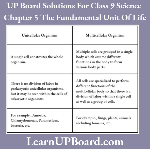
Based on the number of cells present in different organisms, they are classified into two types:
- Unicellular organisms (having single cell)
- Multicellular organisms (having many cells).
Differences Between Unicellular And Multicellular Organisms
Note:
- Every multicellular organism starts its life as a single cell (i.e. zygote), which divides and forms many cells. All cells thus, come from pre-existing cells.
- The invention of magnifying lenses made the discovery of single-celled microscopic organisms possible.
Microscopes
These are high-resolution instruments. They are used for observing the fine details of very minute objects, for example., cells. With the help of a microscope, the size of a small cell can be magnified up to 300-1500 times.
A simple microscope, which is often used in schools is a compound microscope. It uses sunlight for the illumination of objects to be seen, so it is called a light microscope. An electron microscope is used to observe complex structures of the cell.
Shape Of Cells
Some cells have fixed shapes (for example., most plant and animal cells), while some cells like WBCs and Amoeba keep changing their shapes.
Fixed-shaped cells may be of various types like elliptical (for example., fat cell), spherical (for example., ovum), spindle-shaped (for example., smooth muscle cell), knobbed thread (for example., sperm), discoidal (for example., RBC), elongated (for example., nerve cell), etc.
The following figures depict some cells from the human body:

Size Of Cells
The size of the cell varies significantly from the smallest cell of Mycoplasma (0.1-0.5 pm) to very large egg cells of the Ostrich (18 cm). The longest cells of the human body are the nerve cells, which may reach upto a length of 90 cm.
Functions Of Cells
Each living cell can perform some basic functions that characterise living organisms.
- The shape and size of cells are related to the specific function they perform.
- Multicellular organisms like human beings perform these functions by division of labour. Different parts of the human body are specialised to perform different functions such as the heart is made to pump blood, the stomach to digest food, the kidney to filter urine etc.
- Division of labour is also seen within a single cell. Every cell possesses certain specific components known as cell organelles. These enable it to survive and perform special functions.
- Cell organelles together with protoplasm constitute the basic unit of life called the cell. Each kind of cell organelle performs a specific function. For example, obtaining nutrition, respiration, clearing waste material or forming new progeny. Mitochondria is the organelle responsible for providing energy to the cell.
Note: The same cellular organelles are found in all the cells regardless of their function and the type of organism they are found in.
UP Board Class 9 Science Notes For Chapter 5 Structural Organisation Of A Cell
Microscopic studies revealed that every cell possesses three basic features in common, i.e. plasma membrane, nucleus and cytoplasm. Due to the presence of these features, all activities inside the cell and interaction of the cell with its environment are possible.
Plasma Membrane Or Cell Membrane
This is the outermost living, thin and delicate covering of cells. It separates the contents of the cell from its external environment.
- The presence of lipids and proteins (as phospholipids) provides flexibility to the plasma membrane. It enables cells to engulf food and other materials from the external environment.
- This process is called endocytosis, for example., Amoeba acquires food through this process, with the help of finger-like projections called pseudopodia.
Functions Of Plasma Membrane
- It allows the entry and exit of some selective materials in and out of the cell. The cell membrane, therefore, acts as a semipermeable, selectively permeable, partially permeable and differentially permeable membrane.
- It helps to maintain the shape of the cell.
- It acts as a mechanical barrier and protects the internal contents of the cell from leaking out.
- It protects against microbes and foreign substances.
- It gets modified to perform different functions, for example., microvilli in the intestine of human beings for absorption.
- Its semipermeability enables the cell to maintain cellular homeostasis. Amongst all the functions listed above, the transport of substances is the most important function.
It is, therefore, discussed in detail below:
Transport Across The Membrane
Transportation of substances across the membrane may take place with the expenditure of energy (active transport) and without the expenditure of energy (passive transport).
Transport Across The Membrane By Diffusion
The spontaneous movement of a substance (solid, liquid or gas) from a region of its higher concentration to a region of its lower concentration is called diffusion.
- For example, CO2 (cellular waste, which needs to be excreted out) accumulates in higher concentrations inside the cell. In the cell’s external environment, the concentration of CO2 is lowered compared to the inside of the cell.
- Due to this difference in the concentration, CO2 moves out of the cell by the process of diffusion. Similarly, O2 enters the cell by the process of diffusion, when the level or concentration of O2 inside the cell decreases.
- Diffusion is faster in gases than in liquids and solids. It plays an important role in gaseous exchange between the cells and also between the cell and its -external environment.
In addition to gaseous exchange, diffusion also helps an organism in obtaining nutrition from the environment.
Transport Across The Membrane By Osmosis
The movement of water molecules through a selectively permeable membrane along the concentration gradient is called osmosis.
- The movement of water across the plasma membrane is also affected by the amount of substance dissolved in water.
- Osmosis is thus, also defined as the movement of water molecules from a region of its higher concentration to a region of lower concentration through a semipermeable membrane.
- Unicellular freshwater organisms and most plant cells tend to gain water through osmosis.
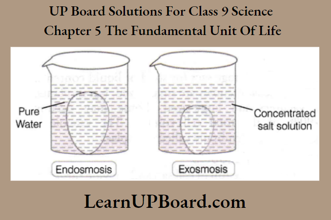
Absorption of water by plant roots is also an example of osmosis. The process of osmosis can be seen in a cell placed in a solution of different concentrations (such as hypotonic, isotonic and hypertonic).
- Hypotonic Solution The medium or solution surrounding the cell has a high water concentration as compared to the inside of the cell (or the outside solution is very diluted).
- The cell gains water and swells up via endosmosis. This happens because the water molecules are free to pass through the cell membrane in both directions. More water however enters the cell than that leaving it.
- Isotonic Solution The medium surrounding a cell has the same concentration of water as that present inside the cell.
- Water crosses the cell membrane in both directions, but the amount moving in remains the same as the amount moving out. So there is no overall movement of water. As a result, no overall change is observed and the cell size remains the same.
- Hypertonic Solution The medium surrounding a cell has a lower concentration of water than the cell (i.e. outside solution is very concentrated).
- Water crosses the cell membrane in both directions, but this time more water leaves the cell than enters it. As a result, the cell protoplasm gets shrunk (exosmosis).
Cell Wall
It is a tough, non-living covering outside the plasma membrane. It is found in plant and fungal cells. It is freely permeable. It is mainly made up of cellulose, a complex substance that provides structural strength to plants.
Functions of Cell Wall
- The cell wall permits the cells of plants, fungi and bacteria to withstand hypotonic conditions without bursting.
- In hypotonic media, the cells tend to take up water by osmosis. The cell swells up, building up pressure against the cell wall. The wall exerts an equal pressure against the swollen cell.
- Cell walls help plant cells to tolerate greater changes in surrounding medium than animal cells.
- It has narrow pores, called pits. Through them, fine strands of cytoplasm (or cytoplasmic bridges) called plasmodesmata can cross the cell walls. Plant cells interact with each other through these cytoplasmic channels.
Plasmolysis
It is the phenomenon, in which a living plant cell loses water through osmosis when kept in a hypertonic solution. It results in either the shrinkage or contraction of protoplasm away from the cell wall.
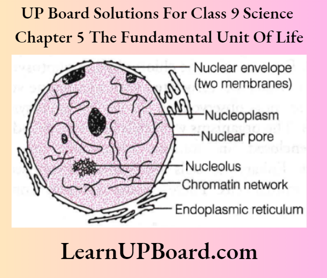
Nucleus Definition
It is popularly called the brain of a cell. It is composed of a double-layered covering called a nuclear membrane. It has numerous pores called nuclear pores. They transfer the materials from inside the nucleus to the cytoplasm.
The nucleus contains chromosomes. They are visible as rod-shaped structures only when the cell is about to divide. It encloses a liquid ground substance called nucleoplasm. It contains nucleolus and chromatin material.
The nucleolus is a more or less round structure found inside the nucleus. It does not have a covering of membranes. It is known as the factory of ribosomes.
Chromatin is an entangled network of long, thread-like structures. It condenses to form chromosomes during cell division.
Chromosomes contain information for the inheritance of features from parents to the next generation in the form of DNA (Deoxyribonucleic Acid). Chromosomes are composed of two components, i.e. DNA and protein.
The DNA molecules contain information necessary for constructing and organising cells. The functional segments of DNA are called genes. The nucleus also contains RNA that directs protein synthesis.
Functions Of Nucleus
- The nucleus plays an important role in cellular reproduction. In this process, a cell divides to form two new cells.
- It determines the cell’s development and maturity by directing the chemical activities of the cell.
- It helps in the transmission of hereditary traits from parents to offspring.
- It controls all metabolic activities of cells. If it is removed, the protoplasm dries up.
In some organisms like bacteria, the nuclear region of the cell is poorly defined because of the absence of a nuclear membrane. The nuclear region in these organisms contains only nucleic acid. Such an undefined nuclear region is called a nucleoid.
Organisms whose cells lack a nuclear membrane are called prokaryotes (pro = primitive, eukaryote ≈ karyon = nucleus).
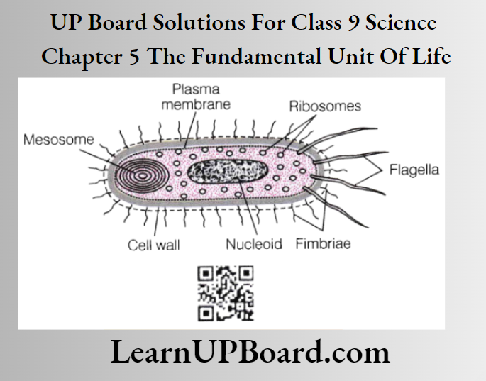
- Prokaryotes also lack cytoplasmic organelles. Most functions are thus performed by poorly developed parts of the cytoplasm.
- For example, the chlorophyll in photosynthetic prokaryotic bacteria is associated with membrane vesicles. Plastids are not observed in it as in photosynthetic eukaryotes.
- The organisms with cells having a well-defined nucleus enclosed in a nuclear membrane are called eukaryotes. Eukaryotic cells are further categorised into plant and animal cells. These are also different from each other in many ways.
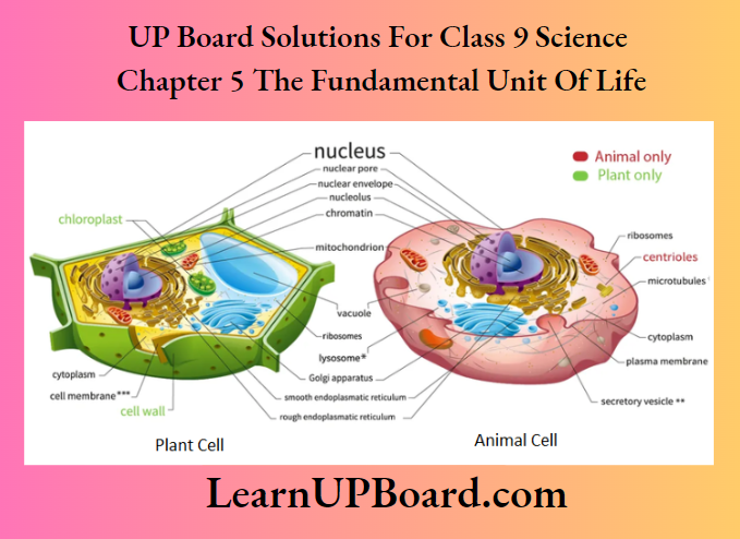
Differences Between Prokaryotic And Eukaryotic:

Cytoplasm
The large region of each cell enclosed by the cell membrane is called cytoplasm. It is the fluid content present inside the plasma membrane. It contains many specialised cell organelles, each of which performs a specific function for the cell.
Fundions Of Cytoplasm
- It helps in the exchange of material between cell organelles.
- It acts as a storehouse of vital molecules such as amino acids, glucose, vitamins, iron etc.
- It acts as the site for certain metabolic pathways such as glycolysis etc.
UP Board Class 9 Science Notes For Chapter 5 Cell Organelles
Large and complex cells need a lot of chemical activities to support their complicated structure and function.
- To keep these activities separated from each other, these cells use membrane-bound structures.
- These structures perform specialised functions within themselves and are called cell organelles. This is the main characteristic feature that differentiates eukaryotic cells from prokaryotic cells.
Endoplasmic Reticulum (ER)
It is a large network of membrane-bound tubes and sheets. It extends from the outer nuclear membrane into the cytoplasm. It looks like long tubules of round and oblong bags (vesicles). The ER membrane is similar in structure to the plasma membrane. lt occurs in three forms, i.e. cisternae, vesicles and tubules. Depending upon the nature of its membrane, ER is of two types:
- Rough Endoplasmic Reticulum (RER) It contains ribosomal particles, due to which its surface is rough. The ribosomes are the site of protein synthesis. RER is mainly formed of cisternae.
- Smooth Endoplasmic Reticulum (SER) It helps in the manufacture of fat molecules or lipids. It is formed of vesicles and tubules. Its surface is smooth due to the absence of ribosomes. ER appears in varying forms in different cells. It always forms a network system of vesicles and tubules.
Functions Of Endoplasmic Reticulum
- Ribosomes present in all active cells act as sites for protein synthesis. Proteins manufactured here are transported throughout the cell by the endoplasmic reticulum.
- Fat and lipid molecules manufactured by SER help in building cell membranes. This process is called membrane biogenesis.
- Some other proteins and lipids synthesised by ER function as enzymes and hormones.
- SER plays a crucial role in the detoxification of poisons and drugs in the liver cells of vertebrates (a group of animals).
- It forms a network system, providing channels for the transport of materials, especially proteins. It transports between various regions of the cytoplasm or between the cytoplasm and the nucleus.
- It functions as a cytoplasmic framework. It provides a surface for some of the biochemical activities of the cell.
- It gives mechanical support to the cells.
Golgi Apparatus
It consists of a system of membrane-bound, fluid-filled vesicles, large spherical vacuoles and smooth, flattened cisternae. These are stacked parallel to each other. Each of these stacks is called a cistern.
The Golgi apparatus (or dictyosomes) arises from the membrane of smooth ER. Therefore, it constitutes another portion of a complex cellular membrane system.
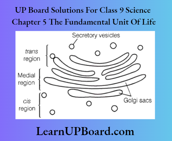
The material that is synthesised near the Endoplasmic Reticulum (ER) is packaged and dispatched to various parts of the cell through the Golgi apparatus.
Functions Of Golgi Apparatus
- Golgi apparatus stores modifies and packs products in vesicles.
- It is involved in the formation of lysosomes.
- It forms complex sugars from simple sugars in some cases.
- It is involved in the synthesis of cell wall and plasma membrane.
Note: The scientist, who described the Golgi apparatus for the first time was Camillo Golgi. Most of his investigations were concerned with the nervous system. His greatest work was a revolutionary method of staining individual nerve and cell structures.
This method is called ‘black reaction’. It uses silver nitrate solution to trace the most delicate ramification of cells. He shared the Nobel Prize in 1906 with Santiago Ramon y Cajal for their work on the structure of the nervous system.
Lysosomes
- These are a kind of waste disposal system of the cell. Lysosomes are membrane-bound sacs that are filled with digestive enzymes. These enzymes are made by the rough endoplasmic reticulum. Lysosomes are also called the suicidal bags of a cell.
- During the disturbance in cellular metabolism or when the cell gets damaged, lysosomes may burst and the enzymes can digest their cell. They are absent in RBCs.
Functions Of Lysosomes
- They help to keep the cell clean by digesting any foreign material that enters the cell as well as worn-out cellular organelles. Hence, called scavengers and cellular housekeepers.
- They remove foreign material by breaking it into small pieces through their powerful digestive enzymes. These enzymes can break down all organic materials.
- During starvation, the lysosomes digest stored food contents by autophagy and supply energy to the cell.
Mitochondria
Mitochondria were first observed by Kolliker in 1880. It is a double membrane-bounded cell organelle. The outer membrane is very porous. The inner membrane is deeply folded into finger-like projections called cristae. It creates a large surface area for ATP-generating chemical reactions.
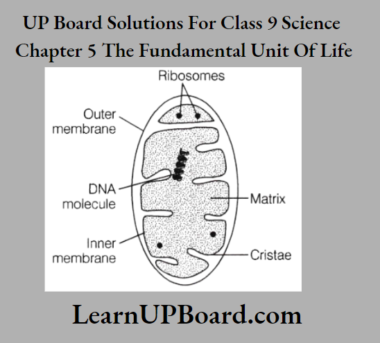
The space between the outer and inner membrane is called intermembranous space. The mitochondrion is a self-replicating (semiautonomous) organelle. It is the largest organelle in animal cells.
Functions Of Mitochondria
- It generates energy for various activities of the cell. It is known as the powerhouse of the cell. Mitochondria are sites of cellular respiration. They release energy required by the cell in the form of ATP (Adenosine Triphosphate). This ATP is known as the energy currency of the cell.
- Whenever the cell requires energy, the ATP molecule breaks down. It generates energy to be used for metabolic activities of the body.
- Mitochondria are strange organelles in the sense that they have their DNA and ribosomes. Hence, they can make some of their proteins.
- They provide intermediates for the synthesis of various chemicals like fatty acids, steroids, amino acids etc.
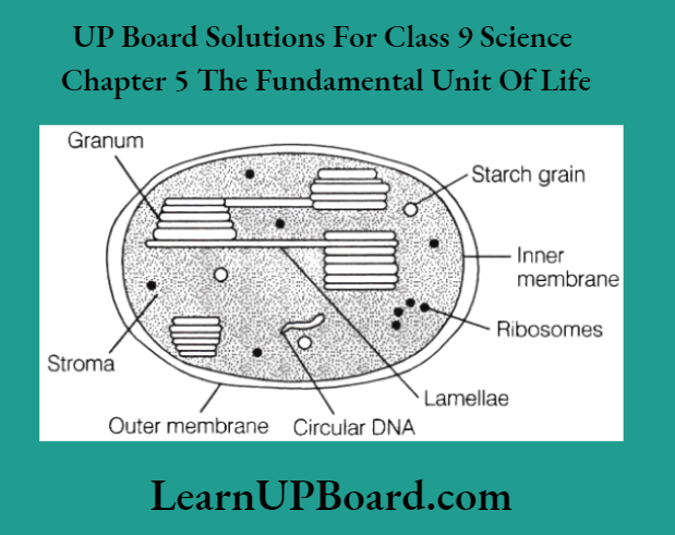
Plastids
- These are found only in plant cells. The internal organisation of plastids contains numerous membrane layers embedded in a material called the stroma.
- Plastids are similar to mitochondria in external structure. They are double-layered. They contain their DNA and ribosomes.
Types Of Plastids
Plastids are of three types:
1. Chloroplasts These are the plastids containing chlorophyll (a green pigment). They give a green colour to the plant. Chloroplasts also contain various yellow or orange pigments in addition to chlorophyll. It is a semiautonomous organelle. Chloroplasts are also known as the kitchen of cells.
Function These are important for photosynthesis in plants.
Note: Photosynthetic bacteria do not contain chloroplasts. They contain light-absorbing pigments and reaction centres, which make them capable of converting light energy into chemical energy.
2. Leucoplasts These are the white or colourless plastids. They can change into other types of plastids.
Function Leucoplasts store materials such as starch (amyloplasts), oils (elaioplasts) and protein granules (aleuroplasts).
3. Chromoplasts These are coloured plastids (except green).
Function Chromoplasts impart colour to flowers and fruits. They are rich in carotenoid pigments and lipids.
Vacuoles
These are the storage sacs for solid or liquid contents. In animal cells, vacuoles are small-sized, but in plants, the vacuoles are large-sized. Some may occupy 50-90% of the total cell volume. The vacuole is bounded by a membrane called the tonoplast.
Functions Of Vacuoles
- Vacuoles are full of cell sap and provide turgidity and rigidity to cells in plants.
- Many substances like amino acids, sugars, organic acids and proteins are stored in vacuoles.
- In Amoeba, consumed food items are stored in food vacuoles.
UP Board Class 9 Science Notes For Chapter 5 Activity 1
Objective
Observation of plant cells.
Materials Required
Onion, knife, forceps, watch glass, water, dilute glycerine, safranine (safranin) solution, thin camel hair paint brush, dropper, mounting needle, glass slide, coverslip, blotting paper and microscope.
Procedure
Cut a small piece from an onion bulb. Separate a fleshy scale with the help of forceps and remove a transparent peel (called epidermis) from the inner concave side of the scale.
- Place it immediately in the watch glass containing a small quantity of water. Pour 2-3 drops of safranin solution into the watch glass. After 5-10 minutes, remove the stain, pour water into the watch glass and wash it again.
- Place a drop of water or dilute glycerine in the middle of a glass slide. Transfer a small piece of stained onion and peel onto it using a fine camel hair paintbrush.
- Place a cover slip over the peel with the help of a mounting needle, avoiding the appearance of air bubbles. Wipe out excess liquid by blotting paper. Now observe it under the compound microscope.
Observation
The peel is found to have several similar elongated rectangular cells joined with each other. Each cell consists of an outer cell wall, a filler material or cytoplasm with a covering of plasma membrane containing a small oval nucleus.
Question 1. Why glycerine is used in this experiment?
Answer:
Glycerine is used in this experiment because it acts as a preservative and has a high refractive index.
Question 2. Why safranin is used in this experiment?
Answer: Safranin is an azo dye commonly used for plant microscopy, especially to stain lignified tissues such as the xylem of plants, etc
Question 3. Why was the sample washed again with water after beeping in safranin solution for 5-10 minutes?
Answer: The sample was washed again with water to remove the extra stain of safranin.
Question 4. What will happen, if air bubbles appear while placing a cover slip?
Answer: If air bubbles appear, then the structure of the sample will not be observed properly.
UP Board Class 9 Science Notes For Chapter 5 Activity 2
Objective
Temporary mounts of cells from different sources. Prepare temporary mounts of onion peel, leaf peel and onion root tip. Study the shapes, sizes and internal structures of the cells.
Materials Required
Onion, knife, forceps, watch glass, water, dilute glycerine, safranine (safranin) solution, thin camel hair paint brush, dropper, mounting needle, glass slide, coverslip, blotting paper and microscope.
Procedure
Repeat the procedure described in Activity 1 for all three types of tissues onion peel, leaf peel and onion root tip.
Observation
The cells from different sources are of different shapes and sizes besides having certain specific structures.
Question 1. What conclusion can be drawn from this activity?
Answer: The cells from different sources are of different shapes and sizes, but have the same structure.
Question 2. Describe briefly the basic structure of cells as observed in this activity.
Answer: Cells consist of an outer cell wall having cytoplasm, plasma membrane and a nucleus.
Question 3. Why blotting paper is used in this activity?
Answer: Blotting paper wipes out excess liquid.
Question 4. What is the shape of cells in onion peel?
Answer: The cells of the onion peel are elongated and rectangular.
UP Board Class 9 Science Notes For Chapter 5 Activity 3
Objective
To observe the phenomenon of osmosis in a typical cell
Skill Developed
Observation skills, problem-solving and critical thinking.
Time Required
1 hour 30 minutes.
Materials Required
Containers dilute hydrochloric acid, eggs, concentrated sugar/salt solution and water.
Procedure
- Place an egg in dilute hydrochloric acid.
- The outer shell starts to dissolve in the acid and the egg membrane appears clearly. A thin outer skin now encloses the egg.
- Place the egg into a container containing distilled water.
- Record the observation.
- Place another similar egg into a container containing a concentrated salt/sugar solution.
- Record the observation.
Observation
- The egg in distilled water swells up because water moves into the egg by osmosis (in this case solution inside the egg is more concentrated).
- The egg in the concentrated solution shrinks because water moves out of the egg, into the concentrated sugar/salt solution (in this case solution outside the egg is more concentrated)
Question 1. Name the process that made the egg swell.
Answer:
The egg swelled up due to the process of endosmosis. The inward flow of a fluid through a semipermeable membrane towards a fluid of greater concentration is called endosmosis.
Question 2. Differentiate between exosmosis and endosmosis.
Answer:
Exosmosis The passage of water molecules out of the cell into a concentrated solution surrounding it. Endosmosis The passage of water molecules into the cytoplasm of a cell from a less concentrated solution surrounding it.
Question 3. What happens to an egg, when it is kept in hydrochloric acid?
Answer: Hydrochloric acid dissolves the outer shell of the egg and the thin semi-permeable membrane of an egg becomes visible.
Question 4. Name the process of diffusion through a semi-permeable membrane that occurs during certain conditions.
Answer: Osmosis is a special case in which diffusion takes place through a selectively permeable membrane.
UP Board Class 9 Science Notes For Chapter 5 Activity 4
Objective
To observe osmosis in raisins or apricots.
Time Required
1 hour 30 minutes.
Materials Required
Water, raisins or apricots, sugar or salt solution and beakers.
Procedure
- Take two beakers and label one as ‘water’ and the other as ‘concentrated’.
- Take some water in the beaker labelled ‘water’ (beaker A)
- Put some raisins or apricots in this beaker A containing water for some time.
- Add salt or sugar-concentrated solution in a beaker marked as ‘concentrated’ (beaker B).
- Now add raisins or apricots to a concentrated beaker.
- Observe the raisins or apricots in both beakers.
Observation

- The raisins in beaker A swell up as water moves inside from outside due to the low concentration of water inside the cell. This process is called endosmosis.
- The raisins in beaker B shrink because water moves out of the cell. Since the solution outside the cell is concentrated and the water concentration is lower than that present inside the cell, the water moves outside the cell and this process is called exosmosis.
Question 1. What does a concentrated solution mean?
Answer:
A concentrated solution is a solution with a high amount of solutes. When a large amount of solute (like sugar) is mixed in a solvent (like pure water) the solution becomes a concentrated solution (for example., sugar solution).
Question 2. Why did the raisins in Beaker A swell?
Answer:
- In beaker A, raisins were kept in water. The concentration of water outside the cell was more than the concentration of water inside the cell.
- Therefore, water moved from the region of high concentration to the region of low concentration (inside raisin) and caused swelling of the raisins.
Question 3. Why did the raisins in Beaker B shrink?
Answer: In beaker B, raisins were kept in a concentrated solution, where water concentration was lower than that of the cell. Hence, water moved from the cell to outside leading to the shrinkage of the cell (exomosis).
Question 4. What is the primary requirement of osmosis?
Answer: For osmosis, the presence of a semi-permeable membrane is the primary requirement.
UP Board Class 9 Science Notes For Chapter 5 Activity 5
Objective
To study plasmolysis using the cells of Rheo leaf.
Time Required
1 hour 30 minutes (approximately).
Materials Required
Microscope, glass slide, cover-slip, Rheo leaf, salt/sugar solution, water and burner.
Procedure
- Mount an epidermal peel of a freshly plucked Rheo leaf in a drop of water on a glass slide.
- Cover it with a cover slip and observe it under a microscope.
- Remove the cover slip and add a drop of concentrated sugar/salt solution. Place the coverslip again onto the slide.
- Observe the slide again under a microscope after 1 or 2 minutes.
- Record your observation.
- Remove the cover-slip and wipe out the salt/sugar solution, add one or two drops of water and observe the slide under a microscope after 1-2 min.
- Record your observation.
- Repeat the same experiment with a leaf that was kept in boiling water for a few minutes.
- Record your observation.
Observation
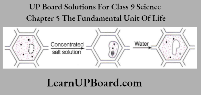
- When the peel is mounted first in water, pink colour is uniformly filling up the cells.
- When sugar/salt solution is added, the pink colour (protoplasm) of the cell appears in small areas of the cell, cell, i.e. it has undergone plasmolysis (shrinking of cell protoplasm due to outward movement of water).
- When water is added to replace the sugar/salt solution, the pink colour/protoplasm fills the cell again.
- In the second leaf, which was kept in boiling water for a few minutes, no effect of sugar/salt solution was seen.
Question 1. Name the phenomenon which occurs due to shrinkage or contraction of the content away from the cell wall.
Answer: During osmosis, when a living plant cell shrinks or contracts with its contents away from the cell wall, the phenomenon is called plasmolysis.
Question 2. The boiled leaf showed no change with the sugar/salt solution. Why?
Answer: The cells of the leaf died when the leaf was kept in boiling water for a few minutes.
Question 3. Can osmosis occur in dead cells?
Answer: No, osmosis can occur only in living cells.
Question 4. What happens to the cell when kept in an isotonic solution?
Answer: In an isotonic solution, the concentration of the medium was the same as that of the cell, there is no net movement of water across the cell membrane. Thus, the cell will stay the same size.
UP Board Class 9 Science Notes For Chapter 5 Activity 6
Objective
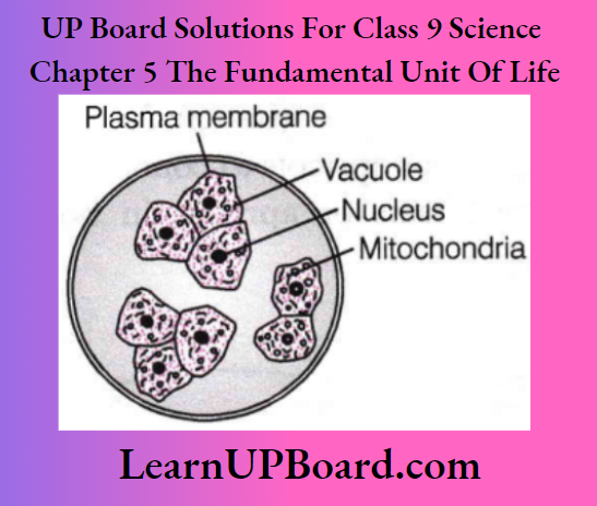
Study of human cheek cells.
Materials Required
Sterilised toothpick or ice-cream spoon, glass slide, needle, cover-slip, water or dilute glycerine, dropper, methylene blue, blotting paper and microscope.
Procedure
- Scrape a small piece of membrane from inside of your cheek lightly with the help of a clean sterilised toothpick or an ice-cream spoon.
- Mount the membranous scraping in a drop of water or dilute glycerine over a clean glass slide. Spread the scraping with the help of a needle.
- Put a drop of methylene blue over it. Wait for two minutes.
- Now put a cover-slip gently over the slide avoiding entry of air bubbles.
- Replace the stain by pouring water drop on one side and soaking the stain from the sides using blotting paper.
- Observe it under the microscope first with normal or low power (1 Ox) and then with high power (45x).
Observation
Several polygonal flat (squamous) cells are present in the scraped membrane. Each cell has a distinct boundary of plasma membrane, a central rounded or oval nucleus, many small dotted mitochondria, small vacuoles and cytoplasm.
Question 1. What is the purpose of putting methylene blue on the cell?
Answer: Methylene blue is poured on the slide to stain the cells.
Question 2. What is the shape of the observed cells?
Answer: The observed cells are polygonal, flat or squamous shaped.
Question 3. Why it is necessary to avoid air bubbles?
Answer: Air bubbles will not give the correct image to the observer.
Question 4. Why after staining, water is poured over the cell?
Answer: Water is poured after staining to remove the excess stain. Blotting paper is used for soaking this stain and water mixture.
The Fundamental Unit Of Life Summary
A cell is the basic structural and functional unit of all living organisms. It was discovered by Robert Hooke in the year 1665.
- Unicellular organisms are those organisms which are made up of a single cell only, for example., Amoeba, Chlamydomonas, bacteria, etc.
- Multicellular organisms are organisms made up of many cells. These cells group and assume different functions in the body to form various body parts, for example., plants and animals.
- Prokaryotic cells are cells lacking a well-defined nucleus enclosed by a nuclear membrane, for example., bacteria and cyanobacteria.
- Eukaryotic cells are those having a well-defined nucleus enclosed in a nuclear membrane, for example., plant cells and animal cells. Plant cells possess a cell wall and a vacuole that occupies most of the space. It lacks centrosome and centrioles.
- Animal cells do not have cell walls, they possess highly complex Golgi bodies, centrioles, etc.
- Structurally, a cell mainly consists of a plasma membrane, cytoplasm and nucleus. Cell organelles such as Golgi bodies, mitochondria, etc, are also present in cytoplasm.
- The plasma membrane is the outermost covering of the cell that is composed of proteins and lipids.
- It permits the entry and exit of some materials. It maintains the shape of the cell, acts as a mechanical barrier and protects the internal contents of a cell. Transport of substances across plasma membrane may take place by diffusion, i.e. process of movement of solutes or osmosis, i.e. process of movement of water.
- The nucleus is properly called as brain of the cells. It controls all functions of a cell. It also determines the development of cells by directing the chemical activities of cells.
- Cytoplasm is the fluid content present inside the plasma membrane that contains many specialised cell organelles and acts as a site for metabolic pathways such as glycolysis.
- The endoplasmic reticulum is a large network of membrane-bound tubules and sheets. It plays an important role in protein and lipid synthesis.
- Mitochondria is known as the powerhouse of cells that releases energy required by the cell in the form of ATP.
- Golgi apparatus consists of a system of membrane-bound vesicles called cisternae. It helps in the formation of lysosomes and in storing and packaging various molecules in a cell.
- Lysosomes are the waste disposal systems of a cell also called suicidal bags of cells.
- Plastids are found in plant cells as chloroplasts, chromoplasts and leucoplasts.
- Vacuoles are storage sacs of solids and liquids.