
UP Board Class 9 Science Notes For Chapter 6 Tissues
All living organisms are composed of cells. In unicellular organism, a single cell performs all the basic functions, but in multicellular organisms, different functions are performed by different cells. Cells specializing in one function group together and form tissues.
- A group of cells similar in structure that work together to achieve a particular function forms a tissue. These cells are arranged and designed, so as to give the highest possible efficiency of the function they perform. All cells of a tissue have a common origin.
- A tissue may be a simple or complex type. Blood, phloem, and muscles are all examples of tissues. The structural and functional organization of cells in plants and animals is different. Plants remain stationary while animals move as per their needs.
- Each pursues different feeding methods and is differently adapted. In this chapter, we will study various types of tissues found in plants and animals along with their respective functions.
UP Board Class 9 Science Notes For Chapter 6 Plant Tissues
Based on dividing capacity, plant tissues can be classified into two fundamental types as follows:
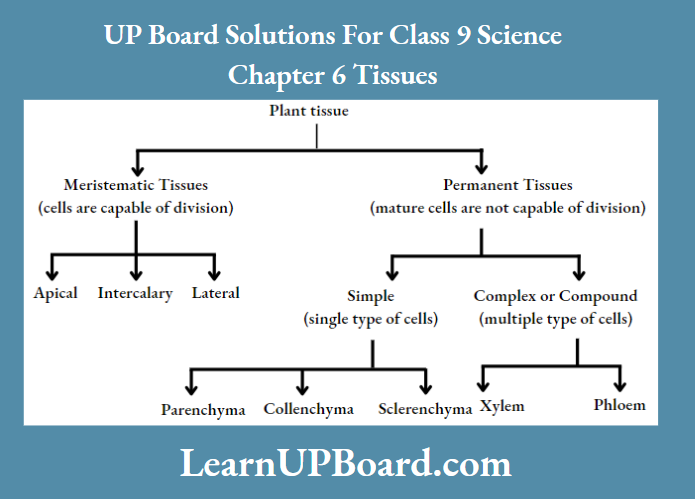
1. Meristematic Tissue Function
The tissues in which cells always keep dividing giving rise to new cells are called meristematic tissues. These tissues are responsible for the growth of plants. Plants grow only in those regions where meristematic tissues are present, for example., root and shoot tips.
Read and Learn More Class 9 Science Notes
- It is also called growth tissue. Cells forming this tissue are very active and have dense cytoplasm, thin cellulose walls, and prominent nuclei. They lack vacuoles. The new cells produced by meristem are initially like those of meristem.
- Their characteristics change once they grow and become differentiated as components of other tissues. Meristematic tissue is classified based on the regions, where they are present.
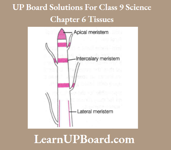
- Apical Meristem
- These are present at the growing tips of stems and roots. This helps increase the length of the stems and the roots. It acts as a pro-meristem having actively dividing cells, giving rise to other meristems.
- Intercalary Meristem
- These are present at the base of the leaves or internodes (on either side of the node) of twigs. It helps in the longitudinal growth (elongation) of plants.
- Lateral Meristem (Cambium)
- These are present on the lateral sides of stems and roots. It helps in increasing the girth of the stem and root.
2. Permanent Tissue
- This tissue is formed from the cells of meristematic tissue when they lose their ability to divide and have attained a permanent shape, size, and function by the process called differentiation.
- As a result of differentiation, the meristematic tissues tend to form different types of permanent tissues as follows:
Simple Permanent Tissue
It is made up of only one type of cells, i.e. the cells forming these tissues are similar in structure and function.
Simple permanent tissue is further classified as:
Parenchyma: A few layers of cells form the basic packing tissue. They are present in the cortex and pith of stems and roots in the mesophyll of leaves.
Characteristics: These are simple living cells with little specialization and thin cell walls.
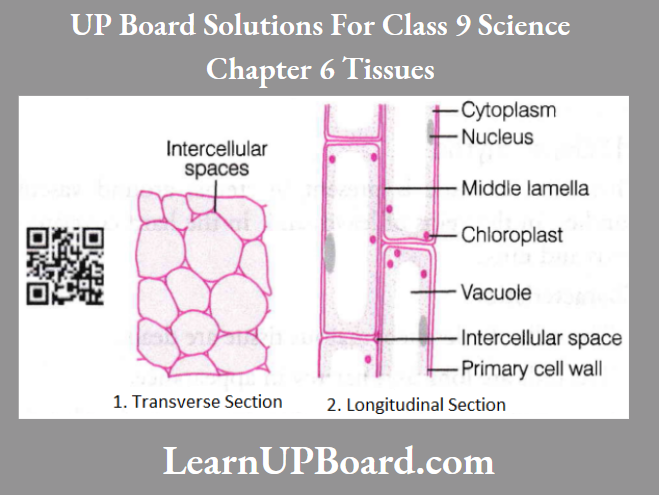
Simple Permanent Tissue Functions
- It serves as a food storage tissue.
- This tissue provides support to plants.
- When the parenchyma cell contains chlorophyll in some situations, it performs photosynthesis. Such type of parenchyma tissue is called chlorenchyma.
- In aquatic plants, large air cavities are present in parenchyma cells to give buoyancy to plants, which helps them to float. Such type of parenchyma tissue is called aerenchyma.
- Parenchyma of stems and roots also stores nutrients and water.
Collenchyma: These tissues are generally found in leaf stalks below the epidermis and leaf midribs.
Collenchyma Characteristics
- Cells are living, elongated, and irregularly thickened at the corners due to the deposition of pectin.
- They have very little intercellular spaces.
Collenchyma Functions
- It provides mechanical support and elasticity (flexibility) to plants.
- It also allows easy bending in various parts of a plant (leaf and stem) without breaking.
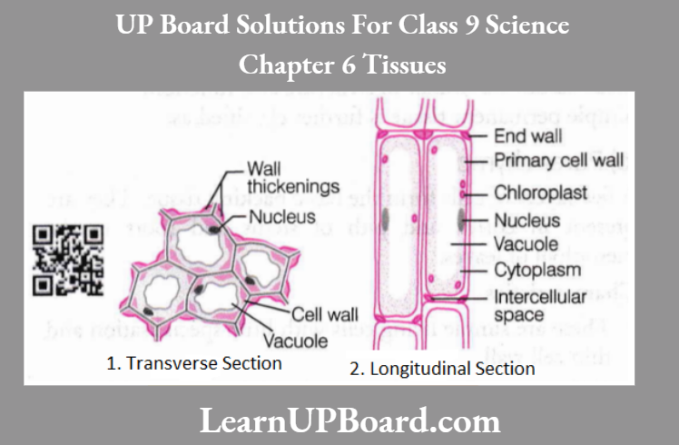
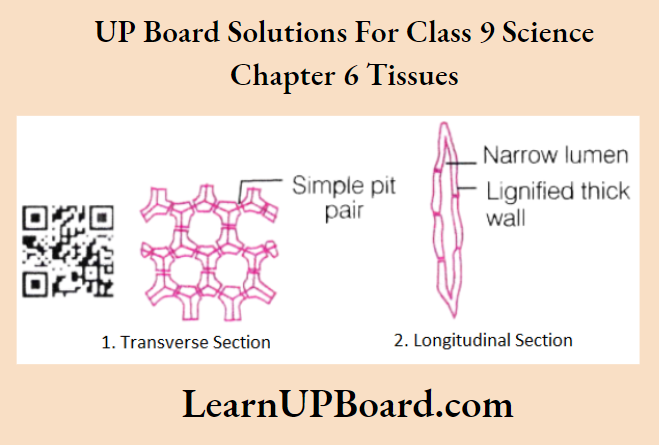
Sclerenchyma Definition
This type of tissue is present in stems, around vascular bundles, in the veins of leaves, and in the hard covering of seeds and nuts.
Sclerenchyma Characteristics
- The cells of sclerenchymatous tissue are dead. The cells are long and narrow in appearance.
- Cell walls are thickened due to lignin (a chemical substance) deposition, which acts as cement and hardens them.
- Due to the presence of thick walls, there is no internal space between the cells.
Sclerenchyma Functions
- It is known to be the chief mechanical tissue, that makes plants hard and stiff, for example., the husk of the coconut is made up of sclerenchymatous tissue.
- It forms a protective covering around seeds and nuts. It gives rigidity, flexibility, and elasticity to the plant body.
Protective Tissues
- The protective tissue i; meant to protect the plants from undue loss of water.
- Thus, they retain adequate water in them. The two types of protective tissues present in plants are:
Protective Tissues Epidermis
- The outermost layer, i.e. epidermis in plants is made up of a single layer of cells. It protects all parts of the plant.
- On the aerial parts of the plant, epidermal cells often secrete a waxy, water-resistant layer on their outer surface. It protects against loss of water, mechanical injury, and invasion by microbes.
- Cells of epidermal tissue form a continuous layer. They have no intercellular spaces due to their protective role. Most epidermal cells are relatively flat. The outer wall and side walls are thicker than the inner wall.
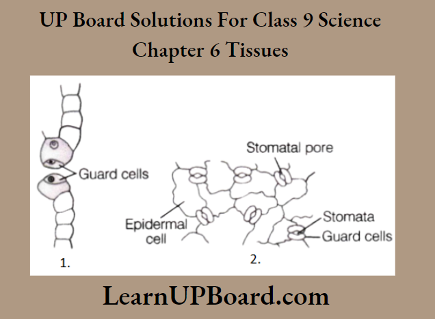
- Epidermal cells of the leaf bear small pores known as stomata. These are enclosed by two kidney-shaped cells called guard cells.
Stomata are responsible for the exchange of gases with the atmosphere and for the process of transpiration (loss of water in the form of water vapors).
- Epidermal cells of the roots bear long hair-like outgrowths called root hairs. They greatly increase the total absorptive surface area and help the roots to absorb water and nutrients from the soil.
- In the case of desert plants, the epidermis of the aerial parts has a thick waxy coating of cutin (a chemical substance with waterproof quality) on its outer surface. It prevents water loss.
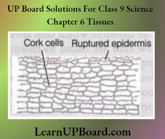
Protective Tissues Cork
It is the strip of secondary meristem, which replaces the epidermis of older stems. Cells of cork are dead, compactly arranged, and have no intercellular spaces. It forms the bark of the tree (several layers thick). A chemical called suberin is present in their walls. It makes them impervious to gases and water.
Complex Permanent Tissue: It is made up of more than one type of cells having a common origin. Regardless of different appearances, all the cells coordinate to perform a common function.
Types of complex permanent tissue are:
- Xylem
- Phloem
Both of them are conducting tissues and constitute vascular bundles. This is a distinctive feature of complex plants. It provides them with the possibility of surviving in the terrestrial environment.
Complex Permanent Tissue Xylem
It is responsible for the transport of water and minerals from roots to other parts of the plant. The cells of the xylem have thick walls and many of them are dead. Xylem consists of various types of elements, which are as follows:
1. Tracheids
- These are dead, long, tubular structures with tapering ends.
- They transport water and minerals vertically.
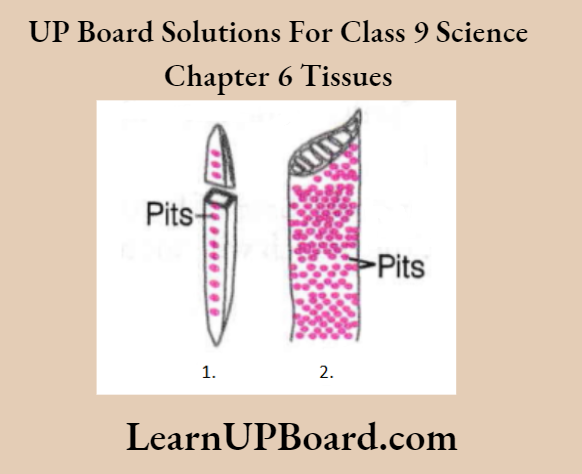
2. Vessels
- Long, tube-like structures, formed by a row of cells, placed end to end.
- These are also dead cells with lignified walls.
- They also help in the conduction of water.
3. Xylem parenchyma
- These are only living cells of the xylem with thin cell walls.
- It stores food and helps in the sideways conduction of water.
4. Xylem fibres
- They are elongated dead cells with tapering ends and thick cell walls.
- These are fibers associated with the xylem and supportive of the functioning of the xylem.
Phloem
It transports food from leaves to other parts of the plant. Materials can move in both directions in it. All phloem cells are living except phloem fibers.
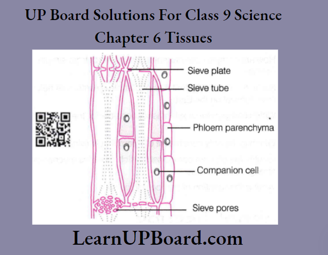
Phloem is made up of the following four types of elements:
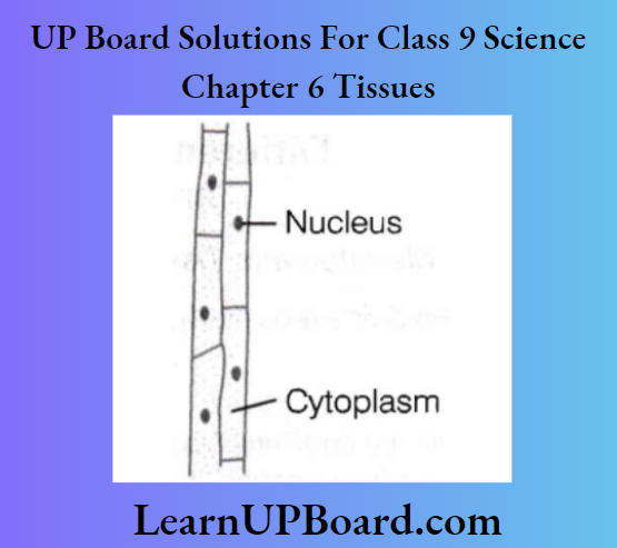
- Sieve tubes
- These are tubular cells with perforated walls.
- They have a thin layer of cytoplasm.
- Companion cells
- These are small elongated cells having thin walls that are not perforated and have active cytoplasm. They help sieve tubes in the translocation of food.
- Phloem fibers
- They are thick-walled sclerenchyma cells that provide mechanical strength to the tissue.
- Phloem parenchyma
- They are thin-walled cells that help in the storage and slow lateral conduction of food.
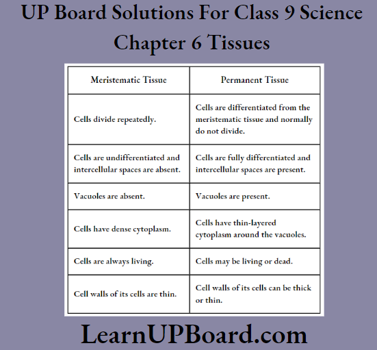
Differences Between Meristematic And Permanent Tissues:
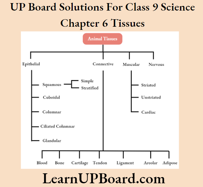
UP Board Class 9 Science Notes For Chapter 6 Animal Tissues
Based on the functions they perform, animal tissues are classified into four basic types namely epithelial, connective, muscular, and nervous tissue.
1. Epithelial Tissue
The covering or protective tissues in the animal body are epithelial tissues. It is the simplest protective tissue of the animal body. It covers most organs and cavities of the body.
- It forms a barrier to keep different body systems separated from each other. In these tissues cells are tightly packed and form a continuous sheet.
- There is almost no intercellular space between them. They have a very small amount of cementing material between them.
- The epithelium is separated from underlying tissue by an extracellular fibrous basement membrane containing collagen.
- Based on the shape of the cells and their arrangement, epithelial tissues are further classified as follows:
Squamous Epithelium
Squamous epithelial tissue constitutes the skin that protects the body. It is further categorized as:
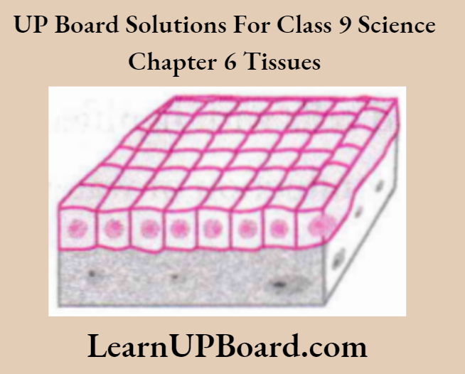
Simple Squamous Epithelium
- It is single-layered and closely fitted. The cells are very thin and flat and appear as tiles over a floor.
- It forms a delicate lining of blood vessels and lung alveoli, where substance transport occurs through a selectively permeable membrane.
- It also covers the esophagus and the lining of the mouth.
Stratified Squamous Epithelium
- It is found on the outer side of the skin as it is highly resistant to mechanical injury and is water-proof.
- Cells are arranged in many layers to prevent their wear and tear
Cuboidal Epithelium
- It is made up of cube-shaped cells, which have round nuclei.
- It forms the lining of kidney tubules and ducts of salivary glands, where it provides mechanical support. It also forms the germinal epithelium of gonads.
- It also helps in absorption, excretion, and secretion.
Columnar Epithelium
- It is made up of tall, pillar-like cells, with elongated nuclei.
- It is usually found in the inner lining of the intestine, where absorption and secretion occur.
- It facilitates movement across the epithelial barrier.
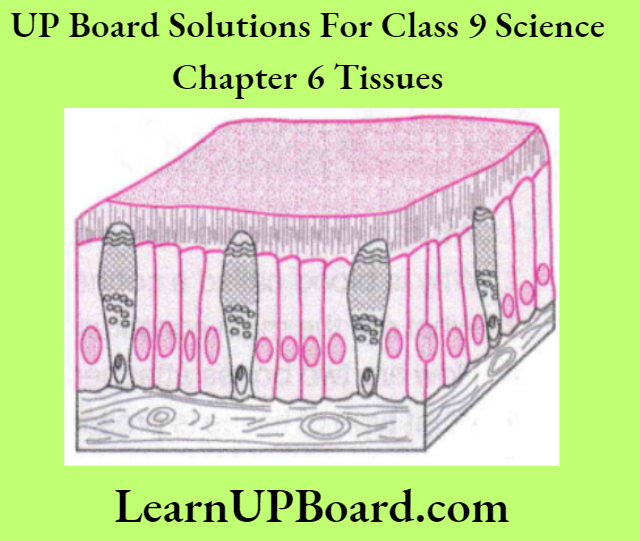
Ciliated Columnar Epithelium
- When columnar epithelial cells possess cilia (hair-like projections), it is called ciliated columnar epithelium.
- The cilia can move. Their movement pushes substances like mucus forward.
- It is found in the respiratory tract and also lines oviducts, sperm ducts, kidney tubules, etc.
Glandular Epithelium
- Gland cells secrete substances at the epithelial surface.
- Sometimes, a portion of epithelial tissue folds inward. This results in the formation of a multicellular gland. Its tissue is called glandular epithelium.
Functions of Epithelial Tissue
- It protects the underlying cells from drying, injury, infections, and also from harmful effects of chemicals.
- It plays a vital role in regulating the exchange of materials between the body and the external environment and between different body parts.
- It helps in the absorption of water and nutrients and in the diffusion of gases.
- It helps in the elimination of waste products from the body.
2. Connective Tissue
- This tissue is specialized to connect various body organs. For example., it connects two or more bones muscles to bones, binds different tissues together, and also gives support to various parts of the body.
- The cells of connective tissue are loosely packed, living, and embedded in an intercellular matrix that may either be jelly-like, fluid, dense, or rigid. The nature of the matrix differs in concordance with the function of the particular connective tissue.
Various types of connective tissues are:
Connective Tissue Blood
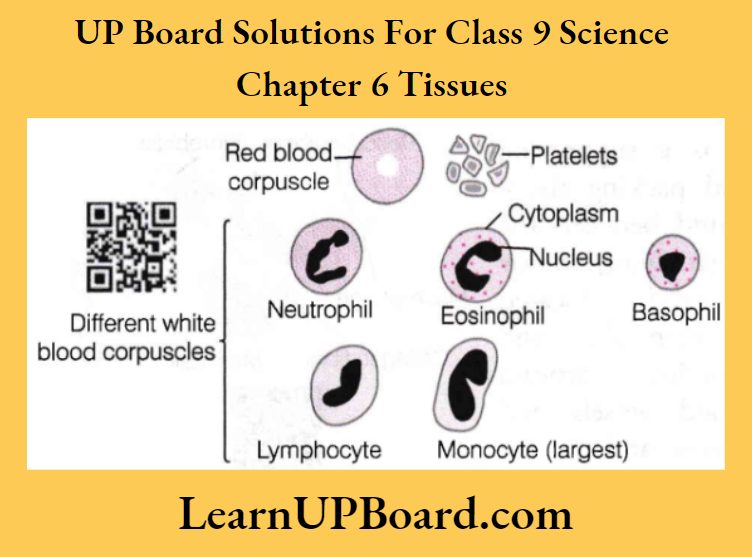
- It is a fluid connective tissue that links different parts of the body. It helps to maintain the continuity of the body. It contains a fluid matrix called plasma and blood cells such as RBCs (Red Blood Corpuscles or Cells), WBCs (White Blood Corpuscles), and platelets suspended in it.
- Plasma also contains proteins, salts, and hormones. Blood transports nutrients, gases, hormones, and vitamins to various tissues of the body. It carries excretory products from tissues to excretory organs. It also conducts heat and regulates body temperature.
Properties shown by different blood cells in the body are as follows:
- RBCs Help in the transport of respiratory gases, oxygen, and carbon dioxide with the help of hemoglobin to and from the various parts of our body. The average lifespan of RBCs is 120 days.
- WBCs Also called leucocytes, fight diseases by producing antibodies.
- Blood platelets Also called thrombocytes, help in the clotting of blood.
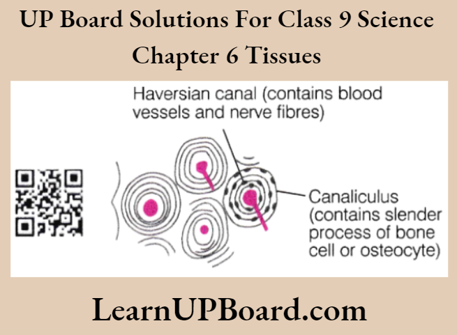
Connective Tissue Bone
- It is a very strong and non-flexible tissue. It is porous, highly vascular, mineralized, hard, and rigid. Its matrix is made up of proteins and is rich in salts of calcium and phosphorus.
- It forms the framework that supports the body. It also anchors the muscles and supports the main organs.
Ligaments: They connect one bone to another bone. A ligament is very elastic and has considerable strength. It contains very little matrix. Ligaments strengthen joints and permit normal movement. Their overstretching leads to sprain.
Tendons: They are strong and inelastic structures, which join skeletal muscles to bones. These are composed of white fibrous tissues with limited flexibility, but great strength.
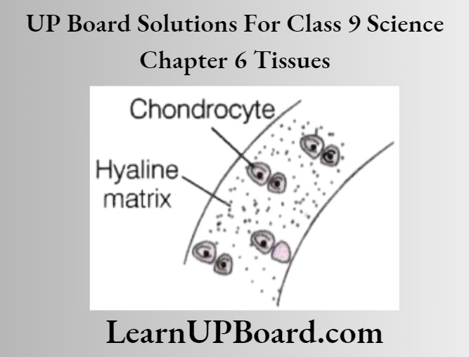
Cartilage: It is a specialized connective tissue having widely spaced cells. It has a solid matrix called chondrin which is composed of proteins and sugars.
Cartilage provides smoothness to the bone surfaces at the joints. It is present in the nose, ear, trachea, and larynx. We can fold the cartilage of the ears, but we cannot bend the bones in our arms.
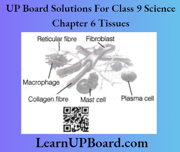
Areolar Tissue: It is a supporting and packing tissue found between the organs lying in the body cavity. It is located between skin and muscles, around blood vessels and nerves, and in the bone marrow.
It is a loose and cellular tissue. It fills the space inside the organs and supports internal organs. It helps in the repair of tissues.
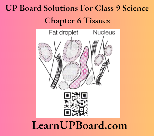
Adipose Tissue: It serves as a fat reservoir, and keeps visceral organs in position. It acts as an insulator due to the storage of fats. It is located below the skin in between the internal organs.
3. Muscular Tissue
It consists of elongated cells, called muscle fibers. This tissue is responsible for the movement in our body. It contains a special type of proteins called contractile proteins which causes the movement of muscles by contraction and relaxation. Different types of muscular tissues are given below:
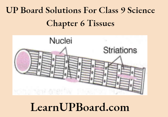
Striated Muscles
- The muscles present in our limbs which move or stop as per our will, are called striated muscles. These are also called as voluntary muscles as we can move them by conscious will. Mostly these are attached to bones and help in body movement, e.g, muscles of limbs.
- Hence, they are also called as skeletal muscles. The cells constituting their muscles are long, cylindrical, unbranched, and multinucleate (having many nuclei). Under microscope, striated muscles show alternate light and dark bands or striations.
Thus, they are also known as striated muscles.
Nuclei
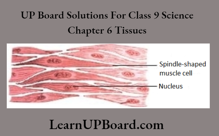
Unstriated Muscles Or Smooth Muscles
- The muscles which we cannot move as per our will are called unstriated or smooth muscles.
- They are also called involuntary muscles. For example, movement of food in the alimentary canal, contraction, and relaxation of blood vessels, iris of the eye, and muscles present in ureters and in bronchi of the lungs.
- The cells constituting these muscles are long, with pointed ends (spindle-shaped) and uninucleate (single nucleus).
- These muscles do not show any dark or light bands. Hence, they are also called unstriated muscles.
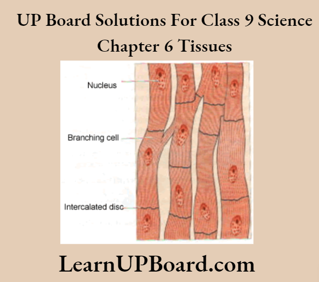
Cardiac Muscles: These are involuntary muscles present only in our heart. They perform rhythmic contraction and relaxation throughout life.
The cells constituting cardiac muscles are cylindrical, uninucleate, and branched. Cardiac muscles have stripes of light and dark bands.
4. Nervous Tissue
- The tissue that receives a stimulus and transmits it from one part of the tissue to another, are nervous tissue.
- The cells that constitute nervous tissue are called nerve cells or neurons. These are highly specialized for receiving a stimulus and then transmitting it very rapidly from one place to another within the body itself.
- The brain, spinal cord, and nerves are composed of nervous tissues.
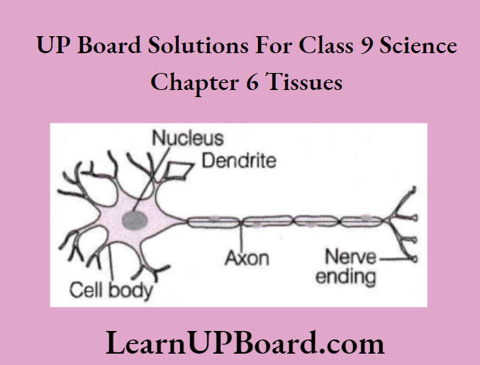
An individual nerve cell or a neuron may be upto a metre long and is composed of three major parts:
- Cell body It consists of cytoplasm, nucleus, and cell membrane.
- An axon is a single long conducting fiber extending from the neuron. It transmits impulses away from the cell body.
- Dendrites These are short-branched fibers of neurons, which receive nerve impulses. Nucleus
Note: Synapse is a region of the union of axons of one neuron with the dendrite of the next. This allows the transfer of nerve impulses generated to and fro in the body.
- Many nerve fibers bound together by connective tissue make up a nerve. Nerve impulse allows us to move our muscles according to our will.
- Combination of nerve and muscle tissue in animals is of fundamental importance as causes rapid movement in response to stimuli.
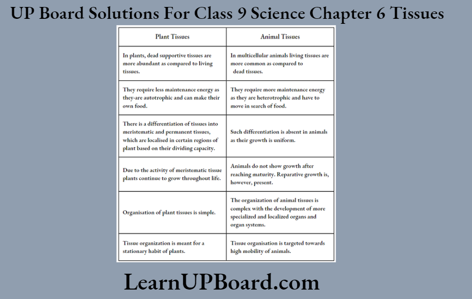
Differences Between Plant Tissues And Animal Tissues
UP Board Class 9 Science Notes For Chapter 6 Activity 1
Objective
Apical meristem causes growth in the length of the plant.
Time Required
Five days or a week.
Materials Required
Two jars of the same size, two onion bulbs, water, scissors, and a measuring scale.
Procedure
- Take two glass jars and fill them with water.
- Now, take two onion bulbs and place one in each jar.
- Observe the growth of roots in both the bulbs for a few days.
- Measure the length of roots on days 1, 2, and 3.
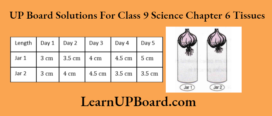
- On day 4, cut the root tips of the onion bulb in jar 2 by about 1 cm. After this, observe the growth of roots in both the jars and measure their lengths each day for five more days, and record the observations in a table.
Observation
Result/Conclusion
When root tips are removed from the onion of jar 2, it is observed that the growth of roots is more in the onion of jar 1, because apical meristem which is responsible for the increase in the length of roots is present only at the growing tips of roots. Thus, when we remove root tips, the apical meristem is also lost and the roots stop growing.
Question 1. What is meristematic tissue?
Answer: Meristematic tissues are those tissues that contain cells that are capable of dividing and forming new cells throughout their life.
Question 2. What will happen if the apical meristem is damaged or cut?
Answer: When apical meristem is damaged or cut, the growth or length of the growing tissue will be retarded.
Question 3. Why did the roots of the bulb of jar 2 stop growing after the fourth day?
Answer: Due to the removal of the apical portion of the roots in jar 2, the growth stops.
Question 4. What is the function of meristematic tissue?
Answer: It is responsible for increasing the length of the plant.
Question 5. Which of the two onions has longer roots? Why?
Answer: The onion in jar 1 has longer roots, as the growth of roots continues in it. This is because apical meristem which is responsible for the increase in the length of roots is present at the growing tips of roots.
UP Board Class 9 Science Notes For Chapter 6 Activity 2
Objective
To understand the structure, location, and arrangement of various types of cells of permanent tissue in plants.
Time Required
30-45 minutes.
Materials Required
A plant stem, blade, safranin, glycerine, coverslip, and a microscope.
Procedure
- Take a plant stem and with the help of your teacher, cut it into very thin slices or sections.
- Now, stain the slices with safranin. Place one needy cut section on a slide and put a drop of glycerine.
- Cover the cut section of the plant stem with a cover slip and observe the arrangement of cells under a microscope.
- Draw the figure you observed and label the parts.
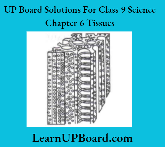
Observation
The section of the stem appears as drawn below:
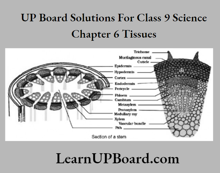
Result/Conclusion
The section of the stem depicts that various types of cells are arranged in specific ways. All cells are different in structure. The epidermis forms the outermost layer followed by the cortex. Vascular bundles are present encircled by the cortex.
Question 1. Which chemical is used to stain the plant sections in this activity?
Answer: Safranin is used to stain the plant sections in this activity.
Question 2. What is the role of glycerine in the above activity?
Answer:
Glycerine forms a protective covering on the plant section. This forms a barrier between the air and the plant section. Thus, the mount does not get dry and remains fresh for some time.
Question 3. Name the tissues that comprise vascular bundles.
Answer: The xylem and phloem comprise vascular bundles.
Question 4. What is the function of a vascular bundle?
Answer: The vascular bundle acts as a conducting tissue, i.e. they conduct water and food to all parts of the plant.
UP Board Class 9 Science Notes For Chapter 6 Activity 3
Objective
To understand the role of epidermis in plants. Or To understand the location and function of stomata.
Time Required
30-45 minutes.
Materials Required
Leaf of Rhoeo, petri dish, water, safranin, slides, coverslip, and microscope.
Procedure
- Take a freshly plucked leaf of the Rhoeo plant.
- Stretch and break it by applying pressure.
- While breaking it, keep it stretched gently so that some peel or skin projects out from the cut.
- Remove this peel and put it in a petri dish filled with water.
- Add a few drops of safranin to it.
- Wait for a couple of minutes and then transfer it onto a slide. Gently place a cover slip over it.
- Observe it under a microscope.
Observation
The cells appear as shown below:

Result/Conclusion
- The section shows the outermost layer called the epidermis. It is a single layer of cells with stomata embedded in it. It protects against water loss and mechanical injury.
- Stomata are small pores present here and there in the epidermis of the leaf. These are enclosed by two kidney-shaped cells called guard cells.
Question 1. Name the outermost layer of cells in old plants.
Answer: The outermost layer of cells in old plants is called epidermis.
Question 2. Name the cells that enclose the stomata.
Answer: Stomata are enclosed by two kidney-shaped cells called guard cells.
Question 3. State the location of stomata in plants.
Answer: Stomata are small pores present in the epidermis of the leaf.
UP Board Class 9 Science Notes For Chapter 6 Tissues Summary
Tissues are group of cells that are similar in structure and work together to achieve a particular function, for example., blood, phloem, and muscles.
- Tissues are broadly classified into plant and animal tissues.
- On the basis of dividing capacity, plant tissues can be classified into two fundamental types, i.e. meristematic tissue and permanent tissue.
- Meristematic tissues divide actively throughout life. They are found in growing regions of plants like root and shoot tips. These tissues are mainly of three types, i.e. apical meristem, intercalary meristem, and lateral meristem.
- Apical meristem are present at the growing tips of stems and roots. These are helpful in increasing the length of the stems and the roots. Intercalary meristems are present at the base of the leaves or internodes of the twigs.
- Lateral meristems are present on the lateral sides of stems and roots. It helps in increasing the girth of the stem or root.
- Permanent tissue is formed from the cells of meristematic tissue when they lose their ability to divide and have attained a permanent shape, size, and function by the process called differentiation. These are mainly of two types, i.e. simple and complex permanent tissue. “
- Parenchyma tissue forms the basic packing tissue. These tissues are present in the cortex, the pith of stem, roots and also in the mesophyll of leaves.
- Collenchyma cells are living, elongated, and irregularly thickened at the corners, generally found in leaf stocks below the epidermis. These provide mechanical support and elasticity to plant tissues.
- Sclerenchyma tissue is present in stems around vascular bundles, in veins of leaves, and in hard covering of seeds and nuts. These provide strength and enable the plant to bear various stresses.
- Complex permanent tissues are made up of more than one type of cell.
- The xylem is a vascular and mechanical conducting tissue. It is responsible for transport of food from roots to other parts of a plant.
- Phloem is a vascular tissue, responsible for transport of food from leaves to other parts of the plant.
- Animal tissues are classified on the basis of the functions they perform, i.e. epithelial, connective, muscular, and nervous tissue. Epithelial tissue is a protective tissue. It is tightly packed. It is present in the skin and lining of the mouth.
- Squamous epithelium cells are flat, it form the delicate lining of the esophagus and mouth. It may be several layers thick as in skin, known as stratified squamous epithelium.
- Columnar epithelium cells are tall, pillar-like, and have elongated nuclei. It is usually found in the inner lining of the intestine, where absorption and secretion occur.
- Ciliated columnar epithelium cells have cilia, hair-like projections found on the outer surface of columnar epithelial cells found in the trachea, bronchi, etc.
- Glandular epithelium cells acquire additional specialization known as gland cells that can secrete substances at the epithelial surface.
- Connective tissue connects various body organs, i.e. blood, bone, tendon, areolar, adipose, cartilage, etc.
- Muscular tissue consists of elongated cells and is responsible for movement. Striated muscles are mostly attached to bones and help in body movement. Unstriated muscles cannot be moved according to will. Cardiac muscles present in the heart, show rhythmic contraction and relaxation throughout life.
- Nervous tissue enables the body to respond to stimuli. They transmit stimuli from one place to another within the body, through neurons.
- Neuron forms the functional unit of nervous tissue.