
UP Board Notes for Class 10 Science Chapter 10 Nervous System Learning Objectives
After completing this chapter, you will be able to:
- State the functions of the nervous system;
- Describe the structure of a neuron, list its various types and state their specific functions;
- Describe the transmission of nerve impulses across a synapse,
- Categorize nervous sytem into central and peripheral nervous systems;
- List the parts of CNS and state their functions;
- Describe the structure of the brain and the spinal cord and explain their functions;
- Describe the structure and functions of peripheral nervous system;
- Differentiate between sympathetic and parasympathetic nervous systems.
| Class 10 Science | Class 11 Chemistry |
| Class 11 Chemistry | Transformation of Sentences |
| Class 8 Maths | Class 8 Science |
We perform many activities consciously or unconsciously, for example, when we eat food, our eyes help to locate the food, the nose smells it, hands bring the food to the mouth, teeth chew and masticate it, the tongue pushes the food inside the alimentary canal, and so on. All these activities occur in a coordinated manner. The organ system in our body, which brings about coordination and integration of body activities is the nervous system. In this chapter, you will learn about the nervous system in the human body.
UP Board Notes for Class 10 Science Chapter 10 Nervous System Major Divisions of Nervous System
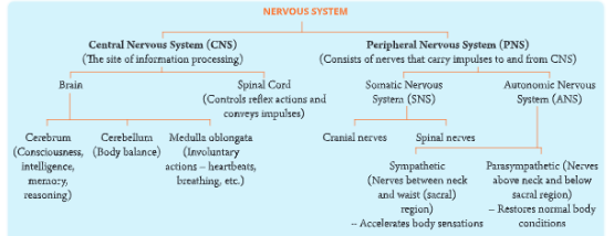
- The nervous system in human beings is divided into two main parts.
- Central nervous system (CNS): It includes the brain and the spinal cord, and is the site of information processing in the nervous system.
- Peripheral nervous system (PNS): It consists of nerves that emerge and enter the brain and spinal cord and run between the CNS and different parts of the body. These nerves are divided into three groups sensory or afferent nerves that transmit information to the CNS, motor or efferent nerves that carry messages from the CNS to the effector organ, and mixed nerves.
- The PNS is further divided into two sub-divisions:
- The somatic nervous system (SNS) regulates voluntary activities and transmits messages to the skeletal muscles.
- The autonomic nervous system (ANS) works independently to regulate involuntary activities. It consists of nerves and ganglia which connect the visceral organs like smooth muscles of heart, lungs, digestive tract and other internal organs and perform a variety of involuntary actions that are not under the control of our will.
- It has two components, viz. sympathetic and parasympathetic, which are antagonistic (opposite) to each other in their functions. You will learn about the autonomic nervous system in a later section.
- The broad organization of nervous system in humans is shown in Let us first learn about nerves and functioning of the nervous system before discussing about the two divisions of nervous system.
UP Board Notes for Class 10 Science Chapter 10 Nervous System Nervous System Nerves
- Nerves are composed of nerve fibers (axons) of separate neurons bundled together like the wires of a telephone cable enclosed by a tubular sheath. These nerve fibers or nerve cells are called neurons which form the basic unit of the nervous system.
- The Structure of Nervous system is shown below.
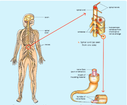
- A neuron is the longest cell in the body. It receives information and transmits it from one part of the body to another part. Thus, a neuron is the structural and functional unit of nervous system and is highly specialized for responding to stimuli.
- Messages are conducted by nerves in the form of electrical impulses.
UP Board Notes for Class 10 Science Chapter 10 Nervous System Types of Nerves
- There are three types of nerves depending upon the direction in which they transmit the impulse.
- Sensory nerves: These are also called afferent nerves (afferent: carry toward). These nerves contain sensory fibers which carry messages (impulses) from sensory receptors (in sense organs) toward the brain or spinal cord. Example: Optic nerve from eye leading to brain.
- Motor nerves: These are also called efferent nerves (referred: to carry away). The nerves contain motor fibers which take messages away from the brain or spinal cord towards the effector organ (such as muscles and glands). Example: Nerves arising in rain and leading to the muscles of the eyeballs for rotating it.
- Mixed nerves: These nerves comprise both sensory and motor nerve fibers. Example: Most cranial and spinal nerves are mixed nerves.
Ganglia (singular: ganglion)
These are the aggregate of nerve cells from where the nerve fibers arise.
UP Board Notes for Class 10 Science Chapter 10 Nervous System Structure Of A Nerve Cell (Neuron)
- Each nerve cell consists of three main parts.
- Cell body or cyton
- Dendrites
- Axon
1. Cell body:
The cell body or cyton (or Perikaryon) has a large, central nucleus surrounded by the granular cytoplasm. The cytoplasm contains (also called neoplasm) Nissl granules and neurofibrils. It has all the cell organelles (like mitochondria, Golgi apparatus, endoplasmic reticulum, microfilaments, and microtubules). There is no centrosome in the cyton because the nerve cells have lost the ability to divide.
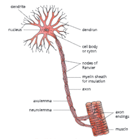
2. Dendrites (dendron: tree): These are short, thread-like branches which arise from the cell body. The dendrites conduct nerve impulses to the cyton.
3. Axon: One of the branches (of dendrites) grows very long in comparison to others. This branch is called the axon. The axon is covered on the outside by three layers.
- Axolemma (the innermost layer)
- Myelin sheath or medullary sheath (the middle layer)
- Neurolemma (the outermost white insulating sheath)
The axolemma and neurotrauma are continuous sheaths, whereas the myelin sheath is not a continuous one. The myelin sheath shows gaps across its length at intervals. These gaps are known as nodes of Ranvier.
The axon ends have swollen bulb-like ends which store acetylcholine (a neurotransmitter). These are called axon endings. Axon endings are closely placed near the dendrites of another neuron but are not connected. Such gaps in between are called synaptic clefts or synapses.
Synapse
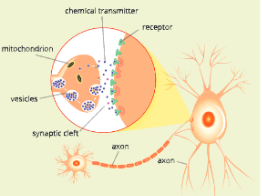
- The neurons are not attached to each other. There is a small gap between them. The fine gap or loose connection between the axon endings of one nerve cell and cyton or dendrite of the next nerve cell is called the synapse. We can also say that the point of contact between two neurons is known as synapse. Signals travel from one neuron to another neuron across these junctions.
Structure of the synapse
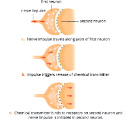
The neurons do not touch at the synapse. The space between adjacent neurons is called the synaptic cleft.
- The axon terminal of a presynaptic neuron has a bulb-like appearance known as a synaptic bulb The transmission.
across synapses is done by chemical means. The synaptic bulb contains chemicals known as neurotransmitters. - When a nerve impulse (action potential) arises at the axon terminal, it causes synaptic bulbs to release neurotransmitters in the synaptic cleft. chemical transmitter
- These neurotransmitters diffuse easily across the synaptic cleft. In this way, the impulse is transmitted from one nerve cell to another nerve cell.
- There are many neurotransmitters but the principal ones are acetylcholine and norepinephrine.
What does a synapse do?
- Allows information to pass from one neuron to another.
- Ensures that the nerve impulse travels in one direction only.
- Allows the adjoining neuron to be excited or inhibited.
- Amplifies a signal (makes it stronger).
- Helps in information processing by adding together the effects of all impulses received.
- Filters out low-leveled stimuli.
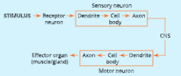
There are three types of neurons:
- Sensory neurons: These carry impulses from sense organs to the brain or spinal cord.
- Motor neurons: These carry impulses from brain or spinal cord to effector organs (muscle or gland).
- Association neurons: These interconnect sensory and motor neurons.
UP Board Notes for Class 10 Science Chapter 10 Nervous System Communication Through The Nerve – Nerve Impulse
The nervous system receives a stimulus through a receptor organ, integrates or coordinates it, and effects a response through the effector organ. Thus, coordinated behavior has six main components – stimulus, receptor, impulse, coordinator, effector, and response.
- In such a coordinated behavior, any stimulus of sound, sight, smell, etc., is perceived by receptor organs like eyes, ears, skin, etc.
- A stimulus is an agent or sudden (external or internal) change that results in a change in the activities of an organism.
- An impulse is a wave of chemical disturbance that travels through the nerve cell.
- Receptors are sensory organs that receive stimulus and send waves in form of impulses towards CNS (coordinator).
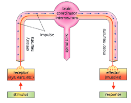
- Effectors are the muscles or organs which show response due to motor nerves. Response is a change that occurs in an organism due to a stimulus.
- The brain and spinal cord are the coordinators that receive information in the form of messages called nerve impulses, from receptor organs via neurons. The information flows to the effector organs, i.e. muscles, which contract or relax or secrete substances to show response.
Transmission of the nerve impulses
- Nerve impulses pass along a neuron in one direction only. At one end, the neuron is connected to a sensory receptor that receives the stimulus and converts it into electrochemical waves which are carried by the neuron. The nerve fiber at this stage is said to be excited.
- The events that take place during the conduction of an impulse along a nerve are given as follows.
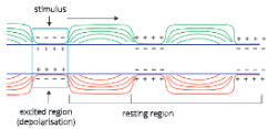
UP Board Notes for Class 10 Science Chapter 10 Nervous System At Resting State – Polarised state
At normal (resting) state, the outer side of the nerve fibers carries more positive (+) charge due to more Na* ions outside the axon membrane. This is called a polarised state.
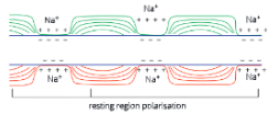
UP Board Notes for Class 10 Science Chapter 10 Nervous System At Stimulated (excited) state – Depolarisation
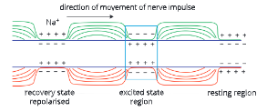
- On receipt of a stimulus, the axon membrane at the place of stimulus becomes more permeable to Na+ ions and as a result, the Na* moves inside causing loss of polarity, i.e. depolarization. This region, thus, becomes an exciting region. This region of depolarisation moves forward to next area which in turn becomes depolarised.
UP Board Notes for Class 10 Science Chapter 10 Nervous System Returning To Normal State – Repolarisation
- The previous area (which has received stimulus) becomes repolarised due to active transport of Nat ions outside. This transport is achieved by sodium pump for which energy in the form of ATP is required. Thus, conduction of nerve impulses is a wave of depolarisation followed by repolarisation.
UP Board Notes for Class 10 Science Chapter 10 Nervous System Central Nervous System
- The central nervous system consists of the brain and The spinal cord.
The human brain
- The human brain is a highly developed organ and is situated in the cranium (brain box) of the skull. In an adult, it weighs about 1200-1400 g, about 2% of body weight.
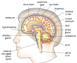
UP Board Notes for Class 10 Science Chapter 10 Nervous System Covering Of Brain (Meninges)
- The brain is covered on the outside by membranes called meninges (singular: menix ). Meninges are protective coverings of the brain which consist of three layers.
Meningitis
The inflammation (swelling) of the meninges is known as meningitis.
- The outer tough, protective layer dura mater is formed of fibrous tissue.
- The middle arachnoid layer is a delicate membrane which gives a web-like cushion.
- The inner thin, transparent, and highly vascular layer the pia mater. It is richly supplied by blood.
The cerebrospinal fluid
- It is the watery fluid which fills the spaces between the meninges and also brain cavities or the ventricles. It also acts like a cushion to protect brain from shocks.
Parts of the brain
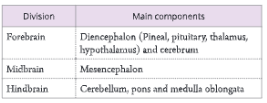
- The human brain is divisible into three major parts
- Forebrain
- Midbrain
- Hindbrain
UP Board Notes for Class 10 Science Chapter 10 Nervous System Forebrain
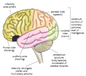
- It is the anterior region of the brain. It has the following parts:
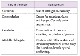
- Cerebrum (seat of intelligence, memory, consciousness, and voluntary action)
- Diencephalon (thalamus, hypothalamus, pineal and pituitary glands)
1. Cerebrum (L. cerebrum: brain)
The cerebrum is the main part of forebrain.
- Cerebrum is the largest and the most prominent part of the brain. It is divided into two halves – the right and left cerebral hemispheres.
- The two hemispheres are connected by a thick band of nerve fibers called corpus callosum. Corpus callosum helps in the transfer of information from one hemisphere to another.
- Each hemisphere is hollow internally and its walls have two regions an outer cortex and an inner medulla. The outer portion or region of the cerebrum (cerebral cortex) contains cell bodies of neurons and is called grey matter (due to its greyish in color).
- The layer of grey matter is highly convoluted in appearance with ridges and grooves. The fold of ridges are called gyri (singular: gyrus) and depressions (grooves) between them are called sulci (singular: sulcus). The ridges and grooves increase the surface area for more nerve cells.
- The number and pattern of convolutions in the cerebrum are associated with the degree of intelligence.
- The inner region of the cerebrum consists of white matter that has axons of nerve cells.
UP Board Notes for Class 10 Science Chapter 10 Nervous System Functions Of Cerebrum
- Because of highly developed grey matter the cerebrum governs mental abilities like thinking, reasoning, learning, memory, and intelligence.
- It also controls all voluntary functions; willpower, emotions, and speech.
- It enables us to observe things around us through sense organs.
- It also controls feelings of love, admiration, and hatred. Centers for subconscious mind are also located in the cerebrum. Overall the cerebrum is the seat of intelligence, memory, and willpower.
2. Diencephalon
- The diencephalon mainly consists of the pineal gland, pituitary gland, thalamus, and hypothalamus. It encloses a cavity called the third ventricle. The thalamus is a relay station for sensory impulses going to the cerebrum. The hypothalamus is situated at the floor of the brain and helps in thermoregulation.
UP Board Notes for Class 10 Science Chapter 10 Nervous System Functions Of Diencephalon
- The diencephalon contains reflex centers for muscular and glandular activities. It also has centers of emotions, hunger, and thirst. It also helps in controlling the body temperature (thermoregulation) and water-salt balance in the body (osmoregulation).
UP Board Notes for Class 10 Science Chapter 10 Nervous System Midbrain
- It is a thick-walled structure and is a smaller portion of the brain. The midbrain or mesencephalon connects the anterior region of the brain to the posterior region and therefore all nerve fibers pass through this region. On the dorsal side of the midbrain lie optic lobes which control vision.
UP Board Notes for Class 10 Science Chapter 10 Nervous System Hindbrain
- The hindbrain has three main parts.
- Cerebellum
- Pons
- Medulla oblongata
1. Cerebellum (Little brain)
- The cerebellum is situated in the dorsal region. of the hindbrain. Cerebellum is a much smaller area and is located at the base under the large cerebrum.
- There are no convolutions, but many furrows.
- It has an outer cortex made of grey matter and an inner section consisting of white matter.
UP Board Notes for Class 10 Science Chapter 10 Nervous System Functions Of Cerebellum
- It maintains body balance and controls muscular activities. It makes the body movements smooth, steady, and coordinated. It regulates and coordinates contraction of skeletal muscles.
2. Pons
- Pons forms the part of the brain stem at the floor of the hindbrain. It is a bridge of transverse nerve tracts extending from the cerebrum to the cerebellum. It also connects the forebrain to the spinal cord.
3. Medulla oblongata
- The medulla oblongata is the third main part of the hindbrain. It is the lowermost part of the brain located at the base of the skull. It is continued as spinal cord in posterior region.
UP Board Notes for Class 10 Science Chapter 10 Nervous System Functions Of Medulla Oblongata
- It contains vital reflex centers, such as cardiac center, respiratory center, and centers for swallowing,\ sneezing, and coughing. Thus, it controls involuntary functions of the body like a heartbeat, swallowing, and breathing.
- Injury to medulla oblongata may lead to death
Medulla oblongata is main part of the hindbrain. It controls vital reflex centers and Involuntary functions like heartbeat, breathing, and swallowing of food. Any Injury to medulla oblongata will affect these functions and may cause death due to stopping of the heartbeat and breathing.
UP Board Notes for Class 10 Science Chapter 10 Nervous System Nervous System Spinal Cord
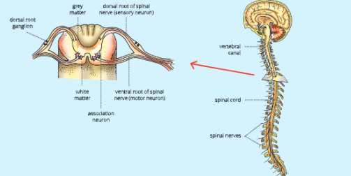
- The spinal cord is a cylindrical, long cord which arises from the medulla oblongata and runs along the vertebral column. It passes through the neural canal of the vertebral column. Like the brain, it is also protected by the three meninges, cerebrospinal fluid, and a cushion of adipose tissue.
- In the transverse section of spinal cord, a central canal can be seen. This canal is filled with cerebrospinal fluid. Immediately surrounding the canal are clusters of cytons which form the grey matter. In the peripheral part, axons are concentrated and this area is called the white matter.
- On each side of the spinal cord, there are two horns, the dorsal horn, and the ventral horn. The dorsal root ganglion contains the cell bodies of sensory neurons. A nerve joined to the dorsal horn or dorsal root ganglion picks up sensations from various organs. It is called the sensory nerve.
- The ventral root contains the axons of large motor neurons. The cell bodies of these motor neurons are located in the spinal cord. Thus, from the ventral horn or ventral root, the motor nerve arises which takes the messages from the spinal cord to the organs concerned.
UP Board Notes for Class 10 Science Chapter 10 Nervous System Functions Of The Spinal Cord
- It is the center for reflex actions. The spinal cord conducts reflexes below the neck.
- It conducts sensory impulses from skin and muscles to the brain.
- It conducts motor responses from brain to the muscles of trunk and limbs.
UP Board Notes for Class 10 Science Chapter 10 Nervous System Reflex Arc And Reflex Action
- There are certain body responses that are immediate and do not require any processing by the brain. These responses or actions are controlled by the spinal cord. These are called reflex actions.
- A reflex action may be defined as a spontaneous, automatic, and mechanical response to a stimulus controlled by the spinal cord without the involvement of the brain.
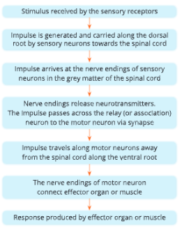
- The pathway followed by sensory and motor nerves in a reflex action is called the reflex arc.
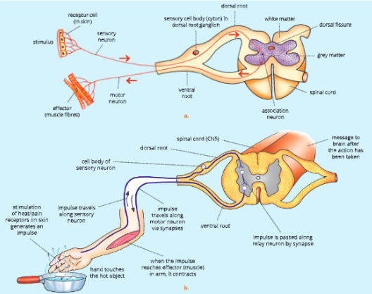
Components of a reflex arc
- A reflex arc has four main components.
- Receptor or sensory organ to perceive the stimulus.
- Sensory or afferent nerve that carries the message from receptor to the spinal cord.
- Relay or association neurons of the spinal cord that transmit impulses from the afferent (sensory) neurons to the efferent (motor) neurons.
- Motor or efferent nerve that carries the message from the spinal cord to the muscles or glands (effector organ).
- The entire sequence of events that constitute a reflex arc are summarized in.
UP Board Notes for Class 10 Science Chapter 10 Nervous System Nervous System Reflexes
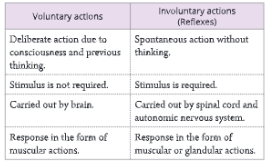
- There are mainly two types of actions – voluntary actions and involuntary actions.
- Voluntary actions are those actions which are performed consciously. For example, if you wish to play, or switch on TV to watch some programs are voluntary actions.
- Involuntary actions are those actions that occur unknowingly without our will. For example, you withdraw your hand when it accidentally touches a hot iron; or you start shivering if it is too cold or you start sweating when it is too hot. All involuntary actions are reflexes and involve some kind of sensory stimulation. Differences between voluntary and involuntary actions are given in Table.
UP Board Notes for Class 10 Science Chapter 10 Nervous System Types Of Reflexes
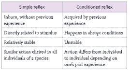
- There are two types of reflexes:
- Simple or natural reflexes
- Conditioned or acquired reflexes
1. Simple or natural reflexes are those reflexes that do not require any previous learning experience. Such reflexes are inborn and inherited from parents.
Some examples of simple reflexes are:
- Blinking of eyelids in response to a foreign particle that approaches the eye.
- Closing of the eyelid in response to a strong beam of light being flashed on the eyes.
- Withdrawal of the leg, if you suddenly step on a nail.
- Knee-jerk response, if a sharp tap is made below the kneecap, then the leg is involuntarily extended.
- Sneezing when any irritant enters the nose.
- Coughing when the swallowed food wrongly enters the windpipe. Immediate withdrawal of hand, if suddenly pricked by a thorn or after touching a hot object.
- Peristaltic reflex to allow movement of food (chyme) when intestine becomes full and distended.
2. Conditioned or acquired reflexes are those which develop due to some previous experience or training. The conditioned reflexes are not inborn and result due to some learning in one’s lifetime.
Some examples of conditioned reflexes are:
- Watering of mouth (salivation) at the sight of tasty food.
- Applying sudden brakes of your bicycle if someone suddenly comes in front.
- Typing on the keyboard of a computer.
- Playing a musical instrument such as guitar.
- Surfing the channels while watching the television.
- Tying one’s shoelace.
- The hand signal automatically shows direction to turn the cycle without thinking.
UP Board Notes for Class 10 Science Chapter 10 Nervous System Peripheral Nervous System
- The peripheral nervous system (PNS) comprises the nerves that connect the central nervous system with different parts of the body.
- Peripheral nervous system is divided into two subdivisions- somatic nervous system and autonomic nervous system.
1. Somatic nervous system
- The somatic nervous system includes both motor neurons and sensory neurons. The fibres of motor and sensory neurons are bundled together into nerves, which are of two types.
- Cranial nerves connected directly to the brain, such as the optic nerve (for eye), auditory nerve (for ears), mixed nerves (for face), etc. There are 12 pairs of cranial nerves.
- Spinal nerves emerge from the spinal cord. There are 31 pairs of spinal nerves. Every spinal nerve is a mixed nerve having both sensory and motor nerves.
- The somatic nervous system regulates voluntary activities while the autonomic nervous system performs a variety of functions that are not under the control of an individual.
2. Autonomic nervous system
The autonomic nervous system (ANS) includes a chain of 22 pairs of ganglia that lie close to the spinal cord and are associated with the organs they control. ANS is primarily a motor system consisting of neurons that control the functioning of many organs.
- Heart muscles
- Glands
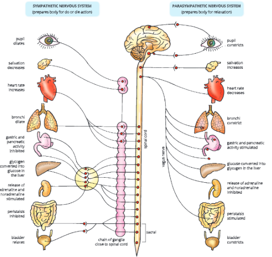
- Smooth muscles (muscles of blood vessels, digestive, respiratory, and reproductive tracts) The ANS can stimulate or inhibit the activity of its target organs. The ANS is divided into two divisions -sympathetic and parasympathetic nervous systems.
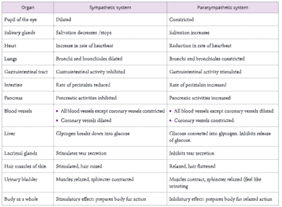
- These two divisions are anatomically and functionally distinct. The sympathetic fibers arise from the thoracic (chest) and lumbar (waist) region of the spinal cord, whereas the parasympathetic fibers arise from the brain and the sacral (pelvic) region of the spinal cord.
- The effect of the two systems is antagonistic. In general, the sympathetic system stimulates a particular function and prepares the body for violent actions against unusual emergency conditions, while the parasympathetic system has an inhibitory or calming down effect, i.e. it re-establishes normal conditions after the violent action is over. The major differences between sympathetic and parasympathetic nervous systems are given in Table.
UP Board Notes for Class 10 Science Chapter 10 Nervous System Nervous System Summary
- The nervous system in humans is divided into two parts: central nervous system and peripheral nervous system.
- The central nervous system includes the brain and the spinal cord and is the site of information processing in the nervous system.
- The peripheral nervous system consists of nerves that run between central nervous system and different parts of the body.
- The structural and functional unit of the nervous system is a highly specialized cell called a nerve cell or neuron. Each neuron has three principal parts – the cell body or cyton, the axon, and dendrites.
- Nerves can be sensory, motor, or mixed.
- The nervous system receives a stimulus through a receptor organ, integrates or coordinates it, and effects a response through the effector organ.
- The human brain has three main divisions the forebrain comprising of cerebrum and diencephalon, the midbrain, and the hindbrain comprising of the cerebellum, pons, and medulla oblongata.
- The cerebrum is the largest and the most prominent part of the brain. It has ridges and grooves (girl and sulci) which increase surface area for nerve cells. The outer cortex of cerebrum contains grey matter. Cerebrum governs mental abilities like thinking, reasoning, learning, memorizing, intelligence, will, and emotions. lotions.
- Cerebellum is the smaller part located at the base under the large cerebrum. It maintains body balance and controls postures and coordinates muscular activities.
- Medulla oblongata is the lowermost part of the brain located at the base of the skull. It contains vital reflex centers and controls the activities of the internal organs.
- The spinal cord is a long cord that arises from the medulla oblongata and runs along the vertebral column.
- Reflex action is a spontaneous, automatic, and mechanical response to a stimulus controlled by the spinal cord without the involvement of the brain. Sympathetic and parasympathetic nervous systems are two divisions of autonomic nervous system and these are antagonistic (opposite to each other) in their functions.