
UP Board Notes for Class 10 Science Chapter 11 Sense Organs The Eye And The Sense Of Sight
Sense Organs Learning Objectives
After completing this chapter, you will be able to:
- Define a receptor and name various kinds of receptors;
- Describe the structure and functions of the eye;
- Trace the pathway of light through the eye to the retina and explain how light is focused for distant and near vision;
- Explain the causes and consequences of various defects of the eye and methods of their correction;
- Describe the structure and general functions of the outer, middle, and Inner ears;
- Explain how the ear serves as an organ for dynamic and static equilibrium.
| Class 10 Science | Class 11 Chemistry |
| Class 11 Chemistry | Transformation of Sentences |
| Class 8 Maths | Class 8 Science |
The eyes, ears, tongue, nose, and skin are the major sense organs in our body, which are sensitive to the sense of light, sound, taste, smell, and touch, respectively. Each of these sense organs is directly connected to the brain. These sense organs have special cells called receptors which receive stimuli from the environment.
UP Board Notes for Class 10 Science Chapter 11 Sense Organs The Eye And The Sense Of Sight Sense Organs Some Key Terms
Stimulus:
A physical event that affects an organism by activating its receptors to bring about a change in its activity. For example, mechanical stimuli (pressure and touch), chemical stimuli (taste and smell), and thermal stimuli (heat and cold).
Sensation:
A general state of awareness of the stimulus.
Receptors:
Sensory cells in tissues that receive stimulus from the environment. Some common specific receptors are:
- Photoreceptors: respond to light rods and cone cells in the retina of the eye.
- Chemoreceptors: respond to chemicals – taste buds on the tongue, smell receptors of the nose.
- Mechanoreceptors: respond to touch – nerve fibers around the hair on the skin.
- Thermoreceptors: respond to changes in temperature – receptors on the skin.
- Photoreceptors: respond to sound/hearing – receptors in the ear.
- Exteroceptors: specialized to detect sensory information from the external environment.
We all respond to the light stimulus. Human beings have two eyes situated in deep sockets or orbits on the front side of the head. Each eye is in the form of a ball called an eyeball that measures about 2.5 cm (1 inch) in diameter. Each eyeball can be rotated with the help of six distinct sets of muscles.
UP Board Notes for Class 10 Science Chapter 11 Sense Organs – The Eye And The Sense Of Sight Accessory Structures Of The Eye
Eyebrows, eyelids, eyelashes, and the tear glands (the lacrimal apparatus) are the accessory structures of the eye.
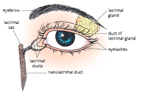
- The eyebrows protect the eyeball from foreign objects, perspiration, and direct rays of sunlight.
Eyelids are folds of skin and muscles. The upper and lower eyelids protect the eyes from excessive light and foreign particles, cover the eye during sleep and spread lubricating secretions over the eyeballs. The upper eyelid is more movable than the lower one. Projecting from the border of each eyelid is a row of thick, short hairs called eyelashes. At the base of the hair follicles of eyelashes, sebaceous glands are found. These glands secrete a lubricating fluid into the hair follicles called sebum. - Tear glands or lacrimal glands (collectively known as lacrimal apparatus) are a group of glands that manufacture and pour tears (L. lacrima: tears). A lacrimal gland is a compound gland located at the upper sideward portion of each eyelid
Each lacrimal gland gives rise to 6 to 12 excretory lacrimal ducts that empty their secretion (tears) onto the surface of the conjunctiva of the upper eyelid. From here it passes to the lacrimal canals and then into the lacrimal sac. From here a - Nasolacrimal duct conducts the secretion into the nasal cavity in the nose. This duct is responsible for the passage of medicine dropped in the eye to the nose or sometimes into the throat.
- Tear or lacrimal secretion is a watery fluid containing salts, some mucus, and a bacteriocidal enzyme called lysozyme.
Functions of tears:
- After being secreted, it spreads over the surface of the eyeball and serves as a lubricant.
- It cleans the front surface of the eyeball by washing away the dust particles.
- It moistens the eyeball.
- The enzyme lysozyme contained in the tears kills the germs.
- Tears help in communicating emotions.
UP Board Notes for Class 10 Science Chapter 11 Sense Organs – The Eye And The Sense Of Sight Structure Of The Eyeball
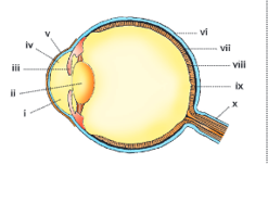
- The eyeball is a spherical structure, measuring about 2.5 cm in diameter. Structurally, the eyeball is composed of three layers – the fibrous tunic (anterior cornea + posterior sclera), the vascular tunic also known as uvea (middle layer of the eyeball) (choroid + ciliary body + iris), and the inner retina.
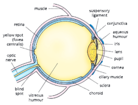
- Sclerotic layer or sclera
- The sclerotic layer or sclera is the outer tough coat of the eyeball made up of mainly collagen fibers. It can be divided into two regions – the posterior region called the sclera, and the anterior region called the cornea.
- The sclera is a white coat of fibrous tissue visible through the conjunctiva, which covers all the eyeballs except the cornea. It is also known as the white of the eye. The sclera gives shape to the eyeball and also protects its inner parts.
- The cornea is a transparent, fibrous coat through which the iris can be seen. The outer surface of the cornea is covered by an epithelial layer which is continuous with the epithelial layer of the conjunctiva. The cornea receives nourishment from tears and aqueous humor.
- Sometimes, the cornea turns white and opaque and can be replaced by a donated eye.
- Uvea
- The choroid layer is the middle layer of the eyeball and is composed of three parts – choroid, ciliary body, and iris.
- The choroid is a thin, dark brown membrane that lines most of the inner surface of the sclera. It contains several blood vessels and a large amount of pigment melanin. The choroid absorbs light rays so that they are not reflected within the eyeball. The numerous blood vessels nourish the retina.
- The choroid expands to form the ciliary body. It consists of circular muscles. The smooth muscles of the ciliary body help in changing the shape of the lens.
- The iris (arid: rainbow) is a clouded part of the choroid around the pupil. It is a clouded part seen through the cornea. There is a hole (round window) in the canter of the iris, known as the pupil. The light enters the eyeball through the pupil. The iris contains radial and circular muscles.
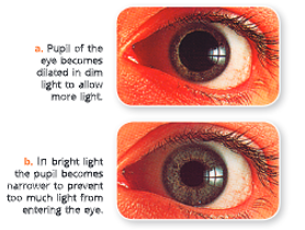
The contraction of these muscles constricts the pupil which regulates the amount of light entering the eyeball through the iris
- In the case of bright light, the circular eye muscles contract, and the size of the pupil is decreased (constriction).
- In case of dim light, the radial muscles contract, and the size of the pupil is increased (dilation).
- Sense Organs Retina – part of the eye where rod and cone cells are located
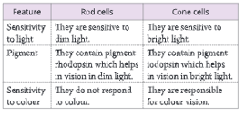
- The retina is the third and the inner layer of the eye. It is located only in the posterior part of the eye. It is the light-sensitive layer. It contains light-sensitive cells called rods and cones.
- The rod cells are sensitive to dim light. They do not respond to color. Rods contain the pigment rhodopsin or visual purple. The rod cells are distributed throughout the retina.
- The cone cells are sensitive to bright light. The cones are responsible for color vision. Cones contain the pigment rodeos in. Cone cells are mostly confined to the yellow spot or fovea centralis or macula.The differences between rod cells and cone cells are given in Table
Yellow spot – the area of the best vision
The yellow spot or macula lutea (Fovea centralis) is a spot located inside the eyeball near the Centre of the retina. It contains a maximum number of light-sensitive cells, especially cone cells. The rest of the retina has lesser cone cells and more rod cells. The yellow spot is the region of color vision and also the brightest, clearest and best vision.
Blind spot – the area of no vision
A blind spot is the region of the retina just below the yellow spot. Since there are no light receptors here, no image is formed here, i.e. blind spot is the area of no vision.

clockwise through 90° and hold it at an arm’s length with the two symbols straight in front of your eyes. Close your left eye and concentrate on the cross with your right eye. Bring the book slowly towards you. Keep looking at the cross on the left: eventually, as the dot falls on the blind spot of your right retina, the image of the dot on the right will disappear.
UP Board Notes for Class 10 Science Chapter 11 Sense Organs – The Eye And The Sense Of Sight Sense Organs Lens
- The main body of the eye is divided into two parts by a biconvex lens which is a transparent crystalline body. The lens lies just behind the pupil and iris. It is flatter at the front than at the back and is soft and slightly yellow in color. It contains transparent lens fibers and an elastic lens capsule made of glycoprotein. There is no blood vessel in the lens.
- The lens is held in position by the suspensory ligaments which attach it to the ciliary body.
The eyeball is divided into two chambers:
- the anterior chamber (aqueous chamber) and
- posterior chamber (vitreous chamber).
- The anterior chamber (between the lens and cornea) is filled with a fluid called aqueous humor. It is a thin and watery fluid. It keeps the lens moist and protects it from physical shock.
- The posterior chamber is called the vitreous chamber. It is a large cavity of the eyeball. It lies between the lens and the retina. It contains a jelly-like substance called vitreous humor. The vitreous humor maintains the shape of the eyeball by preventing the eyeball from collapsing and supporting the retina.
UP Board Notes for Class 10 Science Chapter 11 Sense Organs – The Eye And The Sense Of Sight How Do We See – Working Of The Eye
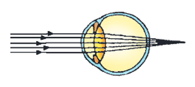
- Light rays enter the eye
The light rays enter the eye through the cornea and then pass through the pupil, aqueous humor, the lens, and the vitreous humor (all transparent structures), before reaching the retina.
The pupil adjusts to different intensities of light. In bright light, the pupil constricts (becomes narrow) and in dim light, it dilates (becomes wider) to allow more light.
- Image formation on the retina
The formation of an image on the retina requires four steps – a. refraction of light rays, b. constriction of the pupil, c. accommodation of the lens, and d. convergence of the rays.
- Refraction of light rays: The eye focuses an image by refracting, or bending the light rays using the cornea and the lens. An upside-down or inverted image is formed at the yellow spot on the retina. Most of the refraction of light occurs in the cornea due to its curved surface.
- Constriction of the pupil: The iris contains radial and circular muscles. Radial muscle widens and circular muscle constricts the pupil. This adjustment of the size of the pupil regulates the amount of light entering the eye.
- Accommodation of the lens: The ability of the eye to focus on different objects placed at a distance by adjusting the curvature of the elastic eye lens is called accommodation. This is brought about by ciliary muscles which change the focal length of the eye lens to focus on near or distant objects on the retina.
(1) In the distant vision (more than 6 meters), the ciliary muscles are relaxed and the lens becomes flatter or thinner due to the stretching of the suspensory ligaments.
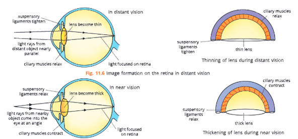
(2) In the near vision (less than 6 meters), the ciliary muscles contract pulling the choroid forward. towards the lens and tension is released on the suspensory ligaments. The lens becomes shortened, more convex, and thickened due to its elastic nature
In normal conditions, the ciliary muscles are relaxed and the lens remains stretched by the suspensory ligaments. Then, it is less convex and best suited for distant vision.
4. Convergence of the rays: We have two eyes
but we see only one image of an object. This is known as binocular vision or stereoscopic vision. While viewing objects, both eyeballs move toward the object. Thus, both images fall on the corresponding points (fovea centralis) of both retinas at the same time and overlap with each other. This is known as the convergence of the rays.
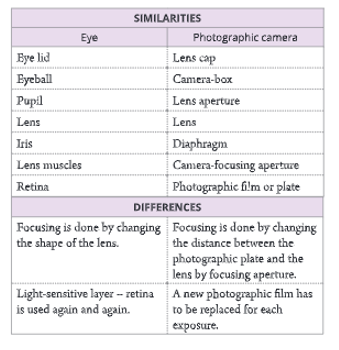
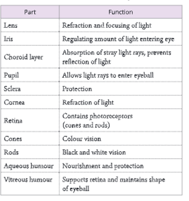
The eye can be compared with a camera. The major similarities and differences between the eye and the camera are given. A summary of various parts and functions of the eyes is given in Table
- Photoreception by brain
The image formed at the retina stimulates photoreceptors. The light energy of the image formed at the retina produces chemical changes in the rods and cones. This generates nerve impulses that travel through the optic nerve from the retina to the visual area of the brain. At the cerebrum (brain), the sensation of sight is interpreted.
How are the eyes adapted to bright light and dark?
- When we are in dark or dim light for a long period, then the rate of photopigment (rhodopsin) formation by rod cells in the retina is much faster than the rate at which they are broken down. The pupil dilates to allow more light to enter through the retina to allow viewing of objects in dim light. This is called dark adaptation.
- When we are in a very bright light for a long period of time, the light-sensitive pigments of the rods are broken down, and the visual purple of the rods gets bleached, reducing their sensitivity. The cone cells in the retina are activated and it forms iodopsin pigment. The further pupil constricts and the size of the pupil is reduced to reduce the amount of light entering the eyes. This is called light adaptation.
Colour vision
- color vision is possible because of cone cells. When all the cone cells are stimulated, we see white color. When very few cone cells are stimulated, we see black color. Stimulation of separate types of cone cells produces red, green, or blue and their combinations.
Why cannot we distinguish color on a moonlit night?
- In dim light, like that during a moonlit night, colors cannot be distinguished because cone cells that detect colors do not work in dim light.
UP Board Notes for Class 10 Science Chapter 11 Sense Organs – The Eye And The Sense Of Sight Common Defects Of The Eye And Their Correction
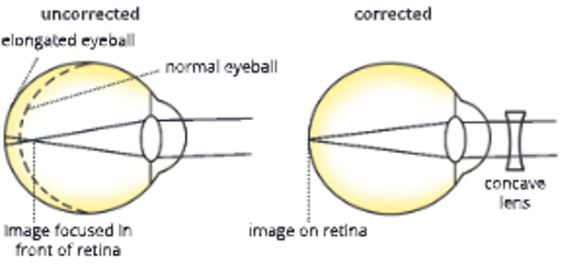
Short-sightedness or Myopia
- In this condition, light is focused in front of the retina, and a blurred image is formed for distant objects. In myopia, the near objects are seen clearly while the distant objects appear blurred. This happens because either the eyeball is elongated or the lens has become too thickened or curved
Long-sightedness or Hyperopia
- In this condition, the light is focused behind the retina (the image is formed behind the retina). As a result, the distant objects are seen clearly, while the near objects appear blurred. Long-sightedness results due to the shortening of the eyeball or the lens have become. too thin
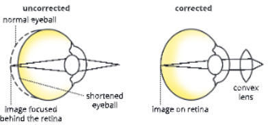
Hyperopia (Hypermetropia) can be corrected by using a convex (converging) lens.
Astigmatism
- This is a more complicated defect in vision. In this, the surface of the cornea becomes irregular and therefore some of the light rays are focused while others are not. As a result, some parts of the object appear blurred while other parts appear clear
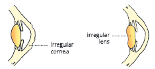
Astigmatism can be corrected by using a cylindrical lens that bends light rays in one direction only.
Presbyopia
- Astigmatism can be corrected by using a cylindrical lens that bends light rays in one direction only. Presbyopia In this condition, the lens loses its flexibility causing long-sightedness (the near objects cannot be seen clearly). This defect normally occurs in older people.
Presbyopia can be corrected by using a convex lens.
Night-blindness
- In this condition, there is difficulty in seeing in dim light (hence the name night blindness). This is because of the non-production of rhodopsin pigment in the rod cells, which function in dim light. In the absence of rhodopsin, these cells cannot function. Thus, there is a lack of normal night vision. It is most often caused due to deficiency of vitamin A.
Night blindness can be cured by having Vitamin A rich diet.
Colour blindness
- In this condition, a person is unable to discriminate between red and green colors. This is a genetic defect which you have already studied in the chapter on heredity and genetics (X-linked inheritance).
Colour blindness cannot be treated during the lifetime of an individual.
Cataract
- In this condition, the lens of the eye becomes opaque and as a result, the vision is cut down. Cataracts can be treated by surgical removal of lenses and using convex lenses in the spectacles which compensate for the removed lens. Nowadays, a small plastic lens is implanted behind the iris to correct the defect.
Squint
- In this condition, either the two eyes somewhat converge (known as cross-eye) or diverge (known as wide-eye). In both these conditions, a person may have double vision. Squint in the eyes can be treated by surgery or by suitable exercises.
UP Board Notes for Class 10 Science Chapter 11 Sense Organs – The Eye And The Sense Of Sight The Ear – Senses Of Hearing And Balance
- Humans have two ears, one on each side of the head. Human ears are organs for the senses of hearing and balance. The ear is a miniature receiver, amplifier, and signal processing system.
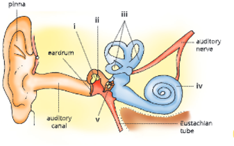
Structure of ear
The human ear is divided into three parts
- External ear
- Middle ear
- Inner ear
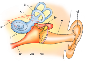
- The external or outer ear
It consists of the externally visible part of the ear called the pinna (or by a thin membrane. auricle) and the internal part, the auditory canal. The auditory canal. the passage leading to the eardrum (also known as the tympanic membrane).
Hair and earwax in the ears
The auditory canal contains fine hairs, which filter the air. In addition, the upper wall of the canal contains modified sebaceous glands which secrete earwax. Earwax prevents the entry of foreign material into the ear.
- Middle ear
The middle ear is a small air-filled cavity. It is separated from the external ear by the eardrum (tympanic membrane) and from the internal ear by thin bony
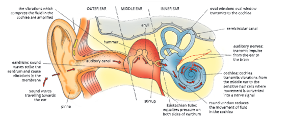
- The partition which contains two small openings namely, an oval window and a round window.
The middle ear contains three tiny bones called ear ossicles. These bones are named malleus (or hammer), incus (or anvil), and stapes (or stirrup). The handle of the malleus is attached to the internal surface of the eardrum (tympanum). Its opposite end is connected with the incus. The incus is the intermediate bone and it articulates with the head of the stapes. - The flat part of the stapes fits into the oval window. Directly below the oval window is another opening called a round window. The round window is covered by a thin membrane.
Ear ossicles (malleus, incus, and stapes) transmit sound vibrations to the internal ear.
- The anterior wall of the middle ear contains an opening that leads directly into the auditory tube (also known as the Eustachian tube). This connects the middle ear with the throat. The Eustachian tube also helps in equalizing air pressure on both sides of the eardrum helping it for free vibration.
- Throat infection can lead to an ear infection.
This is because the Eustachian tube connects the middle ear with the throat. Thus, any infection in the throat can pass to the ear through the Eustachian tube.
- Internal ear
The internal or inner ear is also called the membranous labyrinth. It has two main parts – cochlea and semicircular canals.
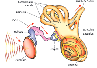
Cochlea: It is a hollow, spiral-shaped (coiled), chamber. It resembles a snail’s shell. It consists of a bony spiral canal that makes about 23%4 turns around a central bony core. Its inner spiral cavity contains three separate channels or canals that run parallel.
These canals are separated by membranes. The median (cochlear) canal is filled with endolymph and the outer two canals are filled with a fluid called perilymph.
- The middle canal contains a spiral organ called the organ of Corti. The organ of Corti is the organ of hearing. It contains a series of nerve cells and hair cells that join the auditory nerve and help in hearing.
- The hair cells of the organ of Corti are very sensitive to sound. They can be damaged by exposure to high-intensity noises such as noise produced by engines of jet planes, loud music, etc.(b) Semicircular canals: The inner ear also contains three semicircular canals. These canals are arranged at right angles to each other in three different planes (one horizontal, two vertical) and are filled with endolymph. One end of each canal is swollen to form an ampulla. The ampulla contains sensory cells which help in the balance of the body
- while moving. The nerve fibers arise from these cells and join the auditory nerve.
- There is a short stem (vestibule) that joins the semicircular canals and cochlea. It contains two small sacs – the utriculus and sacculus. These also contain tiny hair-like sensory cells which help in the static balance of the body while at rest (i.e. position of the head when not in motion like standing).
- The external ear, middle ear, and cochlea help in hearing. The sacculus, utriculus, and semicircular canals help in the sense of balance. A summary of various parts of the human ear and their functions is given in the table.
UP Board Notes for Class 10 Science Chapter 11 Sense Organs – The Eye And The Sense Of Sight Mechanism Of Hearing And Balance
Hearing
Pinna collects and amplifies sound waves which then pass along the auditory canal to the eardrum.
- Sound waves strike the eardrum (tympanum) and cause vibrations in its thin stretched membrane. The Eustachian tube equalizes air pressure on either side of the eardrum which allows a free vibration.
- The vibrations reach ear ossicles in the middle ear. Ear ossicles transmit vibrations from the eardrum to the denser fluid in the inner ear.
- The lever-like action of malleus and incus magnifies the vibrations of the stapes. The vibrating stapes transmit vibrations to the membrane of the oval window.
- Vibrations from oval window get transmitted to cochlea. This leads to vibration in the fluid of cochlear canals. Vibration of fluid in cochlear canals triggers movement of sensory hair cells of organ of Corti in cochlea.
- Movement of sensory hair cells is converted to a nerve impulse.
- Nerve signal or impulse is transmitted to brain via auditory nerve and this results in hearing. Balancing
The following parts of ear are involved with balance.
- Static balance with respect to center of gravity: Sensory cells in vestibule
- Dynamic balance (while the body is in motion): Sensory cells in semi-circular canals
Movement of fluid inside semicircular canals triggers sensory hair cells of ampulla. This sensation passes to nerve cells and then to brain. The three canals are at right angles to each other so that the brain can detect even slightest tilting of the body in any direction.
UP Board Notes for Class 10 Science Chapter 11 Sense Organs – The Eye And The Sense Of Sight Summary
Receptors are the special cells that receive stimuli from the environment. Some common receptors are photoreceptors, chemoreceptors, thermoreceptors, and mechanoreceptors.
- Receptors are contained in sense organs. There are five major sense organs in humans eyes (vision), ears (sound), nose (smell), tongue (taste), and skin (pressure and touch). Each of these sense organs is directly connected to the brain.
- Each eye is in the form of a ball called eyeball that can be rotated by six distinct sets of muscles.
- Eyebrows, eyelids, eyelashes, and tear glands are the accessory structures of the eye.
- Lacrimal glands are a group of glands that manufacture tears. Tears serve as a lubricant and also kill the germs that enter the eye.
- The eyeball has three layers – the sclerotic layer, the choroid, and the retina.
- The sclerotic layer is the outer tough coat of eyeball that is divided into sclera (white coat of dense fibrous tissue) and cornea (transparent fibrous coat through which the iris can be seen).
- The choroid layer is the middle layer of the eyeball and is composed of three parts – choroid, ciliary body, and the iris. There is a hole in the center of iris called the pupil.
- The retina is the third and inner coat of the eye. It contains light-sensitive cells called rods (sensitive to dim light) and cones (sensitive to bright light).
- The lens focuses light rays from outside and a real, inverted image is formed at yellow spot. The yellow spot is the area of best vision, while blind spot is the area of no vision.
- In myopia, the image is formed in front of retina.It can be corrected by using a concave lens.
- Ear is concerned with two functions – hearing and body balance.
- Set eardrum into vibration. It sets malleus, incus and stapes (ear ossicles) into motion. From stapes, vibrations move to oval windows, then to cochlear canal and sensory hairs in cochlea. From here the nerve impulse reaches the brain through the auditory nerve
UP Board Notes for Class 10 Science Chapter 11 Sense Organs – The Eye And The Sense Of Sight Sense Organs Structured/Application/Skill Type Questions
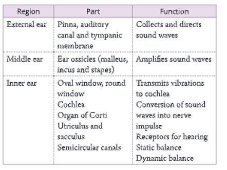
1. The diagram given below depicts a defect of the human eye. Study the same and then answer the following questions.
1. Name the defect shown in the diagram.
2. Give two possible reasons for this defect.
3. Name the parts labeled I to iv.
4. Name the type of lens used to correct this eye defect.
5. Draw a labeled diagram to show how the above-mentioned defect is rectified using the lens named
above
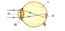
2. The following diagram represents a part of the human ear. Study the same and then answer the following questions.
- Give the biological terms for malleus, incus, and stapes.
- Name the parts labeled I, ii, and iii in the diagram.
- State the functions of the parts labeled I and ii.
- Name the audio receptor region present in the part labeled i.
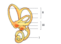
3. The diagram given below represents the vertical section of the human eye.
- Name the parts labeled I to xii.
- What is the function of the part labeled x?
- What would happen if part v is damaged or cut?
4. The diagram of the human ear is given below. Study the same and then answer the questions that follow.
- What role does the eardrum play in hearing?
- What common term is given to the parts labeled i, ii, and v?
- Would there be any difference if these three parts mentioned in Q(2) above were replaced by one big one? Why?
- Give the biological terms for the parts labeled iii and iv.
- Name the fluid which fills the parts mentioned in Q(4) above.
- State the functions of the ear.
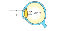
5. The following diagram represents a defect of vision of the human eye.
- Name the defect.
- What is the effect of this defect on man?
- Mention two causes for this defect.
- How can this defect be rectified?
- Draw a neat labeled diagram to show how this defect can be rectified.
- What is the nature of the image that falls on the retina of a normal eye?
6. The diagram given below refers to the vertical section of the eye of a mammal. Label the parts i to x to which the guidelines point.
7. The following diagram given below refers to the ear of mammals.
- Label the parts i to x to which the guidelines point.
- Name the structure which
- converts sound waves into mechanical vibrations.
- converts vibrations into nerve impulses.
- Responds to changes in position.
- Transmits impulses to the brain.
- Equalizes pressure in the ear.
8. The diagram given below represents the parts of the human ear.
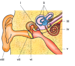
- Name the parts numbered i to viii.
- What is the function of the parts labeled ii and vii?
- Why is it harmful to use a part or any sharp object to remove the wax from the ear?
9. The diagram given below depicts a defect of the human eye. Study the same and then answer the following questions:
- Name the defect shown in the diagram.
- What are the two possible reasons that cause this defect?
- Name the type of lens used to correct this defect.
- With the help of a diagram showing how the defect shown above is rectified using a suitable lens.
10. Draw a diagram of the human eye as seen in a vertical section and label the part which suits the following functions/descriptions:
- The layer which prevents reflection of light.
- The structure alters the focal length of the lens.
- The region of distinct vision.
- The part which transmits the impulse to the brain.
- The outermost transparent layer in front of the eye lens.
- The fluid present in the anterior part of the eye in front of the eye lens.
- The structure is responsible for holding the eye lens in its position.
- The structure maintains the shape of the eyeball and the area of no vision.