
UP Board Notes For Class 10 Science Chapter 13 Reproductive System Learning Objectives
After completing this chapter, you will be able to:
- Define reproduction and differentiate between asexual and sexual reproduction;
- Illustrate male and female reproductive systems in humans and state the functions of each part;
- Draw labelled diagrams of male and female reproductive systems;
- Describe the histology of human testis and ovary;
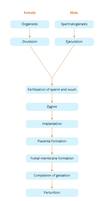
- Describe the main events in the process of reproduction In humans, starting from the production of gametes to pregnancy and childbirth;
- Describe the process of exchange of nutrients and respiratory gases between an embryo and Its mother,
- Explain how twins are produced.
Reproduction is a process by which a living organism is able to produce more of its own
kind. Reproduction involves the transmission of genetic material from the parents to the children, thereby ensuring that characteristics not only of the species but also of the parents, are perpetuated. Reproduction ensures continuity of life and survival of a species on the earth. In this chapter, you will learn about the organs for reproduction, fertilization, menstruation, and development of embryo in human beings.
UP Board Notes For Class 10 Science Chapter 13 Reproductive System Types Of Reproduction
There are two types of reproduction in living organismis – asexual and sexual reproduction.
Asexual reproduction involves the production of offspring from a single organism without the fusion of gametes. It is a common process of reproduction in lower group of plants and lower group of animals.
Sexual reproduction is a type of reproduction in which both sexes, the male and the female, are generally involved. It is the production of offspring by the fusion of the genetic material contained in the sex cells or gametes. As a result of fertilization, the male and the female gametes unite to form a zygote, which develops into an organism. Most animals and higher group of plants multiply by sexual reproduction.
| Class 10 Science | Class 11 Chemistry |
| Class 11 Chemistry | Transformation of Sentences |
| Class 8 Maths | Class 8 Science |
UP Board Notes For Class 10 Science Chapter 13 Reproductive System Sexual Reproduction In Humans
Humans reproduce sexually. The reproduction in humans can be studied in two parts –
- reproductive system in human beings, and
- fertilization, pregnancy and development of the embryo.
UP Board Notes For Class 10 Science Chapter 13 Reproductive System In Human Beings
The reproductive organs in human beings become functional after attaining sexual maturity. The period during adolescence in which the body attains sexual maturity is called puberty. Girls (females) attain puberty earlier than boys (males). In males, sexual maturity is attained at an age of 13-14 years. In females, onset of menstruation takes place around the age of 13 years. This age is known as the age of puberty. During sexual maturity, hormonal changes take place in males and females, and under the influence of these hormones, secondary sexual characteristics are developed.
Secondary sexual characteristics in males develop under the influence of testosterone, a hormone secreted by testes in males. These characteristics Include deepening of voice, widening of shoulders, appearance of beard and moustache, growth of axillary and pubic hair, enlargement of external genital organs and formation of sperms.
Secondary sexual characteristics in females include growth of axillary and pubic hair, widening of pelvis and hip, enlargement of breasts and onset of the menstrual cycle.
Primary reproductive parts and accessory reproductive parts
Primary reproductive parts include gonads (testes in males and ovaries in females). These are the sites of gamete production – sperms in testes and eggs in ovaries.
Accessory reproductive parts include all other structures that help in transfer of sex cells (gametes) and their fusion leading to fertilization and also growth and development of zygote till the birth of the baby.
UP Board Notes For Class 10 Science Chapter 13 Reproductive System Male Reproductive System
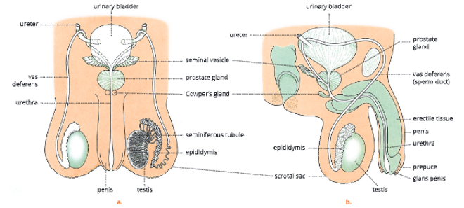
The male reproductive system consists of the following organs – a pair of testes, a pair of epididymis, a pair of sperm duct (vasa deferentia; singular: vas deferens), urethra, penis and accessory glands.
Testes
- Testes (singular: testis) are the male gonads. A pair of testes are present in a human male.
- These are present in a thin pouch made up of skin and connective tissue called scrotal sac or scrotum. Thus, testes are extra-abdominal. The scrotum is divided into right and left compartments by a muscular septum. One testis lies in each compartment.
- In the embryonic stage, the testes are contained within abdomen. They descend down just before birth.
- The high temperature of body does not allow maturation of sperms. Thus, the scrotum acts as thermoregulator, and it helps in maintaining the temperature at about 2-3°C lower than the body temperature. This temperature is suitable for the development of sperms.
UP Board Notes For Class 10 Science Chapter 13 Reproductive System Structure Of Testes
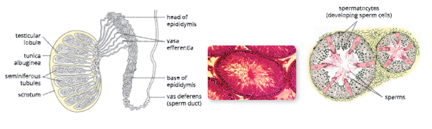
- Each testis is encased in a capsule of white fibrous connective tissue called tunica albuginea. This tissue extends internally as septa, dividing the testis into 15-20 lobules.
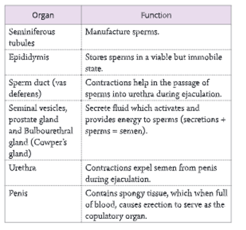
- Each testicular lobule has several highly coiled tubules called seminiferous tubules which are the sites of the maturation of spermatozoa. The process of maturation and differentiation of sperm inside the testes called spermatogenesis.
- Between the seminiferous tubules, clumps of interstitial cells (packing tissue), also called Leydig cells are present. These cells secrete the male sex hormone, testosterone. This hormone regulates the maintenance of primary and secondary sexual characteristics in males.
Epididymis
From each testis arises a network of ducts called efferent ducts. These open into a common tube-like structure. This single, 6 m long, highly coiled tube where sperms are stored, get concentrated and become physiologically (mature) active, is called epididymis. It remains attached to the testis and lies within the scrotal sac.
Vas deferens (Sperm ducts)
- Each epididymis continues from its lower end as a vas deferens. It enters the abdominal cavity through the inguinal canal, passes over the urinary bladder and joins the duct of seminal vesicle to form the ejaculatory duct. The ejaculatory duct opens into the urethra.
- The inguinal canal allows the descent of the testes along with their ducts, nerves and blood vessels from the abdomen to the scrotal sacs just before birth. Sometimes, because of high pressure in the abdomen, the intestine bulges into the scrotum through this inguinal canal and causes inguinal hernia.
Urethra
- The male urethra is about 15-20 cm in length and is differentiated into three parts – an anterior prostatic
- part which passes through the prostate gland, a middle membranous part, and a posterior penile part which passes through the copulatory organ, the penis.
- The male urethra functions as a passage for both semen and urine. The sperm ducts from each testis open into the urethra near its anterior end.
Penis
- Penis is the copulatory organ in males. It is a cylindrical, spongy and a highly vascular organ. It consists of erectile tissue permeated by blood sinuses. The urethra runs through it centrally and serves as a common passage for the exit of urine and semen. It is used to deposit spermatozoa in the female genital tract.
- During sexual excitement, the spongy tissue gets filled up with blood, as the arteries in it dilate. This condition is known as erection. Externally, the penis is covered by skin. The tip of the penis is soft and highly sensitive. It is called glans penis. It is covered by a loose retractile fold of skin called prepuce.
Accessory glands
There are three accessory glands in males. These include seminal vesicles, prostate gland and Cowper’s glands.
1. Seminal vesicles: A pair of seminal vesicles are present at the base of the urinary bladder. The function of the seminal vesicles is to secrete seminal fluid that acts as a medium of transport for the spermatozoa through the sperm duct. The seminal fluid is an alkaline viscous fluid which contains fructose, which provides energy to the sperms. This secretion forms about 60 per cent of the ejaculate. Sperms get active when mixed with seminal fluid.
2. Prostate gland: It is a single gland that surrounds the upper part of the urethra. The alkaline nature. of this fluid neutralizes the acid in the female tract which would otherwise inactivate and kill the sperms.
3. Cowper’s glands or lbourethra glands: These paired glands lie below the prostate gland and join the urethra at a short distance from that of the prostate gland. They secrete a white, viscous, alkaline secretion resembling mucus, which acts as a lubricant.
Spermatozoa and semen
The spermatozoa are minute gametes produced by the testes in males. They are immotile when stored in the epididymis but get activated and motile by the secretions from the accessory reproductive glands in males. The secretions of various accessory glands along with sperms form the semen.
The sperms are released in millions. In one ejaculation about 200,000,000 (2 x 10³) sperms are discharged. Sperms move with the speed of
2 mm/minute in the female tract.
Structurally, a human sperm has three main parts – head, neck and tail. The tip of a sperm is covered by a cap-like structure, acrosome, which helps the sperm to penetrate inside the egg during fertilization.
Acrosome also releases an enzyme (hyaluronidase) which enables the entry of the sperm into the egg by dissolving the proteinaceous wall around the egg. The general structure of a sperm is shown in.
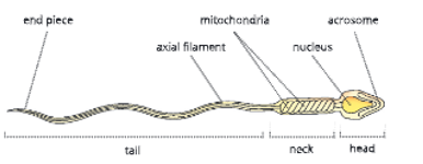
The nucleus of the sperm contains genetic material that is transferred to egg and mixes with the female nucleus during fertilization. The middle piece of the sperm contains mitochondria that provide energy for sperm penetration and sperm motility.
The tail helps the sperm in moving forward through the liquid medium while swimming.
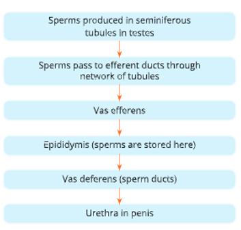
The course of sperm movement from their production in the testes to reach the urethra in penis is given in.
UP Board Notes For Class 10 Science Chapter 13 Reproductive System Female Reproductive system
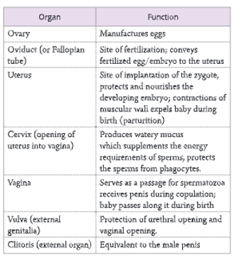
The female reproductive system consists of the following organs – a pair of ovaries, a pair of Fallopian tubes, uterus, vagina and external genitalia. Important functions of the main reproductive organs in the human female are listed in.
Ovaries
A pair of almond-shaped or ovoid-shaped ovaries lie in the lower part of the abdominal cavity, one on each side of the uterus. Each ovary is attached to the

Oogenesis
Eggs can be observed in various stages of development inside the ovary. The egg mother cells or oogonia are diploid cells that undergo meiosis to produce the haploid egg or ovum.
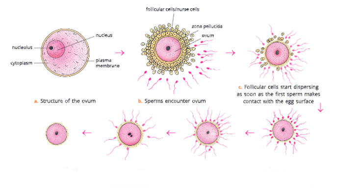
- During the process of development the egg gets surrounded by some special cells called
follicular cells or nurse cells. Nurse cells provide nourishment to the developing egg cell. - The egg cell along with its surrounding nurse cells is called a follicle.
- Towards the end of oogenesis, the follicle develops a fluid-filled cavity amongst the nurse cells and is called the tertiary follicle or Graafian follicle.
- Upon maturation, the follicle ruptures, the ovarian wall ruptures and the ovum is released out of the ovary. This egg is sucked by the funnel of the oviduct or Fallopian tube. The egg is in the secondary oocyte stage as the meiosis is not yet complete. It has a proteinaceous layer around it called the zona pellucida.
Fallopian tubes (Oviducts)
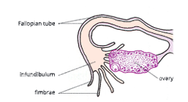
There are two oviducts or Fallopian tubes or uterine tubes in the human female reproductive system. Each oviduct is about 10-15 cm long. The proximal funnel-shaped end of each oviduct lies near the ovary and is called infundibulum.
Its margin bears finger-like projections called fimbrae. Each infundibulum continues as a thin and coiled tube called oviduct or Fallopian tube. Both Fallopian tubes open into the uterus.
The egg released from the ovary is sent into the Fallopian tube by the infundibulum. The Fallopian tube transports the ovum from the ovaries into the uterus by cilia along the wall of funnel and peristaltic contractions of the muscles of the tube wall. Fertilization of ovum and sperm is taking place in Fallopian tube.
Uterus
The uterus is a hollow, pear-shaped, muscular, thick- walled organ located in the pelvic cavity between urinary bladder and rectum. Its upper broader portion is called corpus uteri (body of the womb) and the lower narrow portion is called cervix uteri (neck of the womb).
The innermost wall of the uterus is called endometrium. It is richly supplied with blood vessels as implantation of the embryo takes place in the endometrium. Cervix is mainly a sphincter muscle that closes the lower end of the uterus where it joins the vagina.
Vagina
It is a muscular tube, about 7-10 cm in length. Vagina is the organ where the spermatozoa are deposited during coitus (act of copulation) by the penis. It serves as the birth canal during childbirth (parturition) and also acts as a duct for the passage of uterine secretions and menstrual flow.
The opening of the vagina in young girls is partially closed by a thin membrane called hymen.
External genitalia (Vulva)
The external genitalia in human female (also called vulva) mainly consists of labia majora, labia minora and clitoris.
The labia majora (large lips) is a pair of thick folds of skin containing hair, sweat glands and sebaceous. glands. These folds lie one on either side of the vagina. The labia majora is equivalent to scrotum in males.
The labia minora (small lips) are two small folds of mucous membrane situated on the inner side of labia majora. They are devoid of hair and glands. The two folds of labia majora and labia minora, comprise the vulva. The clitoris is a small erectile organ which
is highly sensitive and is located at the junction of the labia minora. The clitoris corresponds to the penis in males.
The vagina opens to the outside by an opening into the depression between the vulva called vestibulum. In a human female, the urethra and the genital duct have separate openings.
UP Board Notes For Class 10 Science Chapter 13 Reproductive System Menstrual cycle in human females
In a human female, the fertility period extends from the age of puberty, i.e. about 12-13 years up to menopause, i.e. 45-50 years. The stage of puberty is marked by the appearance of secondary sexual
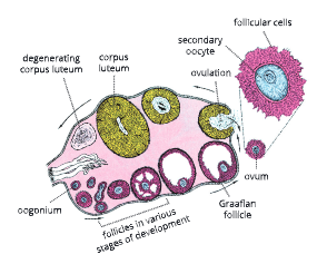
Transverse section through an ovary
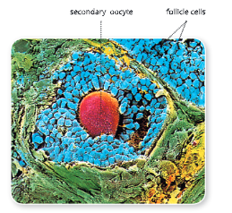
Scanning electron micrograph of egg surrounded by follicle cells
characteristics. Thus, puberty is a period in which reproductive system matures and becomes capable for reproducing. At the time of menopause, ovulation and menstruation stop and the reproductive organs decrease in size.
During each menstrual cycle, an ovum is matured and released once every 28 days. The period of menstrual cycle is counted from the day of onset of menstrual flow to the next onset after about 28 days. There are four phases of menstrual cycle as given below.
Menstrual phase
The menstrual cycle starts with the menstrual flow, during which the cellular lining of the uterus, with blood flow, is shed off. This process continues for 3-5 days. During menstrual phase, the ovary starts to process a new egg in follicle.
Follicular phase
From the 5th day up to the 13th day of the onset of menstrual cycle, growth and maturation of the Graafian follicle take place. Graafian follicle is the final stage in the maturation of an ovum inside the ovary.
It consists of an ovum and a mass of nurse cells or follicular cells surrounding it. The Graafian follicle produces a hormone, oestrogen. This hormone stimulates the uterus to prepare itself to receive the ovum. The cells lining the uterus grow rapidly and develop a dense network of blood vessels.
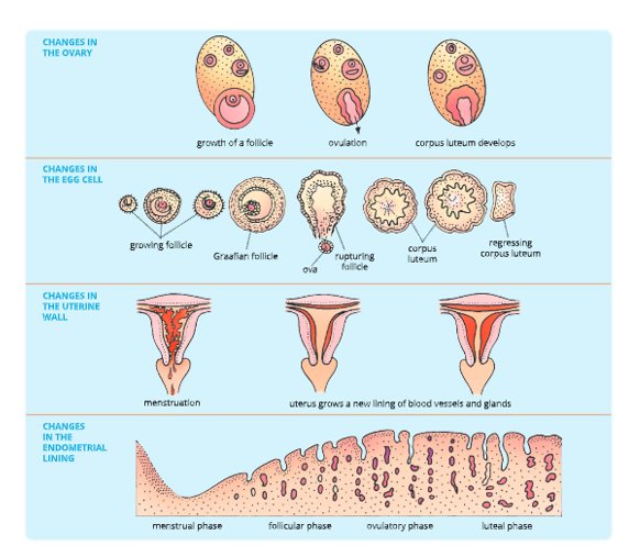
Ovulatory Phase
In this phase, ovulation takes place. The Graafian follicle ruptures to release the ovum. The cells of the ruptured follicle form the corpus luteum that secretes the hormone, progesterone.
The release of the ovum from the ovary is called ovulation. The ovum reaches the uterus via the Fallopian tube on the 13th or 14th day and remains = there up to the 16th day (for 48-72 hours).
Luteal Phase
If the ovum does not get fertilized by any sperm during the ovulatory period then it degenerates. At the end of the 28th day, this ovum is rejected along with the uterine lining.
This marks the onset of the disintegration of the thickened endometrial lining of the uterus – the next menstrual cycle. The remnant corpus luteum develops of the Graafian follicle in the ovary turns into corpus luteum.
If the ovum gets fertilized while in the Fallopian tube, it starts dividing by repeated mitotic divisions called cleavage divisions. The egg is now an early embryo. It reaches the uterus within 5-7 days of fertilization and implants itself in the thick endometrium.
The female is now pregnant and shall give birth to a baby after the development of embryo in 38-40 weeks. This is the gestation period.
What happens to the menstrual cycle if the ovum receives sperm?
If the ovum receives sperm, menstruation (and ovulation) cease for as long as the woman is pregnant. This is because level of progesterone increases by the corpus luteum (which persists in the ovary) and later by the placenta. Progesterone prevents egg maturation in the ovary.
UP Board Notes For Class 10 Science Chapter 13 Reproductive System Fertilization, Pregnancy And Development Of The Embryo
- Fertilization: The sexual intercourse, also known as coitus, is the first step towards pregnancy.
- During intercourse, the semen containing sperms is ejaculated in the vagina. This is known as copulation. A single ejaculation may contains 2-4 hundred million sperms, only one will actually fertilize the egg.
- The spermatozoa are deposited high up in the vagina close to the cervix. They reach the top of the Fallopian tube within 5 minutes of their release due to their motility and contractions in the walls of uterus and Fallopian tube. Spermatozoa remain viable in the female genital tract for 24-72 hours.
- If the ovum receives a sperm during the ovulatory period, the two fuse to form a zygote. This act of fusion of the male gamete (sperm) and the fernale gamete (ovum) to form zygote is called fertilization. About 13-15 days of the menstrual cycle are most favourable for conception. Of the millions of sperms ejaculated in the vagina, the first one to reach the ovum fertilizes it. Only one sperm can fertilize the ovum. This is possible due to a protein coat formed around the ovum soon after the first sperm comes in contact with the ovum.
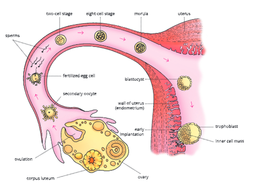
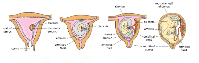
- Implantation: The zygote immediately begins to divide by rapid mitotic divisions called cleavage. The daughter cells produced by cleavage divisions get smaller and smaller progressively. Successive cleavages produce a solid mass of cells called the morula which appears like mulberry on the outside. It’s volume is the same as that of the zygote. With further development, this ball of cells becomes hollow and is called blastocyst. The cells of blastocyst are now differentiated into two types.
- Trophoblast: outer compact layer cells
- Inner Cell Mass (ICM): inner mass of cells located at one end of the hollow ball. Also called embryoblast cells.
The blastocyst is still enclosed in Zona Pellucida which was present around the ovum at ovulation. The blastocyst passes down to the uterus and fixes itself to the endometrium wall of the uterus. The fixing of blastocyst in the endometrium of the uterus is called
implantation and the female is said to be pregnant or in the stage of pregnancy. Implantation takes place about a week after fertilization.
How does the blastocyst implant in the uterine wall?
Site of implantation: Endometrium of the uterine wall Time of implantation: 5-7 days of fertilization
UP Board Notes For Class 10 Science Chapter 13 Reproductive System Process Of Implantation
- Trophoblast cells secrete enzymes that perforate the Zona pellucida and the blastocyst oozes out.
- The special trophoblast cells secrete enzymes that digest and liquefy the endometrial cells at a point creating a pit.
- Blastocyst starts burrowing itself into the pit.
- Eventually, the blastocyst gets completely buried in the endometrium.
- Blastocyst is oriented in such a direction that the ICM faces towards the wall.
- Trophoblast develops further to form a connection between the embryo and the mother called the placenta.
Placenta
The developing embryo is attached to the uterine wall by the placenta. Placenta is an association between maternal and foetal tissue meant for physiological exchange. Umbilical cord is a tough structure that serves as the blood-vascular connection between the foetus and placenta. It develops from embryonic cells.
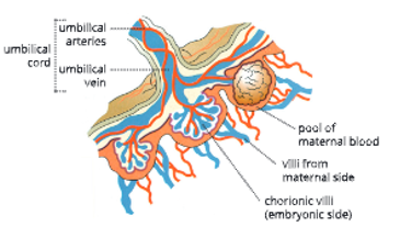
The placenta is formed of fine finger-like processes called villi. There are two sets of villi. One set of villi is given out by the uterine wall which opens up in a pool or sinusoid of blood of mother while the other set is an extension from the embryo.
These two sets of villi are interlocked but they do not open into each other. Thus, although the blood of the mother and the embryo are in close contact with each other, the two do not get mixed.
Oxygen and nutrients diffuse from the mother’s blood into the capillaries of the villi. From here they
are carried by the blood vessels in the umbilical cord to the foetus. Conversely, carbon dioxide and other wastes are carried back by foetal blood and transferred to the mother’s blood through the villi in placenta.
The villi from the embryonic side arise from the trophoblast cells and get lined by chorion and not allantois in humans, which is the outermost lining of the developing embryo.
UP Board Notes For Class 10 Science Chapter 13 Reproductive System Functions Of Placenta
- Nutrition: Placenta serves as a tissue through which oxygen and nutrients (glucose, amino acids and salts) are supplied from the maternal blood to the foetus.
- Excretion: It also transports carbon dioxide and excretory waste from the foetal blood to the maternal blood.
- All these substances move across the placenta by diffusion. Placenta is permeable to respiratory gases, nutrients and antibodies. It prevents harmful material from reaching the embryo. It does not allow the passage of germs from the mother to the embryo. However, certain exceptions like German measles virus and HIV can pass through the mother’s blood to the embryo.
- Endocrine: Placenta also produces two hormones – progesterone and oestrogen. Under the influence of these hormones, neither ovulation nor menstruation takes place till pregnancy continues. However, these phenomena are resumed after childbirth.
Foetal membranes
During development, the embryo gets surrounded by four special membranes which protect and nourish it. These are as follows:
- Yolk sac: contains nutrients
- Allantois: acts as a waste bag
The first two are non-functional in humans as this work is done by placenta.
- Chorion: outermost membrane, takes part in placenta formation from the embryonic side.
- Amnion: a membranous sac filled with amniotic fluid, completely envelops the developing foetus.
UP Board Notes For Class 10 Science Chapter 13 Reproductive System Functions Of Amnion
- Acts as a shock absorber for the developing foetus and protects from any physical damage.
- Assists in regulation of foetal body temperature.
- Prevents adhesion (sticking) of skin of foetus to the surrounding tissues.
- Allows movements of the foetus in the mother’s womb, in a restricted manner.
Amniocentesis Embryonic cells are sloughed off in the amniotic fluid. These cells found in the amniotic fluid are studied by a technique called amniocentesis. In this method, some amount of amniotic fluld is taken by a surgical needle inserted into the amniotic sac through the abdomen.
The embryonic cells are studied for any genetic abnormalities in the child months before birth, by 16-18 weeks of development.
Gestation and parturition
Parturition is the process of childbirth. The sequence of events that occur during childbirth is called labour.
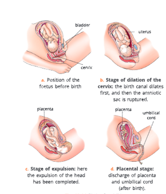
What happens during childbirth?
During Childbirth
- The uterus undergoes rhythmic contractions. The amnion bursts and the amniotic fluid is discharged.
- The uterus contracts powerfully, expelling the baby.
- The baby’s lungs start functioning and the baby takes its first breath.
- The umbilical cord is tied and cut.
- The lung bypasses close so that blood flows to the lungs.
- The placenta is discharged (after birth).
- The breasts start producing milk.
The period of complete development of the foetus from the beginning of the last menstrual period till birth of the baby is called gestation period. It is of about 280 days.
Test tube babies Some women are unable to have babies due to certain reasons. This problem can be overcome by the test tube baby technique.
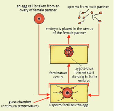
In this technique, one or more ripe ova are sucked from a woman’s ovaries using a special syringe. These ova are placed in a dish containing sperms from her partner. During this time, sperm fertilizes the ovum to form zygote which divides to form an embryo. The embryo is then inserted into the woman’s uterus where there is a chance it will implant and develop into a baby.
The entire process is performed under optimum conditions in a laboratory. The general notion is that people in a laboratory work with test tubes, hence the name test tube baby.
UP Board Notes For Class 10 Science Chapter 13 Reproductive System How Twins Occur?
Occurrence Of Twins
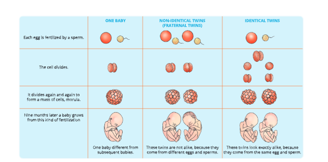
Usually, only one ovum is released by an ovary in every reproductive cycle. If this ovum gets fertilized, one baby is born. But sometimes more than one egg may be released and fertilized. Or an ovum may divide into two or more separate cells after fertilization. This is how twins, triplets, quadruplets, etc. are produced.
Identical twins
Identical twins are the result of a fertilized egg/zygote separating into two cells after the first cleavage division. Both of which continue to divide, so two identical embryos come from the same egg and sperm. Such twins look alike and are of same sex. Since identical twins develop from the same zygote, these are called monozygotic twins.
Non-identical or fraternal twins
Non-identical twins occur when two eggs are produced at the same time and each is fertilized by a different spermatozoa. These twins do not resemble each other physically. They can be of the same sex or different sexes. Since fraternal twins develop from two different zygotes, they are called dizygotic twins.
UP Board Notes For Class 10 Science Chapter 13 Reproductive System Summary
- Reproduction is a process by which a living organism is able to produce more of its own kind.
- In sexual reproduction, both male and female gametes are produced and the process of fertilization takes place. The humans reproduce sexually.
- The age of 13-14 years in males and 12-13 years in females is called puberty. At this age, sex organs get matured and several secondary sexual characteristics appear in them.
- The male reproductive system consists of – a pair of testes, a pair of epididymis, a pair of vasa deferentia, urethra, penis and accessory glands.
- The female reproductive system consists of a pair of ovaries, a pair of Fallopian tubes, uterus, vagina and external genitalia.
- The fusion of male gamete (sperm) and female gamete (egg) to form a zygote is called fertilization.
- The fixing of a fertilized egg in the form of mass of cells (blastocyst) in the uterine wall is called implantation.
- Placenta Is an association between maternal and foetal tissues meant for physiological exchange.
- Amnion contains amniotic fluid which surrounds the foetus in placenta and acts as a shock absorber.
- Twins are of two types – fraternal and identical twins.
1